
Endocrine Pathology — MCQs

On this page
Which of the following is a fibrosing type of thyroiditis?
Which of the following findings is not seen in hyperparathyroidism?
Papillary carcinoma of the thyroid primarily spreads via which route?
Subperiosteal bone resorption is seen in which of the following conditions?
Which of the following is the most characteristic ultrastructural feature of paraganglioma on electron microscopy?
A 21-year-old pregnant woman experiences abruptio placentae at 37 weeks of gestation and develops severe vaginal bleeding that is difficult to control. Five months later, the patient presents with profound lethargy, pallor, muscle weakness, failure of lactation, and amenorrhea. Which of the following best explains the pathogenesis of pituitary insufficiency in this patient?
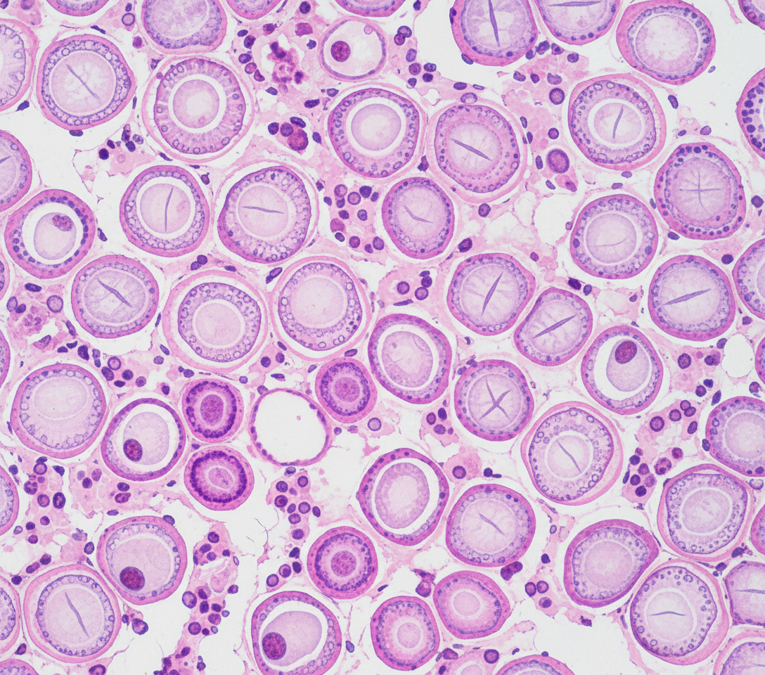
A 24-year-old male presented with a swelling on the anterior aspect of the neck. On examination, the swelling was firm and moved with deglutition. A biopsy from the lesion is shown. What is the typical finding and diagnosis?
Orphan Annie-eye nuclei are seen in which of the following conditions?
What is characteristic of parathyroid carcinoma?
All of the following are true about medullary carcinoma of thyroid except?
Practice by Chapter
Pituitary Gland Disorders
Practice Questions
Thyroid Gland Diseases
Practice Questions
Parathyroid Gland Pathology
Practice Questions
Adrenal Cortical Disorders
Practice Questions
Adrenal Medullary Disorders
Practice Questions
Pancreatic Endocrine Disorders
Practice Questions
Multiple Endocrine Neoplasia Syndromes
Practice Questions
Diffuse Neuroendocrine System
Practice Questions
Pineal Gland Pathology
Practice Questions
Laboratory Diagnosis of Endocrine Diseases
Practice Questions
Want unlimited practice?
Get full access to all questions, explanations, and performance tracking.
Scan to download app