Which of the following is true about the development of thyroid tumors in nodular goiter?
In which condition is thymic hyperplasia commonly observed?
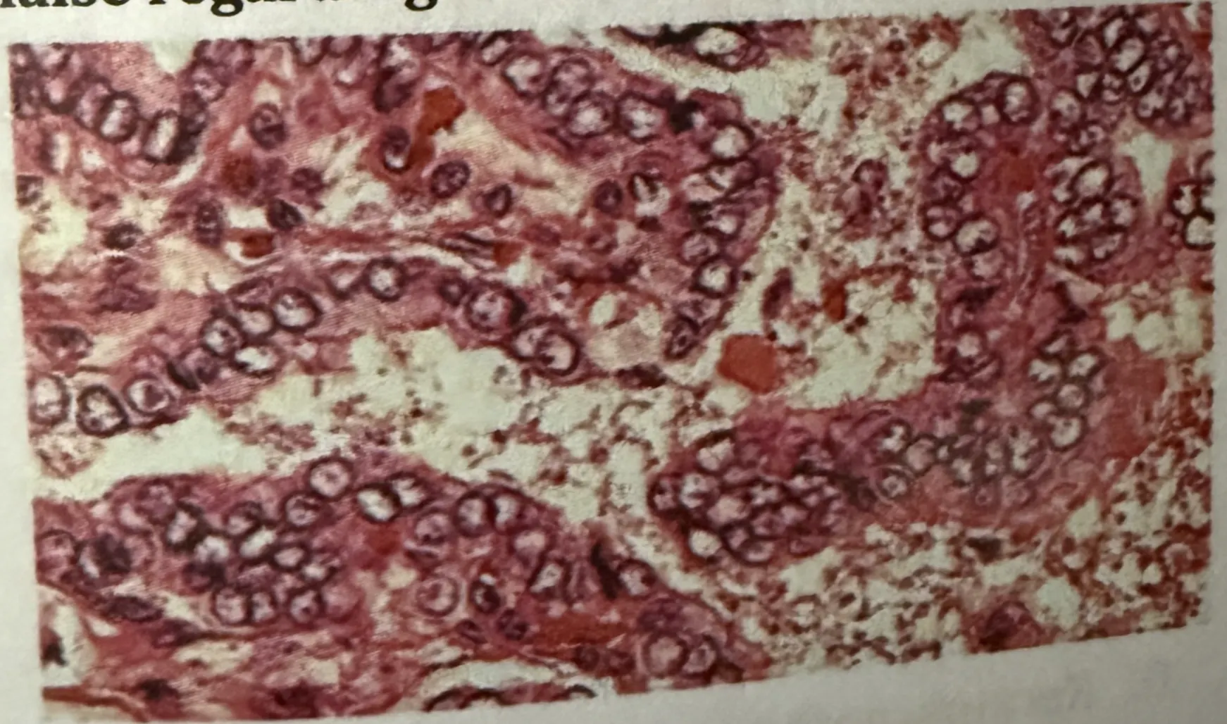
A middle aged male patient presents with painless slow growing neck swelling. On examination, lymph nodes are positive. Surgery is done and biopsy is shown in the image below. Which of the following is false regarding the HPE findings?

A 30-year-old woman presents with thyroid swelling. On investigations, her TSH levels are found to be elevated. Postoperative reports showed lymphocytic infiltration and Hurthle cells. A most probable diagnosis is?
All of the following are features of granulomatous thyroiditis except?
Orphan Annie nuclei are characteristic of which of the following?
Which of the following is a definitive feature of malignant pheochromocytoma?
Which of the following thyroid carcinomas cannot be definitively diagnosed by fine needle aspiration cytology (FNAC)?
What is the most common thyroid tumor associated with multiple endocrine neoplasia (MEN)?
All of the following are histological features of Hashimoto thyroiditis, except which of the following?
Explanation: ***The risk of development of carcinoma is not correlated with the level of TSH*** - Higher levels of **TSH (Thyroid Stimulating Hormone)** can actually stimulate proliferation of follicular cells, thus potentially increasing the risk of **thyroid carcinoma** [1]. - Consequently, lower TSH levels often correlate with lower risks, contradicting the notion that there is **no correlation**. *Both benign and malignant neoplasms can be seen in patients with nodular goiter* - This statement is **true** as nodular goiter can indeed contain both **benign** (e.g., adenomas) and **malignant** lesions. - The occurrence of neoplasms is a recognized complication in the **multinodular goiter** setting. *Prevalence of thyroid carcinoma ranges between 5-15% in the patients with multinodular goiter* - This statement is also **true**; studies show that the prevalence of thyroid cancer in **multinodular goiter** can be in this percentage range. - Routine evaluations often reveal malignancies within this spectrum in patients undergoing surgery for **multinodular goiter**. *Papillary carcinoma is the most common carcinoma developed in patients with nodular goiter* - This statement is **true**, as **papillary thyroid carcinoma** accounts for the majority of thyroid cancers arising in patients with nodular goiter [1]. - Its prevalence in nodular goiter cases supports the need for careful monitoring and management of thyroid nodules. **References:** [1] Kumar V, Abbas AK, et al.. Robbins and Cotran Pathologic Basis of Disease. 9th ed. The Endocrine System, pp. 1093-1094, 1098-1099.
Explanation: ***Myasthenia gravis*** - Thymic hyperplasia is commonly associated with **myasthenia gravis**, particularly in **thymic enlargement** cases [1]. - This autoimmune condition often results in **autoantibody production** against acetylcholine receptors due to changes in the thymus [1]. *Scleroderma* - This condition primarily involves **skin thickening** and is characterized by **autoimmune fibrosis**, with no direct association with thymic hyperplasia. - Thymic changes are not a common finding or related to the pathophysiology of scleroderma. *Thymoma* - While thymoma can cause **thymic enlargement**, it is characterized more by **tumor formation** rather than hyperplasia. - Thymoma is a tumor of the thymus itself, which can be associated with myasthenia gravis, but does not represent **hyperplastic changes**. *Thymic lymphoma* - This is a malignancy of the thymus that presents as a mass rather than **hyperplastic tissue**. - Thymic lymphoma typically results in **destructive processes** rather than dietary overgrowth or hyperplasia of thymic tissue. **References:** [1] Kumar V, Abbas AK, et al.. Robbins and Cotran Pathologic Basis of Disease. 9th ed. Diseases of White Blood Cells, Lymph Nodes, Spleen, and Thymus, p. 634.
Explanation: ***Fine needle aspiration cytology (FNAC) is not diagnostic*** - FNAC can often provide significant insights, but in cases of **specific malignancies** or certain lesions, it may not yield definitive diagnoses [1]. - Diagnostic challenges arise as **cellular architecture** or certain **nuclear features** may not be appreciated in FNAC samples [1]. *It spreads quickly via lymphatics* - This condition can indeed spread via lymphatics, making it **aggressive** in nature [1]. - **Lymphatic spread** is a common pathway for many head and neck conditions, particularly malignancies [1]. *Excellent prognosis is associated with this condition* - While some conditions may have favorable prognoses, many midline neck lesions can have **serious implications** depending on their nature [1]. - Prognosis often varies widely and may not always be classified as **excellent** based solely on initial presentation [1]. *Nuclear characteristics are used for the identification* - Nuclear morphology is critical for identifying various **neoplastic conditions**, aiding in differentiation from benign lesions [1][2]. - Many pathologies, especially those involving **malignancy**, rely heavily on **nuclear features** for accurate diagnosis [1][2]. **References:** [1] Kumar V, Abbas AK, et al.. Robbins and Cotran Pathologic Basis of Disease. 9th ed. The Endocrine System, pp. 1101-1102. [2] Kumar V, Abbas AK, et al.. Robbins and Cotran Pathologic Basis of Disease. 9th ed. The Endocrine System, pp. 1100-1101.
Explanation: ***Hashimoto's thyroiditis*** - The presence of **lymphocytic infiltration** and **Hurthle cells** on postoperative pathology is characteristic of Hashimoto's thyroiditis [1,2]. - Elevated **TSH levels** indicate hypothyroidism, which aligns with the autoimmune nature of Hashimoto's affecting thyroid hormone production [1]. *Graves disease* - Typically presents with **hyperthyroidism**, leading to suppressed TSH levels rather than elevation. - Characterized by **thyroid enlargement** and the presence of **autoantibodies** like TSI, not lymphocytic infiltration. *Follicular carcinoma* - While it can cause **thyroid swelling**, it is usually associated with **malignant characteristics** rather than Hurthle cells and lymphocytic infiltration. - TSH levels can be normal, as it does not principally engage in autoimmune thyroid destruction like Hashimoto's. *Medullary carcinoma thyroid* - Originates from **C cells** producing calcitonin, and typically presents with elevated calcitonin levels, not TSH. - Characteristic findings include **C-cell hyperplasia** or **neoplastic changes**, which do not match the presented lymphocytic infiltration. **References:** [1] Kumar V, Abbas AK, et al.. Robbins and Cotran Pathologic Basis of Disease. 9th ed. The Endocrine System, pp. 1090-1092. [2] Cross SS. Underwood's Pathology: A Clinical Approach. 6th ed. Common Clinical Problems From Liver And Biliary System Disease, pp. 427-428.
Explanation: ***Painless*** - Granulomatous thyroiditis is characterized by **painful** thyroid gland inflammation, which is a distinguishing feature. - Thus, describing it as **painless** contradicts the typical clinical presentation. *Hyperthyroidism* - Granulomatous thyroiditis may lead to **hyperthyroidism** initially due to the release of thyroid hormones from damaged follicles [1]. - However, this condition can also lead to transient thyroid function changes or even permanent hypothyroidism later on [1]. *Hypothyroidism* - While **hypothyroidism** can occur post-thyroiditis, it is not a feature of granulomatous thyroiditis at the outset like the **painless** descriptor. - This condition often starts with hyperthyroid symptoms and may evolve later, differing from primary hypothyroid disorders. *Giant cells on histology* - Histological examination typically reveals **multinucleated giant cells**, a hallmark of granulomatous inflammation, as seen in this thyroid condition. - This significant finding helps in differentiating it from other thyroid disorders. **References:** [1] Kumar V, Abbas AK, et al.. Robbins and Cotran Pathologic Basis of Disease. 9th ed. The Endocrine System, pp. 1091-1092.
Explanation: ***Papillary carcinoma thyroid***[1][2] - Characterized by **Orphan Annie nuclei**[1], which are large and round with a clear or empty appearance due to the presence of intranuclear cytoplasmic inclusions[1]. - Often associated with **thyroid follicular structures** and is the most common type of thyroid cancer[2]. *Meningioma* - Typically presents with **dural-based tumors** and does not exhibit Orphan Annie nuclei. - Histologically, it may demonstrate **whorled patterns** or calcifications instead. *Carcinoma pituitary* - Involves **adenomatous changes** in the pituitary gland but does not demonstrate the characteristic Orphan Annie nuclei. - More commonly shows **varied cellular morphology** depending on the type of secretory cells (e.g., prolactin, ACTH). *Paraganglioma* - Derived from **neuroendocrine cells**, and presents with **zellballen pattern** rather than Orphan Annie nuclei. - Often shows **chromaffin cells** and is typically associated with catecholamine secretion. **References:** [1] Kumar V, Abbas AK, et al.. Robbins and Cotran Pathologic Basis of Disease. 9th ed. The Endocrine System, p. 1099. [2] Cross SS. Underwood's Pathology: A Clinical Approach. 6th ed. Common Clinical Problems From Liver And Biliary System Disease, pp. 429-430.
Explanation: ***Presence of metastases in non-chromaffin tissues*** - The definitive diagnosis of malignant pheochromocytoma is based solely on the presence of **metastases** in sites where chromaffin cells are not normally found, such as bone, liver, lung, or lymph nodes. - Unlike many other cancers, histological features like pleomorphism, mitotic activity, or tumor size do not reliably distinguish between benign and malignant pheochromocytomas. *Capsular invasion* - While **capsular invasion** suggests more aggressive behavior, it is not a definitive criterion for malignancy in pheochromocytoma. - Benign pheochromocytomas can also exhibit some degree of capsular invasion, making it an unreliable indicator of metastatic potential. *Vascular invasion* - Similar to capsular invasion, **vascular invasion** indicates an increased risk of recurrence or more aggressive behavior, but it is not a singular definitive feature of malignancy. - The presence of tumor cells within blood vessels does not automatically confirm metastatic disease without evidence of spread to distant sites. *None of the options are definitive features of malignancy* - This statement is incorrect because the presence of metastases in non-chromaffin tissues is indeed the **definitive feature** for diagnosing malignant pheochromocytoma. - Without evidence of metastasis, distinguishing between benign and malignant pheochromocytoma based on other features is often challenging and unreliable.
Explanation: ***Follicular carcinoma of thyroid*** - The definitive diagnosis of **follicular carcinoma** requires the presence of **capsular or vascular invasion**, which cannot be assessed through **fine needle aspiration cytology (FNAC)** alone [1], [5]. - FNA may show features suggestive of follicular neoplasm (e.g., hypercellularity with microfollicles), but differentiation from **follicular adenoma** requires histological examination of the excised specimen [1], [4]. *Anaplastic carcinoma of thyroid* - **Anaplastic carcinoma** is highly aggressive and characterized by **pleomorphic, bizarre cells** that are easily identifiable on FNAC [2], [5]. - The distinctive cytological features, including **spindle cells, giant cells, and rapid cellular atypia**, allow for a relatively straightforward diagnosis via FNAC [2]. *Medullary carcinoma of thyroid* - **Medullary carcinoma** cells have characteristic cytological features, such as **plasmacytoid appearance**, **amyloid deposition**, and **neuroendocrine granules**, which can be identified on FNAC [5]. - Confirmation can be made by **immunohistochemical staining for calcitonin** on the FNA sample [5]. *Papillary carcinoma of thyroid* - **Papillary carcinoma** has distinct cytological features, including **orphan Annie eye nuclei**, **intranuclear grooves**, **pseudoinclusions**, and **papillary structures**, readily identified by FNAC [3]. - These features are highly specific and often allow for a definitive diagnosis of papillary thyroid carcinoma [3]. **References:** [1] Kumar V, Abbas AK, et al.. Robbins and Cotran Pathologic Basis of Disease. 9th ed. The Endocrine System, pp. 1100-1101. [2] Kumar V, Abbas AK, et al.. Robbins and Cotran Pathologic Basis of Disease. 9th ed. The Endocrine System, pp. 1101-1102. [3] Cross SS. Underwood's Pathology: A Clinical Approach. 6th ed. Common Clinical Problems From Liver And Biliary System Disease, pp. 429-430. [4] Cross SS. Underwood's Pathology: A Clinical Approach. 6th ed. Common Clinical Problems From Liver And Biliary System Disease, pp. 428-429. [5] Cross SS. Underwood's Pathology: A Clinical Approach. 6th ed. Common Clinical Problems From Liver And Biliary System Disease, pp. 430-431.
Explanation: ***Medullary*** - The **commonest thyroid tumor** in Multiple Endocrine Neoplasia (MEN) type 2 is medullary thyroid carcinoma, associated with **calcitonin production** [1]. - It arises from **C cells (parafollicular cells)** and is linked to **RET oncogene mutations** in MEN syndromes [1]. *Papillary* - Papillary thyroid carcinoma is the **most common thyroid cancer overall**, but not specifically associated with MEN syndromes. - It typically presents with **lymphatic spread**, whereas medullary carcinoma has a different genetic association. *Follicular* - Follicular thyroid carcinoma is less common in MEN and usually occurs sporadically. - It primarily arises from **follicular cells** and involves a different mechanism than medullary carcinoma. *Anaplastic* - Anaplastic thyroid carcinoma is a rare and highly aggressive form, not commonly associated with MEN. - It usually arises from **differentiated thyroid cancers** and presents in older patients, which does not align with MEN's typical presentations. **References:** [1] Kumar V, Abbas AK, et al.. Robbins and Cotran Pathologic Basis of Disease. 9th ed. The Endocrine System, pp. 1102-1103.
Explanation: ***Orphan Annie eye nuclei*** - This feature is **not seen** in Hashimoto thyroiditis; it is typically associated with **papillary thyroid carcinoma** [1][2]. - In Hashimoto thyroiditis, the presence of **Orphan Annie eye nuclei** would be considered **abnormal** and indicative of malignancy. *Hurtle cell metaplasia* - **Hurtle cells** can be found in Hashimoto thyroiditis but are not definitive; they are more associated with **thyroid follicular neoplasms**. - Presence of Hurtle cells indicates **metaplasia** but not a hallmark of Hashimoto thyroiditis specifically. *Lymphocytic infiltrate with germinal center formation* - This is a **typical histological feature** of Hashimoto thyroiditis and indicates an **autoimmune response**. - The presence of lymphocytes and germinal centers reflects **chronic inflammation** characteristic of this condition. *Follicular destruction and atrophy* - Follicular destruction is indeed a **key feature** of Hashimoto thyroiditis, leading to hypothyroidism. - Atrophic follicles occur due to **autoimmune-mediated damage**, distinct to Hashimoto thyroiditis. **References:** [1] Kumar V, Abbas AK, et al.. Robbins and Cotran Pathologic Basis of Disease. 9th ed. The Endocrine System, p. 1099. [2] Cross SS. Underwood's Pathology: A Clinical Approach. 6th ed. Common Clinical Problems From Liver And Biliary System Disease, pp. 429-430.
Pituitary Gland Disorders
Practice Questions
Thyroid Gland Diseases
Practice Questions
Parathyroid Gland Pathology
Practice Questions
Adrenal Cortical Disorders
Practice Questions
Adrenal Medullary Disorders
Practice Questions
Pancreatic Endocrine Disorders
Practice Questions
Multiple Endocrine Neoplasia Syndromes
Practice Questions
Diffuse Neuroendocrine System
Practice Questions
Pineal Gland Pathology
Practice Questions
Laboratory Diagnosis of Endocrine Diseases
Practice Questions
Get full access to all questions, explanations, and performance tracking.
Start For Free