
Endocrine Pathology — MCQs

On this page
RET proto-oncogene is associated with the development of
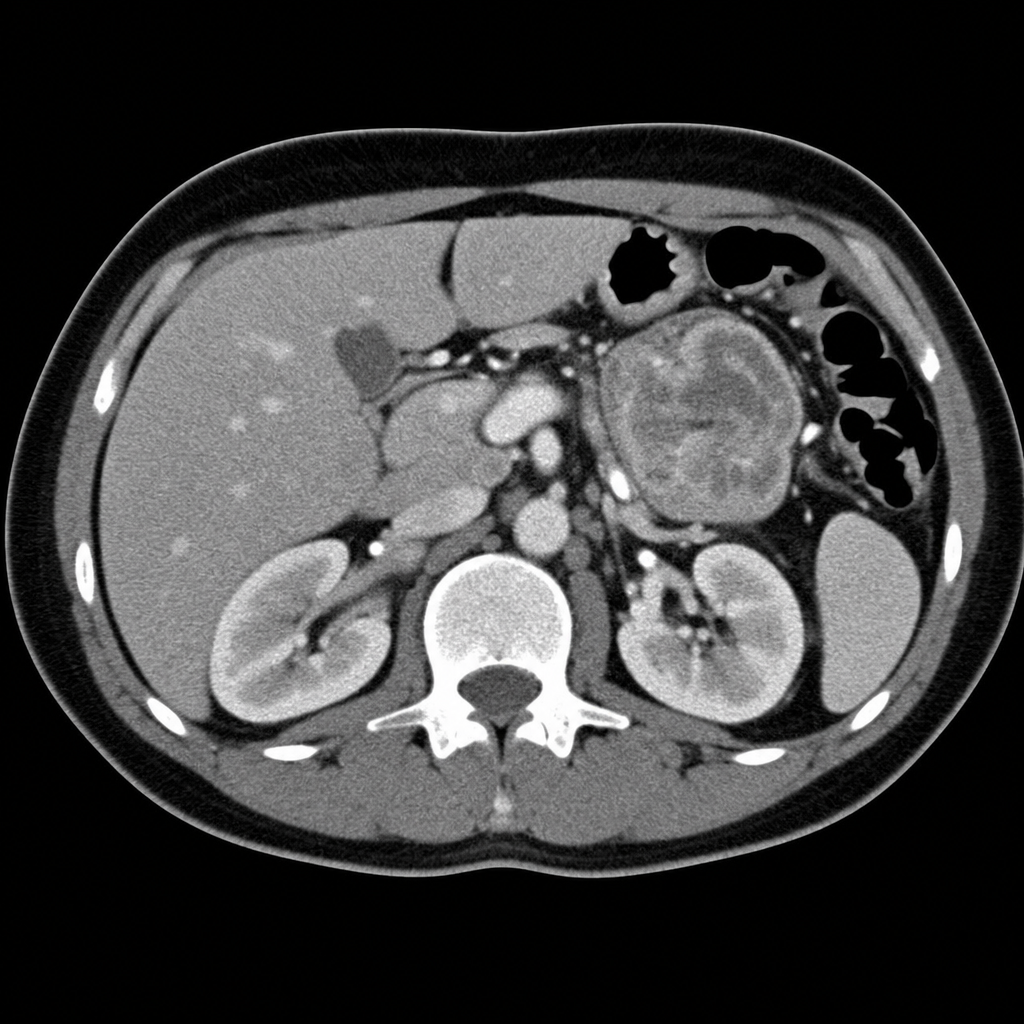
In a patient presenting with an adrenal mass, what is the most common cause of a malignant adrenal mass?
Which chromosomal translocation is associated with follicular thyroid carcinoma?
All of the following are features of granulomatous thyroiditis except?
Calcified pulmonary metastasis is seen in which carcinoma?
All the following are true of craniopharyngioma except:
Hurthle cell carcinoma is a variant of which type of carcinoma?
Orphan Annie nuclei are characteristic of which of the following?
Which of the following is a definitive feature of malignant pheochromocytoma?
Tumor marker for medullary carcinoma of the thyroid is:
Practice by Chapter
Pituitary Gland Disorders
Practice Questions
Thyroid Gland Diseases
Practice Questions
Parathyroid Gland Pathology
Practice Questions
Adrenal Cortical Disorders
Practice Questions
Adrenal Medullary Disorders
Practice Questions
Pancreatic Endocrine Disorders
Practice Questions
Multiple Endocrine Neoplasia Syndromes
Practice Questions
Diffuse Neuroendocrine System
Practice Questions
Pineal Gland Pathology
Practice Questions
Laboratory Diagnosis of Endocrine Diseases
Practice Questions
Want unlimited practice?
Get full access to all questions, explanations, and performance tracking.
Scan to download app