Which of the following cells in the lungs are classified as neuroendocrine cells?
VIPoma is associated with which syndrome?
FDG-PET negative tumor is:
All of the following statements are true about Gleason score, except:
Submucosal neuroma is associated with
Which of the following glands is NOT involved in Type I MEN?
MC location of gastrinoma in MEN-1 syndrome?
Somatostatin is secreted by which type of cells in the pancreas?
What is the most common type of acute myeloid leukemia in patients with Down's syndrome?
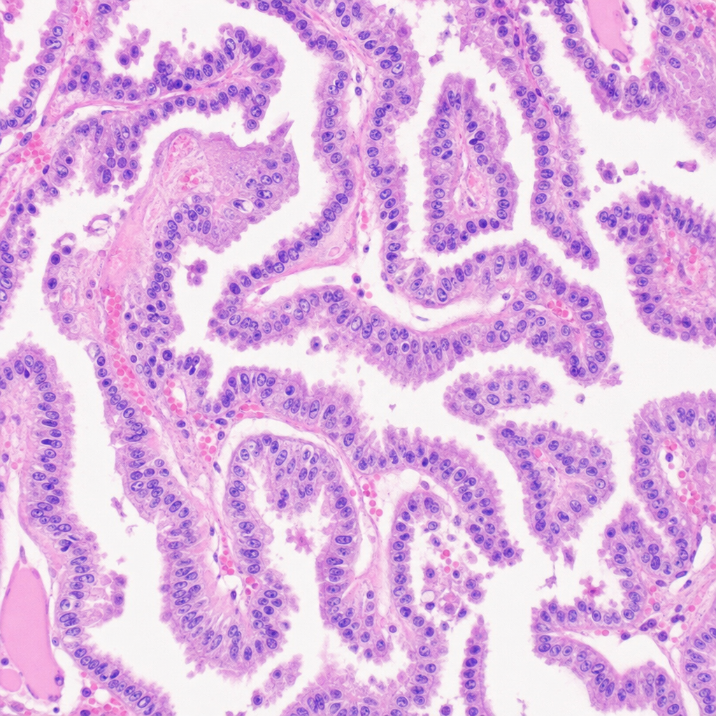
A 25-Year-old male presented with a 2cm thyroid nodule. A thyroidectomy was done. The histology picture is given below. What could be the diagnosis?

Explanation: ***Kulchitsky cells*** - **Kulchitsky cells**, also known as pulmonary neuroendocrine cells (PNECs), are found in the bronchial and bronchiolar epithelium. - They produce various **bioactive peptides and amines**, regulating airway tone and contributing to lung development and pathology. *Dendritic cells* - **Dendritic cells** are **immune cells** that act as antigen-presenting cells, initiating adaptive immune responses. - They are not classified as neuroendocrine cells and primarily function in immune surveillance. *Type I pneumocytes* - **Type I pneumocytes** are **squamous epithelial cells** that form the primary surface for gas exchange in the alveoli [1]. - Their main function is to facilitate **diffusion of gases** (oxygen and carbon dioxide) and they lack endocrine properties [1]. *Type II pneumocytes* - **Type II pneumocytes** are cuboidal cells responsible for producing and secreting **surfactant**, which reduces alveolar surface tension [1]. - They also act as progenitor cells for Type I pneumocytes but are not considered neuroendocrine cells [1].
Explanation: ***Verner-Morrison syndrome*** - **VIPoma** is a neuroendocrine tumor that secretes **vasoactive intestinal peptide (VIP)**, causing symptoms like watery diarrhea, hypokalemia, and achlorhydria (WDHA syndrome) [1]. - This clinical presentation is also known as **Verner-Morrison syndrome**, directly linking the VIPoma to this syndrome [1]. *Cushing's syndrome* - Characterized by excessive **cortisol** production, leading to symptoms like central obesity, moon facies, and hypertension [3]. - This syndrome is not directly associated with VIP excess or the watery diarrhea seen in VIPoma [2]. *Carcinoid syndrome (serotonin syndrome)* - Caused by tumors, typically in the gastrointestinal tract, that produce **serotonin**, leading to flushing, diarrhea, and bronchospasm [1]. - While it involves diarrhea, the primary mediator is serotonin, not VIP, and the other classic VIPoma symptoms (hypokalemia, achlorhydria) are absent [1]. *Zollinger-Ellison syndrome* - Characterized by a **gastrin-producing tumor (gastrinoma)**, which causes excessive gastric acid secretion and severe peptic ulcer disease [4]. - The hormonal excess is **gastrin**, not VIP, and symptoms are related to acid overproduction rather than massive watery diarrhea and hypokalemia [4].
Explanation: ***Typical carcinoid*** - **Typical carcinoid tumors** generally have a low metabolic rate and thus do not avidly take up **FDG (fluorodeoxyglucose)**, appearing **FDG-PET negative**. - These tumors are characterized by low mitotic activity and lack of necrosis, contributing to their low glucose metabolism. *Atypical carcinoid* - **Atypical carcinoid tumors** have a higher proliferative index and increased metabolic activity compared to typical carcinoids. - They tend to be **FDG-PET positive** due to their higher glucose utilization. *Small cell carcinoma* - **Small cell carcinoma** is a highly aggressive tumor type with rapid proliferation and high metabolic activity. - It is typically **FDG-PET positive**, reflecting its significant glucose uptake. *Large cell neuroendocrine carcinoma* - **Large cell neuroendocrine carcinoma (LCNEC)** is also an aggressive tumor with a high mitotic rate and often exhibits necrosis. - Similar to small cell carcinoma, LCNEC is generally **FDG-PET positive** due to its high metabolic demand.
Explanation: ***Scores range from 2-10*** - The **Gleason score** is obtained by summing the two most predominant architectural patterns of tumor growth, with each pattern graded from 1 to 5. The lowest possible sum is 2 (1+1) and the highest is 10 (5+5). However, **Gleason scores are now reported from 6 to 10** for clinical purposes, as grade 1 and 2 patterns are rarely seen in biopsies, and for practical purposes, anything less than a 3+3=6 is considered benign or low-risk. [1] - While mathematically the range is 2-10, in modern clinical practice, a score of 6 is the lowest grade assigned to prostate cancer, making the statement that scores range from 2-10 clinically inaccurate. *Used for grading prostate cancer* - The **Gleason score** is a well-established and widely used histological grading system specifically for prostate adenocarcinoma. [2] - It assesses the **architectural patterns** of glandular differentiation in prostate cancer to predict its aggressiveness. *Higher the score, poorer the prognosis* - A **higher Gleason score** indicates a more poorly differentiated and aggressive tumor, which correlates with a greater likelihood of metastasis and recurrence. [2] - This directly translates to a **poorer prognosis** for the patient. *Helps in planning management* - The **Gleason score** is a critical factor, along with PSA levels and clinical staging, in determining the risk stratification of prostate cancer. - This risk stratification guides treatment decisions, such as whether to pursue active surveillance, radiation therapy, surgery, or systemic therapy. **References:** [1] Kumar V, Abbas AK, et al.. Robbins and Cotran Pathologic Basis of Disease. 9th ed. The Lower Urinary Tract and Male Genital System, pp. 990-992. [2] Kumar V, Abbas AK, et al.. Robbins and Cotran Pathologic Basis of Disease. 9th ed. The Lower Urinary Tract and Male Genital System, pp. 993-994.
Explanation: ***MEN 2B (Multiple Endocrine Neoplasia Type 2B)*** - **Submucosal neuromas** are a distinctive feature of MEN 2B, specifically noticeble as mucosal neuromas on the lips, tongue, and gastrointestinal tract. - This syndrome is characterized by the presence of **medullary thyroid carcinoma**, **pheochromocytoma**, and mucocutaneous neuromas, without hyperparathyroidism. *MEN 2A (Multiple Endocrine Neoplasia Type 2A)* - MEN 2A is characterized by **medullary thyroid carcinoma**, **pheochromocytoma**, and **primary hyperparathyroidism**. - It does not typically feature extensive **submucosal neuromas** as a primary diagnostic criterion. *MEN 1 (Multiple Endocrine Neoplasia Type 1)* - MEN 1 involves tumors of the **parathyroid glands**, **anterior pituitary**, and **pancreatic islet cells** (the '3 Ps'). - **Submucosal neuromas** are not a component of the MEN 1 syndrome. *None of the options* - This option is incorrect because **submucosal neuromas** are a characteristic finding in MEN 2B.
Explanation: ***Adrenal*** - The **adrenal glands** are predominantly involved in **Multiple Endocrine Neoplasia type 2 (MEN2)**, particularly with pheochromocytomas, rather than MEN type 1. - While adrenal lesions (e.g., adenomas, hyperplasia) can occur sporadically or rarely in MEN1, they are not considered a primary or core component of the MEN1 syndrome as defined by the classic "3 Ps." [1] *Pancreas* - The **pancreas** is a primary gland involved in MEN1, frequently developing **neuroendocrine tumors** (e.g., gastrinomas, insulinomas). - These pancreatic tumors are a major cause of morbidity and mortality in MEN1 patients. *Pituitary* - The **pituitary gland** is one of the classic "3 P's" involved in MEN1, commonly developing **adenomas**, especially **prolactinomas**. [1] - These pituitary tumors can cause hormonal imbalances and mass effects within the sella turcica. *Parathyroid* - The **parathyroid glands** are almost universally involved in MEN1, with **hyperplasia** leading to **primary hyperparathyroidism**. [1] - This is often the earliest and most common clinical manifestation of MEN1.
Explanation: ***Pancreas*** - In **Multiple Endocrine Neoplasia type 1 (MEN-1) syndrome**, gastrinomas are most commonly found in the **pancreas**. - While sporadic gastrinomas are frequently duodenal, the **genetic predisposition of MEN-1** shifts the primary location to the pancreas. *Duodenum* - **Sporadic gastrinomas** without MEN-1 syndrome are most frequently located in the **duodenum**, particularly the first and second parts. - However, in the context of **MEN-1**, the pancreas becomes the predominant site for gastrinoma development. *Jejunum* - The jejunum is an **uncommon location** for gastrinomas in both sporadic cases and those associated with MEN-1. - Gastrinomas found in the jejunum are typically **rare** and often associated with more aggressive disease or disseminated metastasis. *Ileum* - The ileum is an **extremely rare site** for gastrinomas. - Gastrinomas developing in the ileum are usually **ectopic** and are not typically the primary location in either sporadic cases or MEN-1 syndrome.
Explanation: ***Delta cells*** - **Delta cells** (δ-cells) of the pancreatic islets are responsible for secreting **somatostatin**. - Somatostatin acts as a paracrine inhibitor, regulating the secretion of other pancreatic hormones like insulin and glucagon. *Gamma cells* - The term "gamma cells" is not a standard classification for pancreatic islet cells. - Pancreatic islet cells are typically categorized as alpha, beta, delta, and PP (pancreatic polypeptide) cells. *Alpha cells* - **Alpha cells** (α-cells) are responsible for secreting **glucagon**. - Glucagon primarily acts to raise blood glucose levels. *Beta cells* - **Beta cells** (β-cells) are the most abundant pancreatic islet cells and produce **insulin**. - Insulin is crucial for lowering blood glucose by promoting glucose uptake into cells.
Explanation: ***Acute megakaryoblastic leukemia M7*** - **Acute megakaryoblastic leukemia (AML M7)** is significantly more common in children with **Down's syndrome (trisomy 21)**, particularly those under 5 years of age. - This association is thought to be due to an increased copy number of certain genes on **chromosome 21** that are involved in hematopoiesis and leukemogenesis. [3] *Acute myeloid leukemia M1* - This subtype, characterized by proliferation of **myeloblasts without maturation**, is not specifically associated with Down's syndrome. [1] - It is a more undifferentiated form of AML. *Acute promyelocytic leukemia M3* - Characterized by the t(15;17) translocation involving the **PML-RARα fusion gene**, resulting in a block in myeloid differentiation at the promyelocyte stage. [2], [4], [5] - This subtype is associated with a specific genetic abnormality and is not preferentially seen in patients with Down's syndrome. *Acute myeloid leukemia M2* - This subtype involves **myeloblasts with maturation** and a characteristic t(8;21) chromosomal translocation. [2] - While it's a common form of AML, it does not show the specific strong association with Down's syndrome that AML M7 does. **References:** [1] Cross SS. Underwood's Pathology: A Clinical Approach. 6th ed. Common Clinical Problems From Blood And Bone Marrow Disease, pp. 607-608. [2] Kumar V, Abbas AK, et al.. Robbins and Cotran Pathologic Basis of Disease. 9th ed. Diseases of White Blood Cells, Lymph Nodes, Spleen, and Thymus, p. 620. [3] Kumar V, Abbas AK, et al.. Robbins and Cotran Pathologic Basis of Disease. 9th ed. Genetic Disorders, pp. 170-171. [4] Kumar V, Abbas AK, et al.. Robbins and Cotran Pathologic Basis of Disease. 9th ed. Diseases of White Blood Cells, Lymph Nodes, Spleen, and Thymus, pp. 621-622. [5] Kumar V, Abbas AK, et al.. Robbins and Cotran Pathologic Basis of Disease. 9th ed. Diseases of White Blood Cells, Lymph Nodes, Spleen, and Thymus, pp. 620-621.
Explanation: ***Papillary carcinoma thyroid*** - Characterized by **papillary structures** and **nuclear features** such as nuclear grooves and overlapping nuclei on histology [1]. - Often presents in young adults and can show **psammoma bodies**, which are indicative of malignancy. *Follicular adenoma* - Generally shows well-circumscribed **follicular structures** without nuclear atypia [2,3]. - Lacks the typical **papillary architecture** and associated aggressive features found in carcinoma. *Graves disease* - Primarily presents with **hyperthyroidism** and diffuse goiter rather than a solitary nodule. - Histologically, it is characterized by **hyperplastic follicles** and does not display features of malignancy. *Adenomatous goitre* - Refers to **nodular enlargement** of the thyroid with benign hyperplastic nodules. - Lacks the **malignant features** present in papillary carcinoma, such as nuclear atypia and invasion. **References:** [1] Kumar V, Abbas AK, et al.. Robbins and Cotran Pathologic Basis of Disease. 9th ed. The Endocrine System, p. 1099. [2] Kumar V, Abbas AK, et al.. Robbins and Cotran Pathologic Basis of Disease. 9th ed. The Endocrine System, pp. 1096-1097. [3] Cross SS. Underwood's Pathology: A Clinical Approach. 6th ed. Common Clinical Problems From Liver And Biliary System Disease, pp. 428-429.
Get full access to all questions, explanations, and performance tracking.
Start For Free