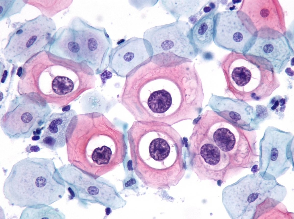
A 15-year-old girl undergoes Pap smear analysis. The smear demonstrates koilocytes, which are approximately the same size as intermediate cells, with enlarged hyperchromatic nuclei and perinuclear halos. These abnormal cells are infected by which of the following viruses?
A 75-year-old woman has experienced increasing dull but constant pain in the back, right chest, left shoulder, and left upper thigh for the past 6 months. She has now developed a sudden, severe, sharp pain in the left thigh. On physical examination, she has intense pain on palpation of the upper thigh, and the left leg is shorter than the right. A radiograph of the left leg shows a fracture through the upper diaphyseal region of the femur in a 5-cm lytic area that extends through the entire thickness of the bone. A bone scan shows multiple areas of increased uptake in the left femur, pelvis, vertebrae, right third and fourth ribs, upper left humerus, and left scapula. Laboratory studies show serum creatinine, 0.9 mg/dL; total protein, 6.7 g/dL; albumin, 4.5 g/dL; total bilirubin, 1 mg/dL; AST, 28 U/L; ALT, 22 U/L; and alkaline phosphatase, 202 U/L. What is the most likely diagnosis?
Based on the characteristic cytological findings on PAP smear examination in a 26-year-old female presented for screening, what is your diagnosis?

A 60-year-old female with a 8-year history of renal failure on hemodialysis develops Carpal Tunnel syndrome. Which of the following types of amyloid will be associated with this condition?
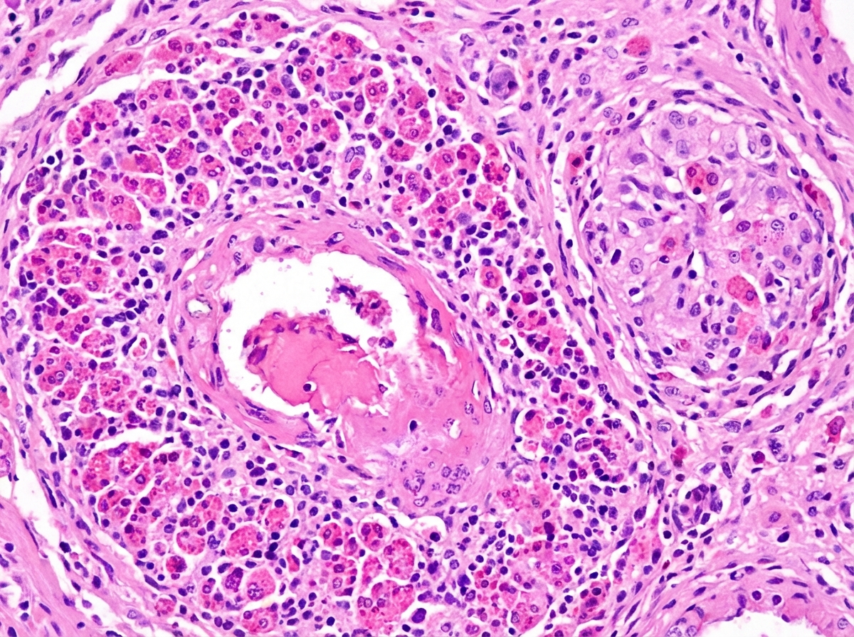
A 35-year-old patient presents with persistent allergic rhinitis, asthmatic episodes, and peripheral hypereosinophilia. Histological findings are shown below. What is your diagnosis?

Acridine orange is a fluorescent dye used to bind which cellular components?
FNAC is useful in diagnosing all the following conditions EXCEPT?
What is the best fixative for a Pap smear?
Regarding the screening of oral cancer, which of the following statements about exfoliative cytology is NOT true?
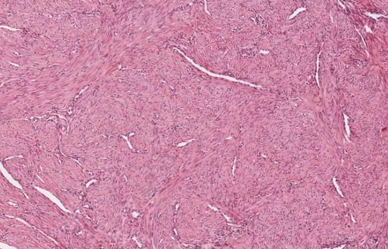
A 45-year-old female underwent hysterectomy for dysfunctional uterine bleeding. Following are the gross and histological findings. Which of the following is the most likely diagnosis?

Explanation: ### Explanation **Correct Answer: D. Human papillomavirus (HPV)** The presence of **koilocytes** on a Pap smear is the pathognomonic cytological hallmark of **Human Papillomavirus (HPV)** infection [1]. Koilocytes are squamous epithelial cells (typically intermediate or superficial) that have undergone specific structural changes due to the E6 and E7 oncoproteins of HPV [1]. **Key features of a Koilocyte:** * **Nuclear changes:** Enlargement (2–3 times the size of a normal intermediate cell nucleus), hyperchromasia, and irregular nuclear contours ("raisinoid" appearance). * **Cytoplasmic changes:** A sharp, well-defined **perinuclear halo** (clear zone) and a dense, "waxy" peripheral rim of cytoplasm. --- ### Why the other options are incorrect: * **A. Human Immunodeficiency Virus (HIV):** While HIV increases the risk and severity of HPV-related lesions due to immunosuppression, it does not cause koilocytic changes directly. * **B. Cytomegalovirus (CMV):** CMV typically presents with large, single basophilic intranuclear inclusions surrounded by a clear halo, giving an **"Owl’s eye"** appearance, usually in glandular or endothelial cells, not squamous koilocytes. * **C. Herpes simplex virus (HSV):** HSV infection is characterized by the **"3 Ms"**: Multinucleation, Margination of chromatin (ground-glass nuclei), and Molding of nuclei. It does not produce perinuclear halos in squamous cells. --- ### High-Yield Clinical Pearls for NEET-PG: * **Koilocytes** are most commonly associated with **Low-grade Squamous Intraepithelial Lesions (LSIL)** [1]. * **HPV Strains:** Types **6 and 11** cause genital warts (Condyloma acuminatum); Types **16 and 18** are high-risk for cervical carcinoma [1]. * The perinuclear halo in koilocytes is caused by the HPV **E4 protein**, which disrupts the cytokeratin network. **References:** [1] Kumar V, Abbas AK, et al.. Robbins and Cotran Pathologic Basis of Disease. 9th ed. The Female Genital Tract, pp. 1006-1008.
Explanation: **Explanation:** The clinical presentation describes an elderly woman with **multifocal lytic bone lesions** and a **pathological fracture** (shortened leg, sudden sharp pain). In an elderly patient, the most common cause of multiple destructive bone lesions is **Metastatic Carcinoma** [1]. **Why Metastatic Carcinoma is correct:** The bone scan shows multiple areas of increased uptake (hot spots) across the axial and appendicular skeleton. The elevated **Alkaline Phosphatase (ALP)** indicates osteoblastic activity (bone remodeling) in response to the tumor. Common primary sites in females that metastasize to bone include the breast, lung, and kidney [1]. **Why other options are incorrect:** * **Multiple Myeloma:** While it causes lytic lesions, it typically presents with a "punched-out" appearance on X-ray [2] and **normal ALP** levels (as there is no osteoblastic activity). Furthermore, the bone scan in myeloma is often **negative** (cold) because it relies on osteoblastic response, which is absent in myeloma. Myeloma typically involves bones containing red marrow like the skull, ribs, and pelvis, but rarely involves distal long bones [2]. * **Hyperparathyroidism:** This can cause "brown tumors" (osteitis fibrosa cystica), but it is usually associated with deranged calcium/phosphate levels and specific radiological signs like subperiosteal resorption of phalanges. * **Osteochondromatosis:** This is a benign condition characterized by multiple cartilage-capped bony outgrowths (exostoses) [1], typically seen in younger patients, and does not cause diffuse lytic destruction. **NEET-PG High-Yield Pearls:** * **Most common bone tumor in adults:** Metastatic Carcinoma (Primary: Prostate/Breast > Lung > Kidney) [1]. * **Bone Scan Sensitivity:** Highly sensitive for metastases (except purely lytic ones like Myeloma) but low specificity. * **Pathological Fracture:** Suspect malignancy when a fracture occurs with minimal trauma in an elderly patient. * **ALP:** A marker of osteoblastic activity; elevated in metastases and Paget’s disease, but characteristically normal in Multiple Myeloma. **References:** [1] Cross SS. Underwood's Pathology: A Clinical Approach. 6th ed. Common Clinical Problems From Osteoarticular And Connective Tissue Disease, pp. 671-672. [2] Cross SS. Underwood's Pathology: A Clinical Approach. 6th ed. Common Clinical Problems From Blood And Bone Marrow Disease, pp. 616-618.
Explanation: ***LSIL (Low-grade squamous intraepithelial lesion)*** - Characterized by **koilocytosis** (cells with enlarged nuclei and perinuclear halos), **nuclear enlargement** 2-3 times normal size, and **mild nuclear atypia** with preserved nuclear-to-cytoplasmic ratio. - Associated with **HPV infection** and shows **superficial cell involvement** with good cytoplasmic maturation, representing reversible changes in a young patient. *HSIL (High-grade squamous intraepithelial lesion)* - Shows **severe nuclear atypia** with nuclear enlargement >3 times normal size and **increased nuclear-to-cytoplasmic ratio**. - Involves **deeper epithelial layers** with **loss of cellular maturation** and more pronounced **chromatin abnormalities**, which are not typical for screening findings in young women. *Squamous cell carcinoma* - Characterized by **invasive malignant cells** with **severe pleomorphism**, **abnormal mitotic figures**, and **loss of cellular cohesion**. - Shows **necrotic background** and **tumor diathesis** with frankly malignant cytological features, which would be extremely rare in a 26-year-old screening patient. *Adenocarcinoma* - Demonstrates **malignant glandular cells** with **enlarged hyperchromatic nuclei**, **prominent nucleoli**, and **three-dimensional cell clusters**. - Originates from **endocervical glands** and shows **mucin production** with different cytomorphological features compared to squamous lesions.
Explanation: **Explanation:** The clinical presentation of a patient on long-term hemodialysis (8+ years) developing Carpal Tunnel Syndrome (CTS) is a classic description of **Dialysis-Related Amyloidosis (DRA)**. **1. Why Beta-2 Microglobulin (Aβ2M) is correct:** In healthy individuals, $\beta_2$-microglobulin (a component of MHC Class I molecules) is filtered by the kidney. In patients with end-stage renal disease, standard hemodialysis membranes cannot effectively filter this protein. Consequently, serum levels rise significantly, leading to the formation of amyloid fibrils [1]. These fibrils have a high affinity for osteoarticular structures, specifically the **synovium and transverse carpal ligament**, leading to compression of the median nerve (Carpal Tunnel Syndrome). **2. Why other options are incorrect:** * **Amyloid Light Chain (AL):** Derived from immunoglobulin light chains; associated with Plasma Cell Dyscrasias (e.g., Multiple Myeloma) [4]. It typically causes systemic amyloidosis affecting the heart, tongue, and kidneys [3]. * **ATTR (Transthyretin):** Seen in Senile Systemic Amyloidosis (wild-type) or Familial Amyloid Polyneuropathy (mutant) [1]. While wild-type ATTR can cause CTS in elderly males, the specific history of **hemodialysis** makes $\beta_2$-microglobulin the definitive answer. * **Serum Amyloid Associated (AA):** Derived from SAA protein (an acute-phase reactant). It is associated with **chronic inflammatory conditions** like Rheumatoid Arthritis, Tuberculosis, or Osteomyelitis [2]. **Clinical Pearls for NEET-PG:** * **Staining:** Like all amyloids, Aβ2M shows **Apple-green birefringence** under polarized light with Congo Red stain [5]. * **Risk Factor:** The duration of dialysis is the strongest risk factor; it rarely occurs in patients on dialysis for less than 5 years. * **Common Sites:** Carpal tunnel, shoulder joint (scapulohumeral periarthritis), and cervical spine. **References:** [1] Kumar V, Abbas AK, et al.. Robbins and Cotran Pathologic Basis of Disease. 9th ed. Diseases of the Immune System, p. 266. [2] Kumar V, Abbas AK, et al.. Robbins and Cotran Pathologic Basis of Disease. 9th ed. Diseases of the Immune System, pp. 267-268. [3] Cross SS. Underwood's Pathology: A Clinical Approach. 6th ed. (Basic Pathology) introduces the student to key general principles of pathology, both as a medical science and as a clinical activity with a vital role in patient care. Part 2 (Disease Mechanisms) provides fundamental knowledge about the cellular and molecular processes involved in diseases, providing the rationale for their treatment. Part 3 (Systematic Pathology) deals in detail with specific diseases, with emphasis on the clinically important aspects., pp. 135-136. [4] Kumar V, Abbas AK, et al.. Robbins and Cotran Pathologic Basis of Disease. 9th ed. Diseases of the Immune System, pp. 266-267. [5] Kumar V, Abbas AK, et al.. Robbins and Cotran Pathologic Basis of Disease. 9th ed. Diseases of the Immune System, pp. 268-269.
Explanation: ***Churg-Strauss syndrome*** - The **clinical triad** of **allergic rhinitis**, **asthma**, and **peripheral hypereosinophilia** is pathognomonic for Churg-Strauss syndrome (EGPA). - Histological findings typically show **necrotizing vasculitis** with **eosinophilic infiltration** and **extravascular granulomas** affecting small to medium vessels. *Behcet Disease* - Characterized by **recurrent oral and genital ulcers**, **uveitis**, and **skin lesions** (erythema nodosum, papulopustular lesions). - Lacks the **eosinophilic component** and **respiratory symptoms** seen in this case. *Kawasaki disease* - Primarily affects **children under 5 years** with **fever**, **conjunctivitis**, and **coronary artery involvement**. - Does not present with **allergic rhinitis**, **asthma**, or **hypereosinophilia** as seen here. *Buerger disease* - **Thromboangiitis obliterans** affecting **small and medium arteries** in **young male smokers**. - Presents with **digital ischemia** and **claudication**, not respiratory or allergic symptoms.
Explanation: **Explanation:** **Acridine Orange (AO)** is a fluorochrome dye with a unique property called **metachromasia**, meaning it can emit different colors depending on the molecule it binds to and the concentration. It is a nucleic acid-selective stain that interacts with DNA and RNA through electrostatic attraction. * **Why DNA and RNA are correct:** Acridine orange intercalates into double-stranded **DNA**, where it fluoresces **green** (maximum emission at 525 nm). Conversely, it binds to single-stranded **RNA** via electrostatic interactions, where it aggregates and fluoresces **orange-red** (maximum emission at 650 nm). This differential staining allows pathologists to assess the metabolic activity and nucleic acid content of cells. **Analysis of Incorrect Options:** * **B. Proteins:** Proteins are typically stained using dyes like Eosin (in H&E) or specific immunohistochemical markers. AO does not have a high affinity for proteins. * **C. Lipids:** Lipids are hydrophobic and require fat-soluble stains like Sudan Black B or Oil Red O. * **D. Carbohydrates:** Carbohydrates (like glycogen or mucin) are demonstrated using Periodic Acid-Schiff (PAS) or Mucicarmine stains. **NEET-PG High-Yield Pearls:** 1. **Clinical Use:** Used in the **Screening of Cervical Cancer** (cytopathology) because malignant cells have increased RNA synthesis (hyperchromasia). 2. **Microbiology:** It is highly sensitive for detecting bacteria in blood cultures (even before they show up on Gram stain) because it binds to bacterial nucleic acids. 3. **Viability Assay:** It is used to differentiate between live, apoptotic, and necrotic cells in research settings. 4. **Requirement:** Observation requires a **Fluorescence Microscope**.
Explanation: **Explanation:** The diagnosis of **Follicular Carcinoma** of the thyroid is the classic "limitation" of Fine Needle Aspiration Cytology (FNAC). [1] **1. Why Follicular Carcinoma is the correct answer:** FNAC evaluates individual cells or small clusters (cytology), but it cannot assess the overall tissue architecture [1]. The definitive diagnosis of Follicular Carcinoma requires the identification of **capsular invasion** or **vascular invasion**. Since FNAC only samples cells from within the lesion and does not provide a view of the intact tumor capsule or surrounding blood vessels, it cannot distinguish between a benign Follicular Adenoma and a malignant Follicular Carcinoma. On FNAC, both are reported under the Bethesda category as "Follicular Neoplasm." **2. Why the other options are incorrect:** * **Papillary Carcinoma:** This is diagnosed based on characteristic **nuclear features** (e.g., Orphan Annie eye nuclei, nuclear grooves, and intranuclear inclusions) and the presence of Psammoma bodies, all of which are easily visible on FNAC [2], [3]. * **Anaplastic Carcinoma:** This presents with highly malignant, pleomorphic cells (giant cells, spindle cells) that are easily identified as high-grade malignancy on cytology [2]. * **Lymphoma:** FNAC is highly effective in identifying the monotonous population of lymphoid cells; when combined with flow cytometry, it can often subtype the lymphoma. **Clinical Pearls for NEET-PG:** * **Gold Standard for Thyroid Nodules:** FNAC is the investigation of choice (IOC) for the initial evaluation of thyroid nodules. * **The "Cannot Diagnose" List:** Besides Follicular Carcinoma, FNAC cannot reliably distinguish between **Lipoma vs. Liposarcoma** or **Leiomyoma vs. Leiomyosarcoma** for the same reason: the need for architectural/invasion assessment. * **Bethesda System:** Remember that Follicular Neoplasm is Bethesda Category IV, necessitating surgical lobectomy for histological confirmation. **References:** [1] Kumar V, Abbas AK, et al.. Robbins and Cotran Pathologic Basis of Disease. 9th ed. The Endocrine System, pp. 1100-1101. [2] Cross SS. Underwood's Pathology: A Clinical Approach. 6th ed. Common Clinical Problems From Liver And Biliary System Disease, pp. 429-430. [3] Kumar V, Abbas AK, et al.. Robbins and Cotran Pathologic Basis of Disease. 9th ed. The Endocrine System, p. 1099.
Explanation: **Explanation:** The gold standard fixative for a Papanicolaou (Pap) smear is a mixture of **95% Ethyl alcohol (ethanol) and ether** in a 1:1 ratio. **Why 95% Ethanol + Ether is the Correct Choice:** Cytopathology requires excellent nuclear detail and transparent cytoplasm to identify dysplastic changes [1]. Alcohol is a **dehydrating fixative** that causes protein precipitation. 95% ethanol provides the ideal balance—it preserves nuclear morphology and chromatin patterns perfectly while preventing cell shrinkage. The addition of **ether** acts as a lipid solvent, enhancing the penetration of the fixative and improving the clarity of the cytoplasmic stain. **Analysis of Incorrect Options:** * **10% Formaldehyde (Option B):** This is the standard fixative for **histopathology** (tissue biopsies). In cytology, formalin causes significant cell shrinkage and alters the staining characteristics of the Pap stain, making it unsuitable. * **80% Isopropyl alcohol (Option C):** While isopropyl alcohol can be used as an alternative (usually at 80-100% concentration), it causes more shrinkage than ethanol and is not the "best" or traditional gold standard. * **90% Ether + alcohol (Option D):** The proportions are incorrect. The traditional "Sander’s fixative" or standard mixture utilizes equal parts or a dominant alcohol base. **High-Yield Clinical Pearls for NEET-PG:** * **Time is Critical:** Smears must be fixed immediately while **wet** (within seconds) [1]. If the smear dries before fixation (**air-drying artifact**), it leads to nuclear swelling and loss of cytoplasmic detail. * **Alternatives:** If ether is unavailable (due to its high inflammability), **95% Ethyl alcohol alone** is the most common practical substitute. * **Carnoy’s Fluid:** Used if the sample is heavily contaminated with blood, as it lyses RBCs. * **Spray Fixatives:** Often contain alcohols and polyethylene glycol (PEG) to protect the cells during transport. **References:** [1] Kumar V, Abbas AK, et al.. Robbins and Cotran Pathologic Basis of Disease. 9th ed. The Female Genital Tract, pp. 1010-1011.
Explanation: ### Explanation **Underlying Concept:** Exfoliative cytology involves the collection of cells that are naturally shed or scraped from the surface of a lesion [1]. In the oral cavity, traditional exfoliative cytology only samples the **superficial layers** of the epithelium. It does **not** sample the full thickness of the epithelium (trans-epithelial). Since dysplastic changes and early carcinomas often begin in the basal and parabasal layers, traditional cytology frequently yields false negatives [2]. To obtain trans-epithelial cells, a specialized "Brush Biopsy" (e.g., OralCDx) is required, which is distinct from standard exfoliative cytology. **Analysis of Options:** * **Option D (Correct - The False Statement):** Standard exfoliative cytology fails to reach the deeper layers; therefore, it does **not** include trans-epithelial cells. This is its primary limitation. * **Option A:** True. Hyperkeratotic (leukoplakic) lesions have a thick layer of keratin on the surface. This acts as a barrier, preventing the spatula from reaching the underlying nucleated cells, often resulting in a non-diagnostic smear. * **Option B:** True. Due to high false-negative rates and the inability to assess tissue architecture (invasion), it is considered a screening aid rather than a definitive diagnostic tool [2]. Histopathology remains the gold standard. * **Option C:** True. In standard scraping, the vast majority of cells collected are mature, superficial squamous cells [1]. **NEET-PG High-Yield Pearls:** * **Gold Standard:** Incisional biopsy is the gold standard for diagnosing oral cancer. * **Brush Biopsy:** Unlike exfoliative cytology, a brush biopsy uses a circular brush to obtain a **full-thickness (trans-epithelial)** sample. * **Toluidine Blue:** A vital stain often used as an adjunct; it binds to DNA in areas of high rapid cell division (dysplasia/cancer). * **Indication:** Cytology is best reserved for monitoring post-radiation patients or when a patient refuses a biopsy. **References:** [1] Cross SS. Underwood's Pathology: A Clinical Approach. 6th ed. (Basic Pathology) introduces the student to key general principles of pathology, both as a medical science and as a clinical activity with a vital role in patient care. Part 2 (Disease Mechanisms) provides fundamental knowledge about the cellular and molecular processes involved in diseases, providing the rationale for their treatment. Part 3 (Systematic Pathology) deals in detail with specific diseases, with emphasis on the clinically important aspects., pp. 237-240. [2] Kumar V, Abbas AK, et al.. Robbins and Cotran Pathologic Basis of Disease. 9th ed. The Lung, pp. 739-741.
Explanation: ***Leiomyoma*** - The **well-circumscribed whorled white-gray mass** with **interlacing bland smooth muscle bundles** and **cigar-shaped nuclei** are classic features of leiomyoma. - **Absence of atypia** and **low mitotic rate** (<10 mitoses/10 HPF) distinguish it from malignant smooth muscle tumors. *Leiomyosarcoma* - Would show **significant nuclear atypia**, **coagulative necrosis**, and **high mitotic rate** (≥10 mitoses/10 HPF). - Often presents as a **poorly circumscribed mass** with irregular borders, unlike the well-demarcated nature described. *Mixed Mullerian tumor* - Contains **biphasic components** with both **epithelial** and **mesenchymal** elements, not pure smooth muscle. - Shows **high-grade malignant features** with significant pleomorphism and mitotic activity. *Vaginal carcinoma* - Represents **squamous** or **glandular malignancy** arising from the **vaginal wall**, not uterine myometrium. - Would not present as a **myometrial mass** removed during hysterectomy for dysfunctional uterine bleeding.
Basic Principles of Cytopathology
Practice Questions
Specimen Collection and Processing
Practice Questions
Gynecologic Cytology
Practice Questions
Respiratory Tract Cytology
Practice Questions
Urinary Tract Cytology
Practice Questions
Effusion Cytology
Practice Questions
Fine Needle Aspiration Cytology
Practice Questions
Gastrointestinal Tract Cytology
Practice Questions
Ancillary Studies in Cytopathology
Practice Questions
Quality Assurance in Cytopathology
Practice Questions
Get full access to all questions, explanations, and performance tracking.
Start For Free