What is the commonly used gauge for Fine Needle Aspiration Cytology (FNAC) needles?
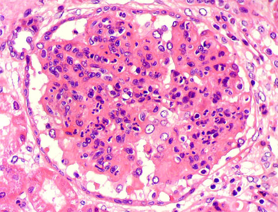
Examine the renal histopathology slide. What is the probable diagnosis?

For the diagnosis of carcinoma of the cervix, Pap smear screening is performed for what purpose?
In Pap smear cytology, which of the following is the most appropriate fixative used to preserve cellular details immediately after smearing?
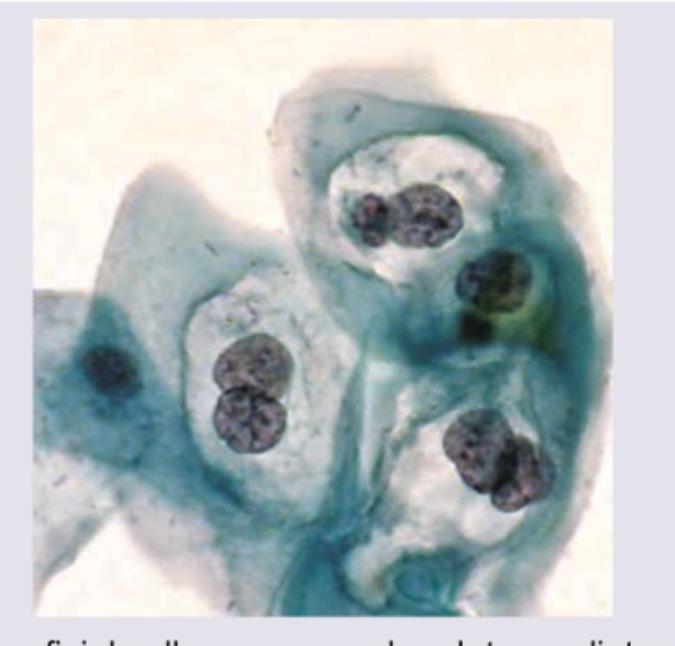
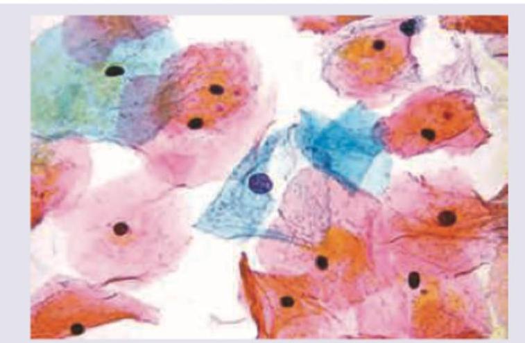
Name the cell shown in the Pap smear image.

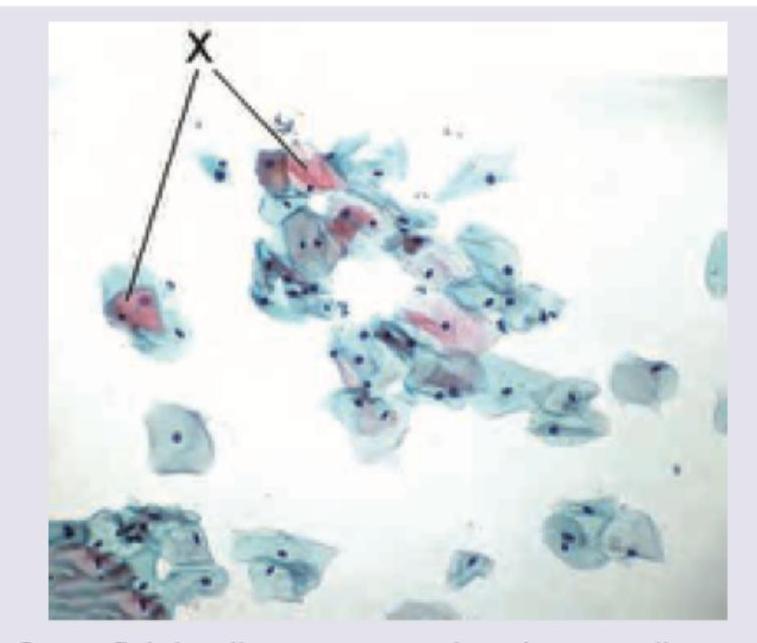
Name the cells marked as X in Pap smear.

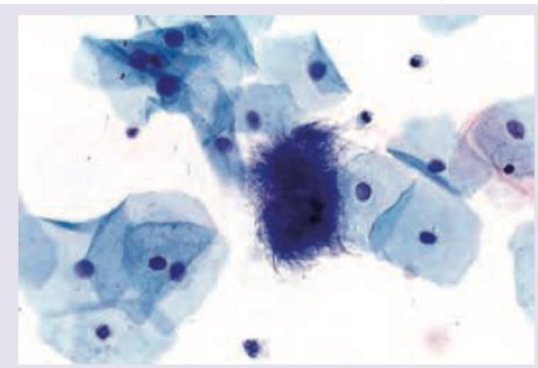
The following pap smear shows infestation by:

The following is the picture of the pap smear of a 45-year-old female with left ovarian tumor in her late menstrual phase showing squamous cells. What is the likely histology of the tumor?

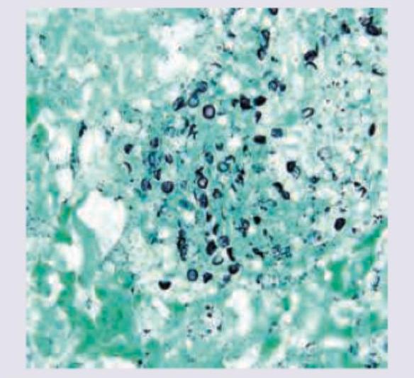
The following image of lung washing in AIDS positive patient is stained with:

Fine Needle Aspiration Cytology (FNAC) is NOT conclusive in which one of the following thyroid swellings?
Explanation: **Explanation:** **1. Why Option B (22-26) is Correct:** Fine Needle Aspiration Cytology (FNAC) is a diagnostic procedure used to collect cellular material from a lesion for microscopic examination. The goal is to obtain a high-quality cellular sample while minimizing trauma and blood contamination. * **22-25/26 Gauge** is the standard range because these needles are thin enough to reduce patient discomfort and prevent significant hemorrhage (which can dilute the sample), yet wide enough to allow cells to pass through the lumen without being crushed or distorted. * In clinical practice, a **23-gauge** needle is the most frequently used "workhorse" for superficial palpable masses (e.g., thyroid, lymph nodes, breast). **2. Why Other Options are Incorrect:** * **Option A (26-29):** These needles are extremely thin (often used for insulin injections or superficial skin anesthesia). While they cause minimal trauma, they frequently yield inadequate cellularity for a definitive diagnosis, especially in fibrotic lesions. * **Option C (18-22):** These are considered "wide-bore" needles in the context of cytology. Using an 18-20 gauge needle often leads to significant bleeding, resulting in a "bloody smear" where red blood cells obscure the diagnostic epithelial cells. * **Option D (16-18):** These are used for **Core Needle Biopsies (CNB)** or fluid aspiration (e.g., pleural tap), not FNAC. They remove a tissue architectural plug rather than individual cells. **3. NEET-PG High-Yield Pearls:** * **The "Non-Aspiration" Technique (Zajdela Technique):** Uses only the needle without a syringe/suction. It is preferred for highly vascular organs (like the thyroid) to minimize blood contamination. * **Fixation:** For Pap stain, smears are **wet-fixed** in 95% ethanol. For Giemsa/Romanowsky stains, smears are **air-dried**. * **Complication:** The most common complication of FNAC is a local hematoma. * **Contraindication:** FNAC is generally avoided in suspected **Phaeochromocytoma** (risk of hypertensive crisis) and **Hydatid cyst** (risk of anaphylaxis).
Explanation: ***Post-streptococcal glomerulonephritis*** - Characterized by **diffuse endocapillary hypercellularity** with **neutrophil infiltration** and enlarged, hypercellular glomeruli on light microscopy. - Electron microscopy shows pathognomonic **subepithelial electron-dense deposits** (humps), and immunofluorescence reveals a **starry sky pattern** with granular C3 and IgG deposits. *Membranoproliferative GN* - Shows **mesangial proliferation** with **double contour** appearance of glomerular basement membrane on light microscopy. - Electron microscopy reveals **subendothelial deposits** and **mesangial interposition**, not subepithelial humps. *Rapidly progressive GN* - Histologically characterized by **crescents** (>50% of glomeruli) composed of proliferating **epithelial cells** and **macrophages**. - Shows **necrotizing glomerulonephritis** with **fibrinoid necrosis** and **segmental sclerosis**, not endocapillary hypercellularity. *Diabetic nephropathy* - Light microscopy shows **nodular glomerulosclerosis** (Kimmelstiel-Wilson nodules) and **diffuse mesangial expansion**. - Electron microscopy reveals **thickened glomerular basement membrane** and **mesangial matrix expansion**, without immune deposits.
Explanation: The primary objective of the **Papanicolaou (Pap) smear** is the early detection of **pre-cancerous lesions** (Cervical Intraepithelial Neoplasia - CIN) [1], which allows for timely intervention before these lesions transform into invasive squamous cell carcinoma [2]. **1. Why Option C is Correct:** Cervical cancer typically follows a slow, predictable progression from low-grade dysplasia to high-grade dysplasia and finally to invasive cancer [3][4]. By identifying cellular atypia (like koilocytic changes or dyskaryosis) at the pre-invasive stage, clinicians can treat the patient (e.g., via LEEP or cryotherapy), thereby **preventing the progression** of the disease to a life-threatening malignancy [2]. **2. Analysis of Incorrect Options:** * **Option A:** No screening test is 100% informative. Pap smears have a significant false-negative rate (approx. 15-20%) due to sampling errors or laboratory interpretation limits [1]. * **Option B:** Pap smears are designed to detect epithelial changes (carcinoma) [1]. **Sarcomas** arise from mesenchymal tissue and are rarely detected or screened for via exfoliative cytology. * **Option C:** While screening intervals vary by guidelines (e.g., WHO, ACOG), the standard recommendation is generally every **3 to 5 years** for low-risk women, not every six months, which would be cost-ineffective and lead to over-treatment. **Clinical Pearls for NEET-PG:** * **Transformation Zone:** The most common site for cervical cancer and the critical area to sample during a Pap smear [2][4]. * **Fixative used:** 95% Ethyl alcohol. * **Stains used:** Papanicolaou method (Hematoxylin, Orange G, and Eosin Azure) [2]. * **Bethesda System:** The standard reporting format for cervical cytology. * **HPV 16 & 18:** The most high-risk types associated with progression to malignancy. **References:** [1] Cross SS. Underwood's Pathology: A Clinical Approach. 6th ed. Common Clinical Problems From Female Genital Tract Disease, pp. 468-470. [2] Kumar V, Abbas AK, et al.. Robbins and Cotran Pathologic Basis of Disease. 9th ed. The Female Genital Tract, pp. 1010-1011. [3] Cross SS. Underwood's Pathology: A Clinical Approach. 6th ed. (Basic Pathology) introduces the student to key general principles of pathology, both as a medical science and as a clinical activity with a vital role in patient care. Part 2 (Disease Mechanisms) provides fundamental knowledge about the cellular and molecular processes involved in diseases, providing the rationale for their treatment. Part 3 (Systematic Pathology) deals in detail with specific diseases, with emphasis on the clinically important aspects., pp. 209-210. [4] Cross SS. Underwood's Pathology: A Clinical Approach. 6th ed. Common Clinical Problems From Female Genital Tract Disease, pp. 467-468.
Explanation: ***95% ethanol*** - This is considered the **gold standard fixative** for conventional **Pap smear** cytology because it acts rapidly to precipitate proteins, effectively preserving cellular and **nuclear detail** crucial for diagnosis. - Immediate fixation in 95% ethanol (or proprietary sprays containing alcohol) is essential to prevent **air-drying artifact**, which can severely distort nuclear morphology and render the smear diagnostically inadequate [1]. *50% formaldehyde* - Formaldehyde is typically used for **tissue fixation** (histopathology) rather than cytology smears, and this concentration is too dilute for effective cellular preservation. - Formaldehyde is an **additive fixative**, which acts differently than the preferred precipitant fixatives (like ethanol) used on thin smears. *70% ethanol* - While an alcohol fixative, **95% ethanol** is the recommended concentration for optimum dehydration and quick stabilization of the cell structures in a Pap smear. - 70% concentration may not fix the cells rapidly enough, potentially leading to suboptimal **chromatin preservation** compared to the higher concentration. *10% buffered formalin* - This is the routine fixative used globally for large **surgical pathology** specimens (biopsies and resections). - Formalin is generally not the preferred immediate fixative for thin-layer cytology slides compared to quick-acting alcohol, which provides superior **nuclear clarity** for Papanicolaou staining. **References:** [1] Kumar V, Abbas AK, et al.. Robbins and Cotran Pathologic Basis of Disease. 9th ed. The Female Genital Tract, pp. 1010-1011.
Explanation: ***Koilocyte*** - A **koilocyte** is a squamous epithelial cell that has undergone a series of structural changes, indicative of **human papillomavirus (HPV) infection** [1][2]. - Key features include a **perinuclear halo** (a clear zone around the nucleus), a **hyperchromatic nucleus** (darkly stained nucleus), and often a slightly irregular nuclear membrane [1][2]. *Superficial cells* - These are mature squamous cells with a **small, pyknotic nucleus** (dense and dark) and abundant, transparent cytoplasm [1]. - They are typically seen in the later stages of the menstrual cycle and do not show the characteristic perinuclear halo of koilocytes. *Intermediate cells* - These are polygonal or oval-shaped squamous cells with a **larger, vesicular nucleus** (less dense) than superficial cells and a moderate amount of cytoplasm. - They are common throughout the menstrual cycle but lack the specific nuclear and cytoplasmic changes associated with HPV infection. *Trichomonas infection* - While **Trichomonas vaginalis** can be identified in a Pap smear, it appears as pear-shaped flagellated protozoa, often associated with a "dirty" background due to inflammatory cells and cellular debris. - It does not present as a specific cell type like a koilocyte, but rather as an infectious agent. **References:** [1] Kumar V, Abbas AK, et al.. Robbins and Cotran Pathologic Basis of Disease. 9th ed. The Female Genital Tract, p. 1010. [2] Kumar V, Abbas AK, et al.. Robbins and Cotran Pathologic Basis of Disease. 9th ed. The Female Genital Tract, pp. 1008-1010.
Explanation: ***Superficial cells*** - These cells are characterized by a **small, pyknotic nucleus** and abundant, clear cytoplasm, which is typical for the cells marked as X in a Pap smear [1]. - They are the most mature cells of the vaginal epithelium and are prominent in the **proliferative phase** of the menstrual cycle. *Intermediate cells* - These cells have a **larger, vesicular nucleus** compared to superficial cells and a more folded cytoplasm. - They are more prominent during the **luteal phase** and pregnancy due to progesterone influence. *Para-basal cells* - These cells are smaller with a **larger nucleus-to-cytoplasm ratio** and are typically seen in atrophic smears or in children and postmenopausal women. - They represent the **immature cells** of the vaginal epithelium. *Basal cells* - These are the **deepest layer** of the squamous epithelium and are rarely seen in a normal Pap smear unless there is significant epithelial damage or sampling from deeper layers. - They have a **large nucleus** and very little cytoplasm. **References:** [1] Kumar V, Abbas AK, et al.. Robbins and Cotran Pathologic Basis of Disease. 9th ed. The Female Genital Tract, p. 1010.
Explanation: ***Actinomyces*** - **Actinomyces** are Gram-positive, anaerobic bacteria that can form characteristic **"sulfur granules"** and appear as tangled, branching filaments on Pap smears, often associated with IUD use. - The image provided (though not visible to me) typically shows these **filamentous, branching organisms** with a "cotton ball" or "starburst" appearance, which is pathognomonic for Actinomyces. *Trichomonas* - **Trichomonas vaginalis** appears as pear-shaped, flagellated protozoa on Pap smears, often associated with a "strawberry cervix" and frothy discharge. - It does not present as filamentous structures or "sulfur granules" on cytology. *Candida* - **Candida albicans** appears as budding yeasts and pseudohyphae on Pap smears, often associated with thick, white, "cottage cheese" like discharge [1]. - It does not form the characteristic branching filaments or "sulfur granules" seen with Actinomyces. *Herpes simplex virus type II* - **Herpes simplex virus (HSV)** infection on Pap smears is characterized by multinucleated giant cells, nuclear molding, and ground-glass chromatin. - It is a viral infection and does not involve bacterial filaments or "sulfur granules." **References:** [1] Kumar V, Abbas AK, et al.. Robbins and Cotran Pathologic Basis of Disease. 9th ed. The Female Genital Tract, p. 998.
Explanation: ***Granulosa cell tumor*** - The Pap smear shows **mature squamous cells with abundant cytoplasm and small nuclei**, indicating a significant **estrogenic effect**. This is consistent with an ovarian tumor that produces estrogen, such as a granulosa cell tumor. - Granulosa cell tumors are **sex cord-stromal tumors** known for their **estrogen production**, leading to endometrial hyperplasia, irregular bleeding, and cytologic changes reflecting hyperestrogenism. *Dysgerminoma* - Dysgerminomas are **germ cell tumors** of the ovary that typically do **not produce hormones** (estrogen or progesterone) in quantities sufficient to cause significant squamous cell maturation or hyperplasia on a Pap smear. - These tumors are often associated with elevated tumor markers like **lactate dehydrogenase (LDH)**, but do not directly influence squamous cell morphology in this manner. *Serous papillary carcinoma* - Serous papillary carcinomas are **epithelial ovarian tumors**, which are common, but primarily present with non-specific symptoms or symptoms related to mass effect or ascites. - These tumors are not typically associated with **estrogen production** that would lead to the widespread maturation of squamous cells seen in the Pap smear. *Mucinous adenocarcinoma* - Mucinous adenocarcinomas are another type of **epithelial ovarian tumor** known for producing mucus. - They also do **not typically produce hormones** in a way that would lead to the hyperestrogenic effect observed in the Pap smear, characterized by mature squamous cells with abundant cytoplasm.
Explanation: ***Grocott's methanamine silver stain*** - This stain is specifically used to visualize fungal organisms, particularly **Pneumocystis jirovecii** (formerly P. carinii), which is a common opportunistic infection in AIDS patients [1,5]. - It stains the **fungal cell walls black**, making them easily identifiable in lung washings or biopsies [1]. *Ziehl-Neelsen stain* - This stain is primarily used for identifying **acid-fast bacilli**, such as **Mycobacterium tuberculosis**. - While tuberculosis can occur in AIDS patients, the image (not provided but implied to show fungi) would not be characteristic of ZN staining. *Auramine Rhodamine* - This is a **fluorescent stain** used for the rapid screening of **acid-fast bacilli** like Mycobacterium tuberculosis. - It is a screening test that requires a fluorescence microscope and is not typically used for fungal identification. *Giemsa stain* - Giemsa stain is commonly used for staining **blood smears** to identify parasites like malaria, and for staining **bone marrow** and **lymph nodes** [2]. - It can also be used to stain some bacteria and fungi, but it is **not the primary or most effective stain** for Pneumocystis jirovecii in lung washings [2]. **References:** [1] Cross SS. Underwood's Pathology: A Clinical Approach. 6th ed. Common Clinical Problems From Respiratory Tract Disease, pp. 318-319. [2] Kumar V, Abbas AK, et al.. Robbins and Cotran Pathologic Basis of Disease. 9th ed. Infectious Diseases, p. 362.
Explanation: ***Follicular carcinoma thyroid*** - FNAC cannot definitively distinguish between a **follicular adenoma** and a **follicular carcinoma** [1]. - This is because the diagnosis of follicular carcinoma relies on the presence of **capsular invasion** or **vascular invasion**, which can only be assessed on **histopathological examination** of the resected specimen, not cytology [1]. *Papillary carcinoma thyroid* - FNAC is highly effective in diagnosing papillary carcinoma due to characteristic **nuclear features** such as **Orphan Annie eye nuclei**, nuclear grooves, and intranuclear inclusions [3]. - These distinct cytological findings allow for a confident diagnosis without needing to assess invasion [3]. *Thyroiditis* - FNAC is typically conclusive for diagnosing various forms of thyroiditis (e.g., Hashimoto's thyroiditis, subacute thyroiditis). - It identifies characteristic inflammatory cells, giant cells, and changes in follicular cells consistent with the diagnosis. *Medullary carcinoma thyroid* - Medullary thyroid carcinoma can be reliably diagnosed by FNAC due to its characteristic **polygonal or spindle-shaped cells**, **amyloid deposition**, and presence of **calcitonin** in the aspirate [2]. - Immunocytochemical staining for calcitonin further confirms the diagnosis. **References:** [1] Kumar V, Abbas AK, et al.. Robbins and Cotran Pathologic Basis of Disease. 9th ed. The Endocrine System, pp. 1100-1101. [2] Cross SS. Underwood's Pathology: A Clinical Approach. 6th ed. Common Clinical Problems From Liver And Biliary System Disease, pp. 429-430. [3] Kumar V, Abbas AK, et al.. Robbins and Cotran Pathologic Basis of Disease. 9th ed. The Endocrine System, p. 1099.
Basic Principles of Cytopathology
Practice Questions
Specimen Collection and Processing
Practice Questions
Gynecologic Cytology
Practice Questions
Respiratory Tract Cytology
Practice Questions
Urinary Tract Cytology
Practice Questions
Effusion Cytology
Practice Questions
Fine Needle Aspiration Cytology
Practice Questions
Gastrointestinal Tract Cytology
Practice Questions
Ancillary Studies in Cytopathology
Practice Questions
Quality Assurance in Cytopathology
Practice Questions
Get full access to all questions, explanations, and performance tracking.
Start For Free