A patient with long-standing, moderately severe anemia dies in an automobile accident. An autopsy is performed. Which of the following cardiac changes will MOST likely be seen when the heart is examined?
What is the most common benign heart tumor?
Which type of endocarditis typically presents with vegetations on both sides of the valves?
What is the pathological finding that results in a tigereye appearance of the heart?
Ascoff's bodies are seen in which condition?
Rupture of the myocardium typically occurs within what time frame after a myocardial infarction?
In a patient with myocardial infarction, which valvular lesion is commonly seen?
Rheumatic activity involves which valves most commonly?
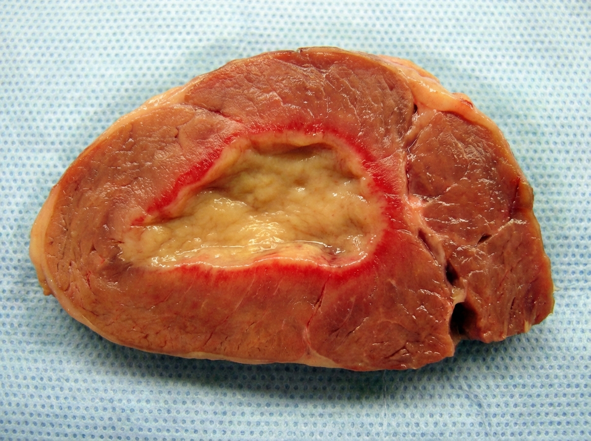
A gross section of the myocardium following myocardial infarction is shown. What is the likely duration following MI?

All are true about myxomas in the heart, EXCEPT:
Explanation: **Explanation:** **Why Option B is Correct:** The heart is highly sensitive to hypoxia. In cases of **long-standing, moderately severe anemia**, there is a chronic reduction in the oxygen-carrying capacity of the blood [1]. This leads to intracellular hypoxia, which impairs the mitochondrial β-oxidation of fatty acids within myocytes. As a result, neutral fats (triglycerides) accumulate as droplets within the cytoplasm—a process known as **Fatty Change (Steatosis)** [1]. Macroscopically, this manifests as the **"Tigroid Heart" or "Tabby Cat Heart"** appearance, characterized by alternating yellow bands of fatty change and reddish-brown bands of normal myocardium, typically seen in the papillary muscles and columns carneae. **Why Other Options are Incorrect:** * **A. Endocarditis:** This is an inflammatory/infectious process of the endocardium, usually triggered by bacteremia or autoimmune conditions (e.g., Rheumatic fever), not by chronic anemia. * **C. Fibrinous Pericarditis:** This occurs due to irritation of the pericardium, commonly seen in uremia, acute myocardial infarction (Dressler syndrome), or viral infections. Anemia does not cause pericardial inflammation. * **D. Fibrosis of valve leaflets:** This is a chronic sequela of inflammation (like Chronic Rheumatic Heart Disease) or age-related degeneration. Anemia affects the myocardium, not the structural integrity of the valves. **NEET-PG High-Yield Pearls:** 1. **Tigroid Heart:** Classic morphological descriptor for cardiac steatosis due to chronic hypoxia/anemia. 2. **Profound Hypoxia vs. Moderate Hypoxia:** While moderate anemia causes the "banded" appearance, profound hypoxia (e.g., severe CO poisoning) causes **diffuse** fatty change. 3. **Reversibility:** Fatty change is a form of **reversible cell injury** [1]; however, if the underlying hypoxia persists, it can progress to cell death. 4. **Stain:** To demonstrate fatty change histologically on frozen sections, use **Sudan IV or Oil Red O**. **References:** [1] Kumar V, Abbas AK, et al.. Robbins and Cotran Pathologic Basis of Disease. 9th ed. Red Blood Cell and Bleeding Disorders, pp. 638-639.
Explanation: **Explanation:** **1. Why Myxoma is the correct answer:** Myxoma is the most common primary cardiac tumor in **adults**, accounting for approximately 50% of all primary heart neoplasms [1]. These are benign mesenchymal tumors, most frequently located in the **left atrium** (75–80%), specifically attached to the interatrial septum near the fossa ovalis [1], [2]. Histologically, they are characterized by "lepidic" cells (stellate or globular cells) embedded in a rich acid mucopolysaccharide (myxoid) stroma [2]. **2. Why the other options are incorrect:** * **Rhabdomyoma:** This is the most common primary cardiac tumor in **infants and children** [1]. It is highly associated with **Tuberous Sclerosis** and often presents as multiple gray-white myocardial masses that may spontaneously regress. * **Hemangioma & Lipoma:** While these are benign primary tumors of the heart, they are significantly rarer than myxomas. Lipomas are most commonly found in the left ventricle, right atrium, or interatrial septum (lipomatous hypertrophy) [1]. **3. Clinical Pearls for NEET-PG:** * **Clinical Presentation:** Myxomas often present with the "Triad": Constitutional symptoms (fever, weight loss due to IL-6 release), Embolic phenomena, and Obstructive symptoms (mimicking mitral stenosis) [1]. * **Physical Exam:** A characteristic **"Tumor Plop"** may be heard during diastole as the pedunculated mass drops into the mitral valve orifice [2]. * **Carney Complex:** An autosomal dominant syndrome (PRKAR1A mutation) featuring atrial myxomas, spotty skin pigmentation (lentigines), and endocrine overactivity. * **Metastatic Disease:** Remember that secondary (metastatic) tumors of the heart are actually **more common** than primary tumors (most common source: Lung, Breast, Melanoma, or Lymphoma). **References:** [1] Cross SS. Underwood's Pathology: A Clinical Approach. 6th ed. Common Clinical Problems From Cardiovascular Disease, pp. 304-306. [2] Kumar V, Abbas AK, et al.. Robbins and Cotran Pathologic Basis of Disease. 9th ed. The Heart, pp. 583-584.
Explanation: **Explanation:** **Libman-Sacks Endocarditis (LSE)** is the correct answer because it is characterized by small, sterile, pinkish-tan vegetations that can occur on **both sides of the valve leaflets** (the surface and the undersurface), as well as on the chordae tendineae and endocardial surfaces [1]. This condition is classically associated with **Systemic Lupus Erythematosus (SLE)** and is a manifestation of immune complex deposition and subsequent inflammation [1]. **Analysis of Incorrect Options:** * **Infective Endocarditis (IE):** Typically presents with large, friable, and destructive vegetations located primarily on the **atrial surface** of AV valves or the **ventricular surface** of semilunar valves (the "flow side") [1]. They do not typically involve both sides of the valve. * **Rheumatic Carditis:** Characterized by small, firm, "wart-like" vegetations called **verrucae** that occur strictly along the **lines of closure** of the valve leaflets [1]. * **Non-Bacterial Thrombotic Endocarditis (NBTE/Marantic):** Presents with small, sterile vegetations along the lines of closure, similar to rheumatic fever, but without the significant underlying inflammation or valve destruction [1]. **NEET-PG High-Yield Pearls:** * **LSE Association:** Strongly linked to SLE and **Antiphospholipid Antibody Syndrome (APS)**. * **Vegetation Nature:** Sterile (non-infective) and composed of fibrin, inflammatory cells, and hematoxylin bodies (the tissue equivalent of LE cells). * **Most Common Valve:** Mitral valve is most frequently involved [1]. * **Key Differentiator:** LSE is the *only* endocarditis where vegetations are commonly found on the undersurface of the valves [1]. **References:** [1] Kumar V, Abbas AK, et al.. Robbins and Cotran Pathologic Basis of Disease. 9th ed. The Heart, pp. 568, 570.
Explanation: ### Explanation The "tigereye" or **"tabby cat" heart** appearance is a classic gross pathological finding associated with **fatty change (steatosis)** of the myocardium. **1. Why Fatty Change is Correct:** Fatty change in the heart occurs due to prolonged, moderate **hypoxia** (often seen in severe anemia). The mechanism involves the deposition of intracellular triglycerides within myocytes [2]. This deposition is not uniform; it occurs in bands. The alternating yellow bands (representing fatty change/steatosis) and reddish-brown bands (representing normal, well-oxygenated myocardium) create a striated appearance resembling the fur of a tiger or a tabby cat. This is most prominently seen in the subendocardial layer and papillary muscles. **2. Why the Other Options are Incorrect:** * **Hyaline change:** This refers to a descriptive histological term for a glassy, pink appearance (e.g., Zenker’s degeneration in skeletal muscle). It does not produce a macroscopically striped pattern in the heart. * **Amyloidosis:** This results in a firm, "waxy" or "lardaceous" appearance [1], . The heart typically becomes enlarged (restrictive cardiomyopathy) and thickened, but not striped. * **Atrophy:** Specifically, "Brown Atrophy" of the heart occurs due to the accumulation of **lipofuscin** (wear-and-tear pigment). This results in a uniformly shrunken, dark brown heart, not a striped appearance. **3. NEET-PG High-Yield Pearls:** * **Tigereye Heart:** Caused by **Chronic Hypoxia** (e.g., profound anemia). * **Diffuse Fatty Change:** Caused by **Severe Toxemia** (e.g., Diphtheria) or profound hypoxia; the heart appears uniformly pale and flabby rather than striped. * **Fatty Infiltration:** Distinct from fatty change; it involves the ingrowth of mature adipose tissue between myocardial fibers (common in the right ventricle). * **Nutmeg Liver:** A similar "mottled" gross appearance, but caused by chronic passive venous congestion (CPVC). **References:** [1] Kumar V, Abbas AK, et al.. Robbins and Cotran Pathologic Basis of Disease. 9th ed. The Heart, pp. 579-580. [2] Kumar V, Abbas AK, et al.. Robbins and Cotran Pathologic Basis of Disease. 9th ed. Cellular Responses to Stress and Toxic Insults: Adaptation, Injury, and Death, p. 73.
Explanation: **Explanation:** **Aschoff bodies** are pathognomonic (diagnostic) histological hallmarks of **Acute Rheumatic Fever (ARF)** [1]. They represent areas of focal interstitial inflammation and are most characteristically found within the **myocardium** [1]. 1. **Why Rheumatic Myocarditis is correct:** Aschoff bodies are granulomatous lesions consisting of a central zone of fibrinoid necrosis surrounded by chronic inflammatory cells (lymphocytes, plasma cells) and characteristic **Anitschkow cells** (caterpillar cells) [1]. While ARF is a pancarditis, these bodies are the classic microscopic finding of rheumatic myocarditis. 2. **Why other options are incorrect:** * **Rheumatic arthritis:** While ARF causes migratory polyarthritis, Aschoff bodies are specific to the heart and are not found in the joints. * **Bacterial endocarditis:** This is characterized by large, friable, destructive **vegetations** containing bacteria and fibrin, not Aschoff bodies. * **Marantic endocarditis (NBTE):** This involves small, sterile, bland thrombi (vegetations) on valve leaflets, typically seen in hypercoagulable states or malignancy. **High-Yield Clinical Pearls for NEET-PG:** * **Anitschkow Cells:** These are activated macrophages found within Aschoff bodies [1]. They feature a "caterpillar-like" chromatin pattern in longitudinal sections and an "owl-eye" appearance in cross-sections [1]. * **McCallum Patch:** A subendocardial thickening, usually in the left atrium, caused by the healing of rheumatic endocarditis. * **Evolution:** Aschoff bodies evolve through three stages: Exudative (early), Proliferative (intermediate/diagnostic), and Healing (fibrotic/polar scar). * **Jones Criteria:** Remember that Carditis is a "Major" criterion for the diagnosis of ARF. **References:** [1] Kumar V, Abbas AK, et al.. Robbins and Cotran Pathologic Basis of Disease. 9th ed. The Heart, pp. 566-567.
Explanation: **Explanation:** **Myocardial rupture** is a catastrophic complication of myocardial infarction (MI) that typically occurs **3 to 7 days** after the initial event, placing it firmly within the **first week** [1]. **Why the First Week is Correct:** The timing of rupture correlates with the peak of the inflammatory response and tissue remodeling. By days 3–7, **neutrophils and macrophages** have infiltrated the necrotic area to clear dead myocytes. This process involves the release of powerful **proteolytic enzymes and metalloproteinases**, which degrade the extracellular matrix. At this stage, the infarcted wall is composed of soft, friable "yellow-tan" necrotic tissue that has not yet been replaced by strong collagen (granulation tissue). The mechanical stress of ventricular pressure against this weakened wall leads to rupture [1]. **Why Other Options are Incorrect:** * **Second Week:** By the second week, **granulation tissue** (characterized by neovascularization and fibroblast proliferation) is well-established. This tissue is structurally more stable than necrotic debris, significantly reducing the risk of rupture. * **Third & Fourth Weeks:** During this period, **collagen scarring (fibrosis)** occurs. The myocardium is replaced by a firm, contracted scar, making rupture virtually impossible. Complications at this stage are more likely to be chronic heart failure or ventricular aneurysms. **High-Yield Clinical Pearls for NEET-PG:** * **Risk Factors:** Most common in first-time MIs (no pre-existing fibrosis/collateral circulation), elderly patients, females, and those with hypertension [1]. * **Types of Rupture:** 1. **Free Wall Rupture:** Leads to hemopericardium and **cardiac tamponade** (most common cause of death in this context). 2. **Interventricular Septal Rupture:** Leads to a Left-to-Right shunt and acute VSD [1]. 3. **Papillary Muscle Rupture:** Leads to acute, severe **mitral regurgitation** [1]. * **Morphology:** Look for "Coagulative necrosis" in the first 24 hours and "Macrophage infiltration" during the rupture window (Days 3-7). **References:** [1] Kumar V, Abbas AK, et al.. Robbins and Cotran Pathologic Basis of Disease. 9th ed. The Heart, pp. 556-557.
Explanation: **Explanation:** **Why Mitral Regurgitation (MR) is the correct answer:** Myocardial Infarction (MI) commonly leads to Mitral Regurgitation through two primary mechanisms: 1. **Papillary Muscle Dysfunction/Rupture:** The papillary muscles (especially the posteromedial muscle, which has a single blood supply from the RCA) are highly susceptible to ischemia. If they become dysfunctional or rupture (typically 3–5 days post-MI), the mitral valve leaflets fail to coapt, leading to acute, severe MR [1]. 2. **Ventricular Remodeling:** Chronic MI leads to left ventricular dilatation. This "stretches" the mitral valve annulus and displaces the papillary muscles, causing functional MR. **Analysis of Incorrect Options:** * **A. Aortic Stenosis:** This is typically a chronic degenerative or congenital process (e.g., senile calcification or bicuspid valve) and is not caused by acute ischemic events [1]. * **C. Aortic Regurgitation:** This usually results from aortic root dilation (e.g., syphilis, Marfan syndrome) or valve damage (e.g., endocarditis), not myocardial ischemia. * **D. Septal Defect:** While a **Ventricular Septal Rupture (VSR)** is a known complication of MI, it is a structural defect of the wall, not a "valvular lesion" [1]. **High-Yield NEET-PG Pearls:** * **Posteromedial Papillary Muscle:** Most common to rupture because it is supplied solely by the **Right Coronary Artery (RCA)**. * **Timeline:** Papillary muscle rupture typically occurs **3 to 5 days** after an MI [1]. * **Clinical Sign:** A new-onset **holosystolic murmur** at the apex radiating to the axilla post-MI should immediately raise suspicion of MR. * **Dressler Syndrome:** An autoimmune pericarditis occurring weeks after MI (Late complication). **References:** [1] Kumar V, Abbas AK, et al.. Robbins and Cotran Pathologic Basis of Disease. 9th ed. The Heart, pp. 556-557.
Explanation: **Explanation:** Rheumatic Heart Disease (RHD) is a sequela of Rheumatic Fever caused by molecular mimicry between Streptococcal M-proteins and human cardiac antigens [1]. The frequency of valvular involvement in RHD is directly related to the **hemodynamic stress** (pressure) placed on the valves. **1. Why Mitral and Aortic are correct:** The **Mitral valve** is the most commonly affected valve in RHD (involved in nearly all cases), followed by the **Aortic valve** [5]. This is because the left side of the heart operates under significantly higher pressures than the right side. High mechanical stress leads to greater endothelial trauma, facilitating the deposition of fibrin and the formation of characteristic small, friable vegetations (verrucae) along the lines of closure [2]. **2. Analysis of Incorrect Options:** * **Tricuspid and Pulmonary valves (Options A, B, & C):** Right-sided valves are rarely involved in RHD. When they are affected, it is almost always in association with severe multivallular involvement of the mitral and aortic valves [4]. Isolated right-sided rheumatic disease is clinically rare. * **Mitral and Tricuspid (Option C):** While the mitral valve is #1, the tricuspid valve is only the 3rd most common [4]. Therefore, the "Mitral and Aortic" combination is statistically more frequent. **High-Yield Clinical Pearls for NEET-PG:** * **Order of frequency:** Mitral > Aortic > Tricuspid > Pulmonary (M > A > T > P) [4]. * **Pathognomonic lesion:** **Aschoff bodies** (interstitial myocardial inflammation containing Anitschkow "caterpillar" cells) [1][4]. * **Macroscopic hallmark:** "Fish-mouth" or "Button-hole" stenosis due to commissural fusion and thickening of chordae tendineae [3][4]. * **McCallum Patch:** An area of endocardial thickening, usually in the posterior wall of the left atrium, caused by regurgitant jets. **References:** [1] Kumar V, Abbas AK, et al.. Robbins and Cotran Pathologic Basis of Disease. 9th ed. The Heart, p. 566. [2] Kumar V, Abbas AK, et al.. Robbins and Cotran Pathologic Basis of Disease. 9th ed. The Heart, p. 568. [3] Cross SS. Underwood's Pathology: A Clinical Approach. 6th ed. Common Clinical Problems From Cardiovascular Disease, pp. 293-294. [4] Kumar V, Abbas AK, et al.. Robbins and Cotran Pathologic Basis of Disease. 9th ed. The Heart, pp. 566-567. [5] Kumar V, Abbas AK, et al.. Robbins and Cotran Pathologic Basis of Disease. 9th ed. The Heart, p. 572.
Explanation: ***2 weeks*** - At **2 weeks post-MI**, the gross appearance shows a characteristic **yellow-tan depressed area** with a **hyperemic rim**, representing organized granulation tissue and early scar formation. - This timeframe corresponds to the **organization phase** where necrotic tissue is being replaced by **fibrous connective tissue** and **capillary proliferation**. *Immediate MI* - **Immediate MI** shows **no gross changes** as cellular death and coagulative necrosis have not yet become visible macroscopically. - The myocardium appears **normal** or may show only subtle **congestion** without distinct color changes or depression. *2 days* - At **2 days post-MI**, the infarct appears as a **pale, gray-white area** without the yellow coloration or hyperemic rim seen later. - This represents **early coagulative necrosis** with **neutrophilic infiltration** but lacks the organized granulation tissue of later stages. *Postmortem infarct artifact* - **Postmortem changes** produce **diffuse pallor** without the **well-demarcated borders** and **hyperemic rim** characteristic of true infarction. - These artifacts lack the **inflammatory response** and **organized tissue changes** that develop in living tissue following MI.
Explanation: ### **Explanation** Cardiac myxomas are the most common primary cardiac neoplasms in adults. This question tests the specific anatomical and genetic characteristics of these tumors. **Why Option D is the correct answer (The Exception):** While myxomas are predominantly found in the atrial chambers, they are **not strictly limited** to them. Although rare, myxomas can arise from the ventricular endocardium or directly from the **heart valves**. Therefore, the statement that they "never" occur on valves is factually incorrect. (Note: Papillary fibroelastomas are the most common tumors specifically involving valves, but myxomas can occur there too). **Analysis of Other Options:** * **Option A:** True. Myxomas are the **most common primary heart tumor** in adults [1]. (Rhabdomyomas are the most common in children [1]). * **Option B:** True. Cytogenetic studies have identified recurrent **clonal abnormalities of chromosomes 12 and 17** in both sporadic and familial cases. * **Option C:** True. Approximately **90% of myxomas occur in the atria** [2], with a strong predilection for the **left atrium (75-80%)** [1], typically attached to the fossa ovalis in the interatrial septum [2]. **High-Yield Clinical Pearls for NEET-PG:** * **Morphology:** They can be "sessile" or "pedunculated" [2]. Pedunculated myxomas can cause a **"wrecking ball" effect** [2], damaging valve leaflets or causing intermittent AV valve obstruction (mimicking mitral stenosis) [1]. * **Microscopy:** Characterized by **"Lepidic" cells** (myxoma cells) embedded in an abundant acid mucopolysaccharide ground substance [2]. * **Carney Complex:** An autosomal dominant syndrome (PRKAR1A gene mutation) featuring multiple cardiac/extracardiac myxomas, spotty skin pigmentation (lentigines), and endocrine overactivity. * **Clinical Sign:** A characteristic **"Tumor Plop"** may be heard on auscultation during diastole. **References:** [1] Cross SS. Underwood's Pathology: A Clinical Approach. 6th ed. Common Clinical Problems From Cardiovascular Disease, pp. 304-306. [2] Kumar V, Abbas AK, et al.. Robbins and Cotran Pathologic Basis of Disease. 9th ed. The Heart, pp. 583-584.
Congenital Heart Disease
Practice Questions
Ischemic Heart Disease
Practice Questions
Hypertensive Heart Disease
Practice Questions
Valvular Heart Disease
Practice Questions
Myocarditis and Cardiomyopathies
Practice Questions
Pericardial Disease
Practice Questions
Cardiac Tumors
Practice Questions
Heart Failure Pathophysiology
Practice Questions
Cardiac Transplantation Pathology
Practice Questions
Endocarditis
Practice Questions
Get full access to all questions, explanations, and performance tracking.
Start For Free