A four-month-old child develops cardiac failure and dies three months later. At autopsy, the heart has no obvious congenital defects, but the cardiac chambers are small and covered with thick, white endocardium. Histological sections of the heart demonstrate regular bands of elastic tissue in the thick fibrous endocardium. Which of the following is the most likely diagnosis?
Which of the following is the commonest cardiac neoplasm in adults?
What is the characteristic pathological finding in carcinoid heart disease?
Left ventricular hypertrophy is caused by all EXCEPT?
What is the most characteristic histological finding of acute rheumatic carditis?
What are the essential hypertrophy changes seen in the heart?
Carcinoid heart disease affects which part of the heart?
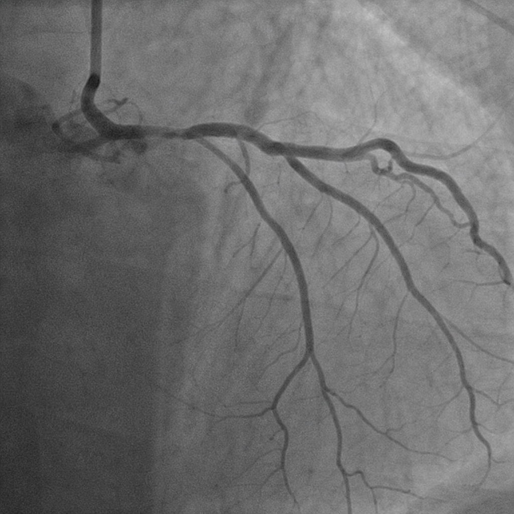
Aschoff's bodies are seen in which of the following conditions?
The light brown perinuclear pigment seen on H&E staining of the cardiac muscle fibres in the grossly normal appearing heart of an 83-year-old man at autopsy is due to deposition of?
Hemorrhagic pericarditis can be caused by which of the following?
Explanation: ### Explanation **Correct Answer: C. Endocardial fibroelastosis (EFE)** **1. Why it is correct:** Endocardial fibroelastosis is a rare cause of restrictive cardiomyopathy in infants, typically occurring within the first two years of life. The hallmark pathological finding is **diffuse, pearly-white thickening** of the endocardium, most commonly involving the left ventricle. Histologically, this thickening is due to the proliferation of **collagen and elastic fibers** (forming "regular bands" as mentioned in the question). This rigid endocardial layer impairs ventricular filling (diastolic dysfunction) and contraction, leading to rapid-onset heart failure and death in infancy. **2. Why incorrect options are wrong:** * **A. Coxsackievirus myocarditis:** While a common cause of pediatric heart failure, it presents histologically with **lymphocytic infiltration** and myocyte necrosis, not organized fibroelastic thickening of the endocardium. * **B. Cardiac amyloidosis:** This is a restrictive cardiomyopathy typically seen in **elderly patients** (AL or ATTR amyloid). Histology shows extracellular eosinophilic deposits that stain with **Congo Red** [1] (apple-green birefringence), not elastic tissue bands. * **C. Idiopathic subaortic stenosis:** Now more commonly known as **Hypertrophic Obstructive Cardiomyopathy (HOCM)**, it is characterized by asymmetrical septal hypertrophy and "myocyte disarray" [2] on histology, rather than endocardial thickening. **3. NEET-PG High-Yield Pearls:** * **Primary EFE:** Often associated with **mumps virus** infection during pregnancy or genetic factors. * **Secondary EFE:** Often associated with congenital heart defects, most notably **Aortic Stenosis** or **Hypoplastic Left Heart Syndrome**. * **Key Histological Stain:** **Verhoeff-Van Gieson (VVG) stain** is used to highlight the black-staining elastic fibers in EFE. * **Clinical Presentation:** Sudden onset of dyspnea, wheezing, and cyanosis in a previously healthy infant (often misdiagnosed as pneumonia). **References:** [1] Kumar V, Abbas AK, et al.. Robbins and Cotran Pathologic Basis of Disease. 9th ed. The Heart, pp. 580-581. [2] Kumar V, Abbas AK, et al.. Robbins and Cotran Pathologic Basis of Disease. 9th ed. The Heart, pp. 577-578.
Explanation: **Explanation:** The correct answer is **C. Metastatic tumour**. In clinical practice, **secondary (metastatic) tumors** are significantly more common than primary cardiac tumors, occurring approximately **20 to 40 times** more frequently [1]. The most common primary sites that metastasize to the heart include the lungs, breast, melanoma, and hematologic malignancies (lymphoma/leukemia) [1]. These typically reach the heart via lymphatic spread, hematogenous seeding, or direct extension [1]. **Analysis of Incorrect Options:** * **A. Ventricular rhabdomyoma:** This is the most common **primary** cardiac tumor in **infants and children**, often associated with Tuberous Sclerosis [2]. It is not common in adults. * **B. Atrial myxoma:** This is the most common **primary** cardiac tumor in **adults** [2]. While frequently tested, it is still rarer than metastatic disease. About 90% occur in the left atrium, often in the region of the fossa ovalis [1]. * **D. Angiosarcoma:** This is the most common **primary malignant** tumor of the heart in adults, typically arising in the right atrium [1]. However, overall metastatic involvement remains more prevalent. **High-Yield Clinical Pearls for NEET-PG:** * **Most common tumor of the heart (overall):** Metastatic tumor. * **Most common primary tumor (adults):** Myxoma (usually "Fossa Ovalis" in the Left Atrium) [1]. * **Most common primary tumor (children):** Rhabdomyoma [2]. * **Most common primary malignant tumor (adults):** Angiosarcoma. * **Carcinoid Heart Disease:** Typically affects the **right side** (tricuspid/pulmonary valves) because the lungs contain monoamine oxidase (MAO) which inactivates serotonin before it reaches the left heart. **References:** [1] Kumar V, Abbas AK, et al.. Robbins and Cotran Pathologic Basis of Disease. 9th ed. The Heart, pp. 582-586. [2] Cross SS. Underwood's Pathology: A Clinical Approach. 6th ed. Common Clinical Problems From Cardiovascular Disease, pp. 304-306.
Explanation: **Explanation:** **Carcinoid Heart Disease** occurs in approximately 50% of patients with systemic carcinoid syndrome (usually secondary to metastatic neuroendocrine tumors in the liver). **1. Why Option A is Correct:** The hallmark of carcinoid heart disease is the formation of **glistening, white, plaque-like fibrous thickenings** on the endocardium [1]. These plaques are composed of smooth muscle cells and sparse collagen embedded in an acid mucopolysaccharide-rich matrix [1]. Crucially, these lesions primarily affect the **right side of the heart** (right ventricle, tricuspid, and pulmonary valves) because the vasoactive substances (serotonin, bradykinin) are inactivated by the lungs (monoamine oxidase) before reaching the left heart [1]. **2. Analysis of Incorrect Options:** * **Option B:** While the tricuspid valve is involved, the functional consequence is typically **tricuspid regurgitation** rather than isolated severe stenosis [1]. The plaques cause the leaflets to become thickened and retracted (fixed in an open position). * **Option C:** Carcinoid plaques are characteristically **devoid of elastic fibers**, which helps pathologically distinguish them from other types of endocardial fibroelastosis [1]. * **Option D:** Unlike rheumatic heart disease or calcific aortic stenosis, the valvular dysfunction in carcinoid heart disease is due to fibrous plaque deposition, **not calcification** [1]. **3. NEET-PG High-Yield Pearls:** * **Biomarker:** Elevated urinary **5-HIAA** (5-hydroxyindoleacetic acid) is the diagnostic gold standard. * **Left-sided involvement:** Occurs only in the presence of a **Right-to-Left shunt** (e.g., Patent Foramen Ovale) or if the primary tumor is in the **lung** (bronchial carcinoid). * **Morphology:** "Plaque-like" is the keyword. These plaques do not extend into the underlying myocardium [1]. **References:** [1] Kumar V, Abbas AK, et al.. Robbins and Cotran Pathologic Basis of Disease. 9th ed. The Heart, pp. 570-572.
Explanation: **Explanation:** The correct answer is **Chronic cor pulmonale**. **1. Why Chronic Cor Pulmonale is the correct answer:** Left ventricular hypertrophy (LVH) is a compensatory response to increased pressure or volume load on the left side of the heart. **Cor pulmonale** is defined as hypertrophy and dilation of the **right ventricle** resulting from pulmonary hypertension caused by primary disorders of the lung parenchyma or pulmonary vasculature (e.g., COPD, interstitial lung disease) [1]. Since the pathology is isolated to the pulmonary circuit, it leads to Right Ventricular Hypertrophy (RVH), not LVH [1]. **2. Why the other options are incorrect:** * **Aortic Stenosis:** This creates a significant pressure gradient across the aortic valve. To maintain cardiac output against this high resistance (increased afterload), the left ventricle undergoes **concentric hypertrophy** [2,5]. * **Chronic Kidney Disease (CKD):** CKD is a classic cause of LVH through multiple mechanisms: chronic fluid overload (volume stress), activation of the Renin-Angiotensin-Aldosterone System (RAAS), and secondary hypertension [3]. * **Essential Hypertension:** This is the most common cause of LVH [4]. Persistent high systemic vascular resistance forces the left ventricle to contract more forcefully, leading to the thickening of the ventricular wall [3,4]. **Clinical Pearls for NEET-PG:** * **Concentric Hypertrophy:** Caused by **pressure overload** (e.g., Hypertension, Aortic Stenosis). New sarcomeres are added in **parallel** [2]. * **Eccentric Hypertrophy:** Caused by **volume overload** (e.g., Aortic Regurgitation, Dilated Cardiomyopathy). New sarcomeres are added in **series**, leading to chamber dilation [2]. * **Steell’s Law:** In pure cor pulmonale, the left ventricle remains normal in size and function unless there is a secondary systemic cause [1]. **References:** [1] Cross SS. Underwood's Pathology: A Clinical Approach. 6th ed. Common Clinical Problems From Cardiovascular Disease, pp. 284-285. [2] Kumar V, Abbas AK, et al.. Robbins and Cotran Pathologic Basis of Disease. 9th ed. The Heart, p. 536. [3] Cross SS. Underwood's Pathology: A Clinical Approach. 6th ed. Common Clinical Problems From Cardiovascular Disease, pp. 276-277. [4] Kumar V, Abbas AK, et al.. Robbins and Cotran Pathologic Basis of Disease. 9th ed. The Heart, pp. 560-562. [5] Kumar V, Abbas AK, et al.. Robbins and Cotran Pathologic Basis of Disease. 9th ed. The Heart, pp. 541-542.
Explanation: **Explanation:** **Aschoff bodies** are the pathognomonic (most characteristic) histological hallmark of acute rheumatic fever [3]. These are focal areas of interstitial inflammation consisting of a central zone of fibrinoid necrosis surrounded by a collection of T lymphocytes, plasma cells, and characteristic **Anitschkow cells** (activated macrophages with "caterpillar-like" nuclei) [1]. When these cells become multinucleated, they are known as **Aschoff giant cells**. While rheumatic fever is a pancarditis, Aschoff bodies are most distinctively found within the myocardium [3]. **Analysis of Incorrect Options:** * **A. Fibrinous pericarditis:** Often described as a "bread and butter" appearance, this is a common feature of acute rheumatic carditis but is non-specific; it can occur in uremia, post-MI (Dressler syndrome), or viral infections [4]. * **B. Vegetations on mitral valve leaflets:** Small, sterile friable vegetations (verrucae) along the lines of closure are characteristic of rheumatic heart disease, but they are not as specific as the microscopic Aschoff body [2]. * **D. Increased vascularity of the valves:** Chronic or recurrent inflammation leads to neovascularization of the normally avascular valve leaflets, but this is a secondary change rather than the primary diagnostic histological finding [1]. **NEET-PG High-Yield Pearls:** * **Anitschkow Cells:** The most characteristic cell within the Aschoff body; look for "caterpillar nuclei" (longitudinal) or "owl-eye" appearance (cross-section) [1]. * **MacCallum Patch:** Subendocardial thickening, usually in the left atrium, caused by regurgitant jets. * **Valve Involvement:** Mitral valve is most commonly affected (Mitral > Aortic > Tricuspid > Parker) [1]. * **Molecular Mimicry:** The pathogenesis involves Type II hypersensitivity where antibodies against Group A Streptococcal M-protein cross-react with cardiac myosin [3]. **References:** [1] Kumar V, Abbas AK, et al.. Robbins and Cotran Pathologic Basis of Disease. 9th ed. The Heart, pp. 566-567. [2] Kumar V, Abbas AK, et al.. Robbins and Cotran Pathologic Basis of Disease. 9th ed. The Heart, p. 568. [3] Kumar V, Abbas AK, et al.. Robbins and Cotran Pathologic Basis of Disease. 9th ed. The Heart, p. 566. [4] Cross SS. Underwood's Pathology: A Clinical Approach. 6th ed. Common Clinical Problems From Cardiovascular Disease, pp. 297-298.
Explanation: **Explanation:** **1. Why Option B is Correct:** Cardiac muscle cells are **permanent cells**, meaning they have lost their capacity for cell division (mitosis) in postnatal life [1]. When the heart is subjected to increased hemodynamic overload (e.g., hypertension or valvular stenosis), it cannot adapt by increasing the number of cells. Instead, it adapts through **hypertrophy**—an increase in the size of individual myocytes [1], [2]. This is driven by increased protein synthesis and the assembly of additional sarcomeres, leading to an increase in the overall muscle mass and wall thickness [3]. **2. Why Other Options are Incorrect:** * **Option A (Hyperplasia):** This refers to an increase in the number of cells. As mentioned, adult cardiomyocytes are non-dividing cells; therefore, hyperplasia is not a feature of adult cardiac adaptation [1]. * **Option C & D (Mitochondrial changes):** While there is an increase in metabolic demand, the hallmark of cellular hypertrophy is the synthesis of structural proteins (actin, myosin) and new myofibrils. While mitochondrial mass may increase to meet energy demands, it is a secondary organelle adaptation and not the "essential" defining pathological change of cardiac hypertrophy. **3. NEET-PG High-Yield Pearls:** * **Gene Expression:** Cardiac hypertrophy involves a "fetal gene program" switch, where adult isoforms of proteins (e.g., α-MHC) are replaced by fetal isoforms (e.g., β-MHC) which are more energy-efficient [2]. * **ANP/BNP:** Hypertrophic myocytes secrete **Atrial Natriuretic Peptide (ANP)** and **Brain Natriuretic Peptide (BNP)** to reduce blood volume and pressure. * **Concentric vs. Eccentric:** Pressure overload (Hypertension) leads to **concentric hypertrophy** (sarcomeres added in parallel), while volume overload (Regurgitation) leads to **eccentric hypertrophy** (sarcomeres added in series) [3]. **References:** [1] Kumar V, Abbas AK, et al.. Robbins and Cotran Pathologic Basis of Disease. 9th ed. Cellular Responses to Stress and Toxic Insults: Adaptation, Injury, and Death, pp. 45-46. [2] Kumar V, Abbas AK, et al.. Robbins and Cotran Pathologic Basis of Disease. 9th ed. Diseases of Infancy and Childhood, pp. 535-536. [3] Kumar V, Abbas AK, et al.. Robbins and Cotran Pathologic Basis of Disease. 9th ed. The Heart, p. 536.
Explanation: **Explanation:** **Carcinoid Heart Disease** is a manifestation of systemic carcinoid syndrome, occurring in approximately 50% of patients with the syndrome. **Why Valvular Endocardium is correct:** The primary pathology involves the deposition of **fibrous, plaque-like thickenings** on the endocardial surfaces of the heart [1]. These plaques are composed of smooth muscle cells and collagen embedded in an acid mucopolysaccharide-rich matrix [1]. The lesions characteristically involve the **valvular endocardium**, leading to thickening, shortening, and rigidity of the valve leaflets [1]. This typically results in **Tricuspid Regurgitation** and **Pulmonary Stenosis** [1]. **Why other options are incorrect:** * **Pericardium & Epicardium:** These are the outer layers of the heart. Carcinoid heart disease is a luminal (internal) pathology mediated by circulating humoral factors; it does not involve the external lining or the pericardial space. [1] * **Myocardium:** While chronic valvular dysfunction can lead to secondary right ventricular hypertrophy or failure, the primary site of the carcinoid-induced lesion is the endocardium, not the cardiac muscle fibers [1]. **High-Yield Clinical Pearls for NEET-PG:** * **Right-sided Predominance:** It primarily affects the **Right Heart** (Tricuspid and Pulmonary valves) [1]. * **Left Heart Sparing:** The Left Heart is usually spared because the lungs contain **Monoamine Oxidase (MAO)**, which inactivates the circulating serotonin (5-HT) and bradykinin before they reach the left atrium. * **Exception:** Left-sided involvement occurs only in the presence of a **Right-to-Left shunt** (e.g., Patent Foramen Ovale) or **Primary Bronchial Carcinoid**. * **Biomarker:** Elevated urinary **5-HIAA** (5-hydroxyindoleacetic acid) is the diagnostic hallmark. **References:** [1] Kumar V, Abbas AK, et al.. Robbins and Cotran Pathologic Basis of Disease. 9th ed. The Heart, pp. 570-572.
Explanation: **Explanation:** **Aschoff bodies** are the pathognomonic histological hallmark of **Acute Rheumatic Heart Disease (RHD)** [1]. They represent areas of focal interstitial inflammation consisting of a central zone of fibrinoid necrosis surrounded by mononuclear inflammatory cells (lymphocytes and plasma cells) and characteristic **Anitschkow cells** [1]. Anitschkow cells are specialized macrophages with "caterpillar-like" nuclei due to condensed chromatin [1]. When these cells become multinucleated, they are termed **Aschoff giant cells**. **Analysis of Options:** * **Rheumatic Heart Disease (Correct):** Aschoff bodies can be found in all three layers of the heart (pancarditis)—pericardium, myocardium, and endocardium [1]. * **Libman-Sacks Endocarditis:** Associated with **Systemic Lupus Erythematosus (SLE)** [2]. It is characterized by small, sterile, "verrucous" vegetations on both sides of the valve leaflets [2]. It does not feature Aschoff bodies. * **Bacterial Endocarditis:** Characterized by large, friable, destructive vegetations containing bacteria and inflammatory cells (neutrophils), leading to valvular perforation or abscesses [2]. * **Marantic Endocarditis (NBTE):** Occurs in wasted (cachectic) patients or those with hypercoagulable states (e.g., Trousseau syndrome). It features small, sterile thrombi along the lines of closure without significant inflammation or Aschoff bodies [2]. **High-Yield Pearls for NEET-PG:** * **Anitschkow Cells:** "Caterpillar cells" (longitudinal section) or "Owl-eye appearance" (cross-section) [1]. * **MacCallum Patch:** Subendocardial thickening, usually in the left atrium, caused by regurgitant jets in RHD. * **Jones Criteria:** Used for the clinical diagnosis of Acute Rheumatic Fever. * **Molecular Mimicry:** The pathogenesis involves Type II hypersensitivity where antibodies against Group A Streptococcal M-protein cross-react with cardiac self-antigens [1]. **References:** [1] Kumar V, Abbas AK, et al.. Robbins and Cotran Pathologic Basis of Disease. 9th ed. The Heart, pp. 566-567. [2] Kumar V, Abbas AK, et al.. Robbins and Cotran Pathologic Basis of Disease. 9th ed. The Heart, p. 568.
Explanation: **Explanation:** The correct answer is **Lipochrome** (also known as **Lipofuscin**). **1. Why Lipochrome is Correct:** Lipofuscin is an insoluble, brownish-yellow granular intracellular pigment [1]. It is known as the **"wear-and-tear"** or **aging pigment**. It is composed of polymers of lipids and phospholipids complexed with protein, derived through the **peroxidation of polyunsaturated lipids** of subcellular membranes [3]. In the heart of an elderly individual (as in this 83-year-old), it typically accumulates in the **perinuclear** region of cardiac myocytes [1]. When associated with tissue shrinkage/atrophy, the condition is termed **"Brown Atrophy."** **2. Why Other Options are Incorrect:** * **Hemosiderin:** This is a golden-yellow to brown pigment derived from hemoglobin (iron) [4]. While it looks similar to lipofuscin, it is usually associated with iron overload (hemosiderosis) and is identified using the **Prussian Blue stain**, which would be negative for lipofuscin. * **Cholesterol metabolite:** Cholesterol typically appears as clear, needle-like "clefts" in tissue sections (e.g., in atherosclerosis) rather than granular brown pigment [2]. * **Anthracotic pigment:** This is exogenous carbon/dust pigment commonly found in the lungs and hilar lymph nodes of smokers or city dwellers. It appears **jet black**, not light brown [2]. **3. High-Yield Clinical Pearls for NEET-PG:** * **Nature:** Lipofuscin is a hallmark of **free radical injury** and lipid peroxidation [3]. * **Staining:** It is **PAS positive** and can be visualized with Sudan Black B (though it is not a true fat). * **Key Association:** It is not toxic to the cell itself but serves as a marker of past oxidative stress and aging. * **Differential:** Always distinguish from Melanin (brown-black) and Hemosiderin (golden-brown) based on clinical context and special stains. **References:** [1] Kumar V, Abbas AK, et al.. Robbins and Cotran Pathologic Basis of Disease. 9th ed. Cellular Responses to Stress and Toxic Insults: Adaptation, Injury, and Death, p. 75. [2] Kumar V, Abbas AK, et al.. Robbins and Cotran Pathologic Basis of Disease. 9th ed. Cellular Responses to Stress and Toxic Insults: Adaptation, Injury, and Death, p. 77. [3] Cross SS. Underwood's Pathology: A Clinical Approach. 6th ed. (Basic Pathology) introduces the student to key general principles of pathology, both as a medical science and as a clinical activity with a vital role in patient care. Part 2 (Disease Mechanisms) provides fundamental knowledge about the cellular and molecular processes involved in diseases, providing the rationale for their treatment. Part 3 (Systematic Pathology) deals in detail with specific diseases, with emphasis on the clinically important aspects., pp. 241-242. [4] Kumar V, Abbas AK, et al.. Robbins and Cotran Pathologic Basis of Disease. 9th ed. Cellular Responses to Stress and Toxic Insults: Adaptation, Injury, and Death, pp. 75-76.
Explanation: **Explanation:** Hemorrhagic pericarditis is characterized by an inflammatory exudate composed of blood mixed with a fibrinous or suppurative effusion. It is fundamentally caused by processes that lead to significant vascular damage or the erosion of epicardial and pericardial surfaces. **Analysis of Options:** * **Tuberculosis (A):** While TB classically causes "bread and butter" (fibrinous) or caseous pericarditis, it is a frequent cause of hemorrhagic effusions, especially in chronic stages where granulomatous inflammation erodes small vessels [1]. * **After Cardiac Surgery (B):** Post-operative pericarditis (including Post-pericardiotomy syndrome) often involves surgical trauma to the pericardial vessels and the use of anticoagulants, leading to blood-stained effusions [1]. * **Metastatic Disease (C):** Malignancy is the **most common cause** of persistent hemorrhagic pericarditis [1]. Tumor infiltration (most commonly from Lung or Breast cancer, or Lymphoma) causes direct invasion and rupture of blood vessels within the pericardial space. **Why "All the Above" is correct:** All three conditions share the common pathophysiological mechanism of disrupting the integrity of the pericardial microvasculature, leading to the leakage of red blood cells into the pericardial sac [1]. **High-Yield NEET-PG Pearls:** * **Most common cause of Hemorrhagic Pericarditis:** Metastatic Malignancy. * **Other causes:** Uremia, Acute Myocardial Infarction (Dressler Syndrome), and bleeding diathesis [1]. * **Clinical Significance:** If the accumulation of hemorrhagic fluid is rapid, it can lead to **Cardiac Tamponade**, a medical emergency characterized by Beck’s Triad (Hypotension, JVD, and muffled heart sounds). * **Distinction:** It must be differentiated from **Hemopericardium** (pure blood in the sac), which is usually caused by a ruptured MI, aortic dissection, or penetrating trauma. **References:** [1] Kumar V, Abbas AK, et al.. Robbins and Cotran Pathologic Basis of Disease. 9th ed. The Heart, pp. 582-583.
Congenital Heart Disease
Practice Questions
Ischemic Heart Disease
Practice Questions
Hypertensive Heart Disease
Practice Questions
Valvular Heart Disease
Practice Questions
Myocarditis and Cardiomyopathies
Practice Questions
Pericardial Disease
Practice Questions
Cardiac Tumors
Practice Questions
Heart Failure Pathophysiology
Practice Questions
Cardiac Transplantation Pathology
Practice Questions
Endocarditis
Practice Questions
Get full access to all questions, explanations, and performance tracking.
Start For Free