Calcification of the aortic valve is seen in which of the following conditions?
A 66-year-old woman collapses and expires suddenly of cardiac arrest. Her past medical history is significant for long-standing type 2 diabetes mellitus. Her relatives note that she had complained of chest heaviness and shortness of breath for the past 2 weeks. Sterile fibrinous pericarditis and pericardial effusion are observed at autopsy. What additional finding would be expected during autopsy of this patient?
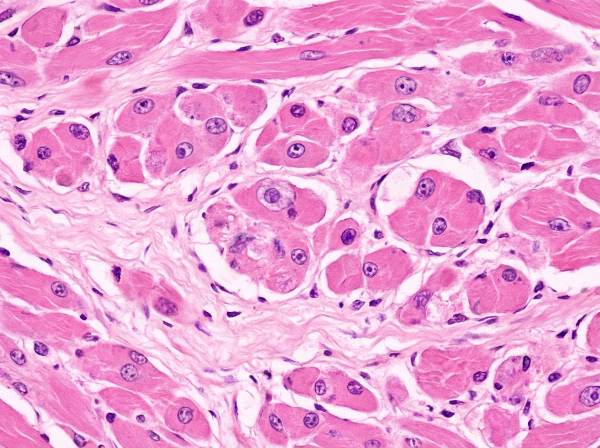
A 25-year-old bodybuilder was using anabolic steroids and started having puffy feet for the last 2 months. He died suddenly during a workout. A post-mortem heart biopsy specimen is shown below. What is the diagnosis?

What is the most common tumor of the heart?
A 59-year-old man experiences acute chest pain and is rushed to the emergency room. Laboratory studies and ECG demonstrate an acute myocardial infarction; however, coronary angiography performed 2 hours later does not show evidence of thrombosis. Which of the following mediators of inflammation causes relaxation of vascular smooth muscle cells and vasodilation of arterioles at the site of myocardial infarction in this patient?
Carcinoid of the heart typically involves which structure?
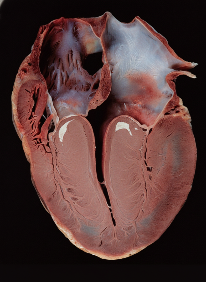
A 29-year-old male athlete suddenly collapsed and died during a football game. Autopsy revealed a specific gross examination finding. He had a history of two similar deaths in the family. What is the most likely cause of death?

Dilated cardiomyopathy is due to which of the following?
Which of the following dissection methods of the heart is most useful for demonstrating the distribution of infarction?
A 50-year-old woman presents with fatigue and shortness of breath. Physical examination shows evidence of pulmonary edema, enlargement of the left atrium, and calcification of the mitral valve. A CT scan demonstrates a large obstructing mass in the left atrium. Before open heart surgery can be performed to remove the tumor, the patient suffers a stroke and expires. Which of the following hemodynamic disorders best explains the pathogenesis of stroke in this patient?
Explanation: **Explanation:** **Calcific Aortic Stenosis** is the most common cause of aortic stenosis in the elderly [1]. The underlying mechanism is **dystrophic calcification**, where calcium salts deposit in injured or aging tissues despite normal serum calcium levels [2]. Chronic "wear and tear" leads to valvular injury, lipid accumulation, and inflammation, eventually resulting in the formation of large, stony-hard calcified nodules within the Sinuses of Valsalva [1]. These nodules prevent the cusps from opening fully, leading to left ventricular outflow obstruction. **Analysis of Options:** * **Aortic Stenosis (Correct):** Calcification is the hallmark of "Senile Calcific Aortic Stenosis" (occurring in the 7th–9th decades) and "Congenital Bicuspid Aortic Valve" (occurring earlier, in the 5th–6th decades) [1]. * **Aortic Regurgitation:** While severe stenosis can have a regurgitant component, pure aortic regurgitation is typically caused by aortic root dilation (e.g., syphilis, hypertension) or cusp destruction (e.g., endocarditis), rather than primary calcification. * **Marfan’s Syndrome:** This is characterized by **cystic medial necrosis** of the aorta, leading to aortic root dilation and mitral valve prolapse (myxomatous degeneration), not valvular calcification. * **Hurler’s Syndrome:** This mucopolysaccharidosis leads to the accumulation of dermatan and heparan sulfate, causing **valvular thickening** due to metabolite deposits, but not primary calcification. **NEET-PG High-Yield Pearls:** * **Dystrophic Calcification:** Occurs in dead/dying tissues; Serum Calcium is **Normal** [2]. * **Bicuspid Aortic Valve:** The most common congenital heart defect; it predisposes patients to calcific stenosis much earlier than tricuspid valves [1]. * **Clinical Triad of AS:** Dyspnea, Angina, and Syncope (SAD). * **Morphology:** In calcific AS, the free edges of the cusps are usually not involved (unlike Rheumatic Heart Disease, where commissural fusion occurs) [1]. **References:** [1] Kumar V, Abbas AK, et al.. Robbins and Cotran Pathologic Basis of Disease. 9th ed. The Heart, pp. 562-563. [2] Kumar V, Abbas AK, et al.. Robbins and Cotran Pathologic Basis of Disease. 9th ed. The Heart, p. 572.
Explanation: **Explanation:** The patient’s clinical presentation of sudden cardiac arrest, history of diabetes mellitus (a major risk factor for atherosclerosis), and symptoms of chest heaviness/shortness of breath for two weeks strongly suggest an **Acute Myocardial Infarction (MI)** [3], [4]. The key diagnostic clue at autopsy is **sterile fibrinous pericarditis** [1]. In the context of an MI, this typically occurs during two distinct timeframes [2]: 1. **Early (2–4 days post-MI):** Localized fibrinous pericarditis occurs due to transmural inflammation extending to the epicardium [1]. 2. **Late (2–10 weeks post-MI):** Known as **Dressler Syndrome**, an autoimmune-mediated pericarditis. Given the 2-week history of symptoms, the pericarditis is a direct complication of a recent transmural myocardial infarct. **Analysis of Incorrect Options:** * **A. Endocardial fibroelastosis:** Characterized by diffuse thickening of the endocardium (porcelain-like), typically seen in infants and associated with restrictive cardiomyopathy, not acute cardiac events in adults. * **B. Marantic endocarditis (NBTE):** Non-bacterial thrombotic endocarditis involves sterile vegetations on valves, usually associated with advanced malignancy or wasting diseases, not pericarditis. * **C. Mitral valve prolapse:** While it can cause sudden death, it does not typically present with fibrinous pericarditis or a 2-week prodrome of "heaviness" in a diabetic patient. **High-Yield Pearls for NEET-PG:** * **Silent MI:** Diabetic patients often experience "painless" or silent MIs due to autonomic neuropathy [3]. * **Bread and Butter Appearance:** The gross appearance of fibrinous pericarditis is often described as "bread and butter" due to the shaggy exudate [2]. * **Post-MI Timeline:** * *1–3 days:* Neutrophilic infiltrate, fibrinous pericarditis [1]. * *3–7 days:* Macrophage infiltration, risk of free wall rupture [1]. * *7–14 days:* Granulation tissue formation [1]. **References:** [1] Kumar V, Abbas AK, et al.. Robbins and Cotran Pathologic Basis of Disease. 9th ed. The Heart, pp. 556-557. [2] Cross SS. Underwood's Pathology: A Clinical Approach. 6th ed. Common Clinical Problems From Cardiovascular Disease, pp. 297-298. [3] Kumar V, Abbas AK, et al.. Robbins and Cotran Pathologic Basis of Disease. 9th ed. The Heart, p. 556. [4] Cross SS. Underwood's Pathology: A Clinical Approach. 6th ed. Common Clinical Problems From Cardiovascular Disease, pp. 286-288.
Explanation: ***Dilated cardiomyopathy*** - **Anabolic steroids** cause **myocyte hypertrophy** followed by **myocyte degeneration** and **interstitial fibrosis**, leading to dilated cardiomyopathy with reduced ejection fraction and sudden cardiac death. - The **puffy feet** indicate **peripheral edema** from heart failure, and the biopsy would show **dilated ventricles** with **thin walls** and **myocardial fibrosis**. *Restrictive cardiomyopathy* - Characterized by **impaired ventricular filling** with **preserved systolic function**, not the systolic dysfunction seen with anabolic steroid toxicity. - Would show **increased wall thickness** with **stiff myocardium**, not the dilated thin-walled appearance of steroid-induced cardiomyopathy. *Hypertrophic cardiomyopathy* - Features **asymmetric septal hypertrophy** and **preserved or hyperdynamic systolic function**, contrasting with the dilated failing heart from steroid use. - While it can cause sudden death in athletes, it doesn't typically present with **peripheral edema** or the **systolic heart failure** pattern seen here. *Athlete's heart* - Represents **physiologic adaptation** to exercise with **mild ventricular enlargement** and **normal systolic function** without heart failure symptoms. - Would not cause **peripheral edema**, **sudden cardiac death**, or the **pathologic myocardial changes** (fibrosis, degeneration) associated with anabolic steroid toxicity.
Explanation: **Explanation:** The correct answer is **Myxoma**. **Why Myxoma is correct:** Primary tumors of the heart are rare; however, among them, **Myxoma** is the most common primary cardiac tumor in adults [1]. It is a benign mesenchymal tumor, most frequently located in the **left atrium** (approx. 75%), specifically attached to the interatrial septum near the fossa ovalis [1],[2]. Histologically, it is characterized by "lepidic" cells (stellate or globular cells) embedded in a glycosaminoglycan-rich myxoid stroma [2]. **Analysis of Incorrect Options:** * **B. Rhabdomyosarcoma:** This is the most common primary **malignant** cardiac tumor in adults, but it is far less common than the benign myxoma. (Note: In children, *Rhabdomyoma* is the most common primary tumor [1]). * **C. Fibroma:** This is a benign connective tissue tumor. While it is the second most common primary cardiac tumor in infants and children, it is rare in adults. * **D. Leiomyosarcoma:** This is a rare malignant tumor of smooth muscle origin that can occur in the heart but is significantly less frequent than myxomas or rhabdomyosarcomas. **High-Yield Clinical Pearls for NEET-PG:** 1. **Metastatic Tumors:** While Myxoma is the most common *primary* tumor, **metastatic secondary tumors** (from lung, breast, or melanoma) are actually 20-40 times more common than primary cardiac tumors. 2. **"Wrecking Ball" Effect:** Myxomas are often pedunculated and mobile; they can physically obstruct the mitral valve, mimicking mitral stenosis symptoms that change with body position [2]. 3. **Carney Complex:** An autosomal dominant syndrome (PRKAR1A mutation) characterized by cardiac myxomas, skin pigmentation (lentigines), and endocrine overactivity. 4. **Auscultation:** A characteristic "tumor plop" may be heard during diastole as the tumor drops into the valve orifice. **References:** [1] Cross SS. Underwood's Pathology: A Clinical Approach. 6th ed. Common Clinical Problems From Cardiovascular Disease, pp. 304-306. [2] Kumar V, Abbas AK, et al.. Robbins and Cotran Pathologic Basis of Disease. 9th ed. The Heart, pp. 583-584.
Explanation: **Explanation:** The correct answer is **Nitric Oxide (NO)**. In the setting of an acute myocardial infarction (MI), the body initiates an inflammatory response. Nitric oxide is a potent endogenous gas produced by endothelial cells (eNOs) and macrophages (iNOs). It acts on vascular smooth muscle cells by activating **guanylate cyclase**, increasing **cGMP** levels, which leads to dephosphorylation of myosin light chains and subsequent **vasodilation**. In this clinical scenario, the absence of a visible thrombus on angiography despite evidence of MI suggests **coronary artery spasm** (Prinzmetal angina) [2] or spontaneous thrombolysis; however, the question specifically asks for the mediator responsible for arteriolar relaxation and vasodilation at the site of injury. **Analysis of Incorrect Options:** * **Bradykinin:** While it causes vasodilation and increases vascular permeability, its primary role in inflammation is the mediation of **pain** via the kinin system [1], [2]. * **Histamine:** Released by mast cells, it causes vasodilation [1] and increased venular permeability (leading to edema), but it is primarily involved in the **immediate phase** of acute inflammation and type I hypersensitivity, rather than the sustained vascular regulation post-MI. * **Leukotrienes:** Specifically $LTC_4, LTD_4,$ and $LTE_4$ are potent **vasoconstrictors** and bronchoconstrictors [1]. $LTB_4$ is primarily a chemotactic agent for neutrophils. **NEET-PG High-Yield Pearls:** * **Nitric Oxide Mechanism:** L-Arginine $\xrightarrow{NOS}$ Nitric Oxide + Citrulline. * **Vasodilation Site:** NO acts primarily on **arterioles**, whereas histamine acts primarily on **post-capillary venules** to increase permeability. * **Clinical Correlation:** The lack of thrombus on angiography 2 hours post-MI can occur due to **spontaneous fibrinolysis** or **coronary vasospasm**. * **Nitroglycerin:** Works by being converted into Nitric Oxide, mimicking this endogenous relaxation pathway to relieve angina. **References:** [1] Kumar V, Abbas AK, et al.. Robbins and Cotran Pathologic Basis of Disease. 9th ed. Inflammation and Repair, p. 101. [2] Kumar V, Abbas AK, et al.. Robbins and Cotran Pathologic Basis of Disease. 9th ed. The Heart, pp. 547-548.
Explanation: **Explanation:** **Carcinoid Heart Disease** is a manifestation of systemic carcinoid syndrome, occurring in approximately 50% of patients with metastatic neuroendocrine tumors (usually from the ileum) [2]. **1. Why Option A is Correct:** The pathology is driven by high circulating levels of **serotonin (5-HT)** and other vasoactive substances. These mediators cause fibrous, plaque-like thickenings of the **endocardium**, primarily affecting the **valves of the right heart** (tricuspid and pulmonary valves) and the endocardial surfaces of the **right atrium and ventricle** [1]. The right side is targeted because serotonin is inactivated in the lungs by monoamine oxidase (MAO) before it can reach the left heart. **2. Why Incorrect Options are Wrong:** * **Option B:** The **left heart** is typically spared because the lungs act as a metabolic filter, degrading serotonin into the inactive metabolite 5-HIAA. Left-sided involvement only occurs in rare cases of right-to-left shunts (e.g., Patent Foramen Ovale) or primary bronchial carcinoids. * **Option C & D:** While the mural endocardium can be involved, the hallmark and most clinically significant lesions of carcinoid heart disease are **valvular** [1]. The myocardium is not the primary site of deposition or injury in this syndrome [1]. **3. High-Yield Clinical Pearls for NEET-PG:** * **Pathognomonic Lesion:** Glistening, white, pearly fibrous plaques composed of smooth muscle cells and collagen in a glycosaminoglycan-rich matrix [1]. * **Valve Lesions:** Typically causes **Tricuspid Regurgitation** and **Pulmonary Stenosis** [1]. * **Diagnostic Marker:** Elevated urinary **5-HIAA** (5-hydroxyindoleacetic acid). * **Treatment:** Somatostatin analogues (Octreotide) to reduce mediator release. **References:** [1] Kumar V, Abbas AK, et al.. Robbins and Cotran Pathologic Basis of Disease. 9th ed. The Heart, pp. 570-572. [2] Kumar V, Abbas AK, et al.. Robbins and Cotran Pathologic Basis of Disease. 9th ed. The Gastrointestinal Tract, pp. 781-782.
Explanation: ***Hypertrophic cardiomyopathy*** - **Sudden cardiac death** in a young athlete with **family history** strongly suggests hypertrophic cardiomyopathy, which is the **leading cause of sudden death** in athletes under 35. - Autopsy reveals characteristic **asymmetric septal hypertrophy** with **small left ventricular cavity**, and the condition follows **autosomal dominant inheritance** explaining the familial deaths. *Rheumatic heart disease* - Typically results from **group A streptococcal infection** causing **valvular damage**, particularly mitral and aortic stenosis. - More common in **developing countries** and rarely causes sudden death in young athletes without prior symptoms of valvular disease. *Coronary heart disease* - Predominantly affects **older individuals** (>40 years) with **atherosclerotic risk factors** like diabetes, hypertension, and smoking. - Uncommon in **healthy young athletes** and lacks the **familial clustering** pattern described in this case. *Dilated cardiomyopathy* - Characterized by **enlarged heart chambers** with **reduced ejection fraction** and typically presents with **heart failure symptoms**. - While it can cause sudden death, it's less common in **asymptomatic young athletes** and doesn't show the specific **septal hypertrophy** pattern seen at autopsy.
Explanation: **Explanation:** **Dilated Cardiomyopathy (DCM)** is characterized by ventricular dilation and systolic dysfunction [1]. It is the most common form of cardiomyopathy, and approximately 30–50% of cases are genetic. 1. **Why Option C is Correct:** **Titin (TTN gene)** is the largest protein in the human body and acts as a molecular spring, maintaining the structural integrity and passive stiffness of the sarcomere [1]. Mutations in the *TTN* gene (specifically truncating mutations) are the **most common genetic cause of DCM**, accounting for approximately 20–25% of familial cases [1]. 2. **Why Other Options are Incorrect:** * **Option A (Defective desmosomes):** This is the hallmark of **Arrhythmogenic Right Ventricular Cardiomyopathy (ARVC)**. Mutations in proteins like desmoplakin or plakoglobin lead to fibrofatty replacement of the right ventricular myocardium [3]. * **Option B (Mutated ̢-myosin heavy chain):** This is the most common genetic cause of **Hypertrophic Cardiomyopathy (HCM)** [3]. HCM is characterized by a "nondilated" thick ventricle and myofiber disarray. * **Option D (Mutated osteoglycin):** While osteoglycin is involved in collagen fibrillogenesis and left ventricular mass regulation, it is not a primary recognized cause of DCM in clinical pathology exams. **High-Yield Clinical Pearls for NEET-PG:** * **DCM Morphology:** Characterized by "ballooning" of the heart (globular shape) and all four chambers being dilated [4]. * **Most common non-genetic cause:** Chronic Alcoholism [1]. * **Other causes:** Viral myocarditis (Coxsackie B), Pregnancy (Peripartum cardiomyopathy) [2], and Drugs (Doxorubicin/Adriamycin) [2]. * **Key Complication:** Mural thrombi due to stasis of blood in dilated chambers, leading to systemic embolism [4]. **References:** [1] Kumar V, Abbas AK, et al.. Robbins and Cotran Pathologic Basis of Disease. 9th ed. The Heart, p. 574. [2] Cross SS. Underwood's Pathology: A Clinical Approach. 6th ed. Common Clinical Problems From Cardiovascular Disease, pp. 302-303. [3] Kumar V, Abbas AK, et al.. Robbins and Cotran Pathologic Basis of Disease. 9th ed. The Heart, pp. 576-577. [4] Kumar V, Abbas AK, et al.. Robbins and Cotran Pathologic Basis of Disease. 9th ed. The Heart, p. 576.
Explanation: ### Explanation **1. Why the Ventricular Slicing Method is Correct:** The **Ventricular Slicing Method** (also known as the Bread-loafing technique) is the gold standard for evaluating Ischemic Heart Disease (IHD). In this method, the heart is sliced transversely (short-axis) at 1 cm intervals starting from the apex toward the base, stopping just below the mitral valve. This approach allows for a direct, circumferential comparison of the left and right ventricles, making it the most effective way to visualize the **extent and distribution of myocardial infarction** (e.g., transmural vs. subendocardial) and to correlate the damage with specific coronary artery territories [1]. **2. Analysis of Incorrect Options:** * **Inflow-Outflow Method (Virchow’s Method):** This is the standard autopsy technique where the heart is opened in the direction of blood flow. While excellent for visualizing valves and chambers, it disrupts the myocardium, making it difficult to assess the full circumferential extent of an infarct. * **Sandwich Method:** This is a variation used primarily for studying the conduction system or specific septal defects, rather than routine infarct mapping. * **Perfusion Method:** This involves injecting radio-opaque dyes or resins into coronary arteries. It is used to study coronary anatomy and blockages (angiographic correlation) rather than the parenchymal distribution of the dead muscle (infarct) itself. **3. High-Yield Clinical Pearls for NEET-PG:** * **TTC Stain (Triphenyl Tetrazolium Chloride):** Used on fresh heart slices. Viable myocardium turns **brick red** (due to lactate dehydrogenase activity), while infarcted areas remain **pale/white** [1]. * **Earliest Gross Change:** An MI is usually not visible grossly until **12–24 hours** (appears as dark mottling) [1]. * **Most Common Site of MI:** Left Anterior Descending (LAD) artery, affecting the anterior wall of the LV and the anterior 2/3rd of the septum. **References:** [1] Kumar V, Abbas AK, et al.. Robbins and Cotran Pathologic Basis of Disease. 9th ed. The Heart, pp. 552-554.
Explanation: **Explanation:** The clinical presentation of a large obstructing mass in the left atrium, combined with mitral valve calcification and symptoms of pulmonary congestion, is highly suggestive of a **Left Atrial Myxoma**. [1] **1. Why Arterial Embolism is correct:** Atrial myxomas are the most common primary cardiac tumors in adults. They are often pedunculated and friable (crumbly) in nature [1]. The pathogenesis of stroke in this patient involves **systemic arterial embolism**, where either a fragment of the tumor itself or an overlying thrombus (formed due to blood stasis in the enlarged left atrium) detaches [2]. This embolus enters the systemic circulation, travels through the aorta, and lodges in a cerebral artery, causing an ischemic stroke [3]. **2. Why the other options are incorrect:** * **Atherosclerosis:** While a common cause of stroke, it is a chronic degenerative process of the arterial wall. The acute presentation of a cardiac mass makes embolism the more direct and likely cause. * **Cardiogenic shock:** This would cause global cerebral hypoperfusion (watershed infarcts) rather than a focal stroke, and typically presents with profound hypotension. * **Hypertensive hemorrhage:** This results from the rupture of small penetrating arteries (e.g., Charcot-Bouchard aneurysms). There is no evidence of chronic hypertension or intracranial bleeding provided in the history. **High-Yield Clinical Pearls for NEET-PG:** * **Atrial Myxoma:** Most common primary heart tumor; 75-80% occur in the **left atrium** near the fossa ovalis [1]. * **"Wrecking Ball" Effect:** The tumor can physically damage or obstruct the mitral valve, mimicking mitral stenosis (diastolic murmur) [1]. * **Constitutional Symptoms:** Patients may present with fever and weight loss due to the release of **Interleukin-6 (IL-6)**. * **Diagnosis:** Echocardiography is the gold standard for initial identification. **References:** [1] Kumar V, Abbas AK, et al.. Robbins and Cotran Pathologic Basis of Disease. 9th ed. The Heart, pp. 583-584. [2] Cross SS. Underwood's Pathology: A Clinical Approach. 6th ed. (Basic Pathology) introduces the student to key general principles of pathology, both as a medical science and as a clinical activity with a vital role in patient care. Part 2 (Disease Mechanisms) provides fundamental knowledge about the cellular and molecular processes involved in diseases, providing the rationale for their treatment. Part 3 (Systematic Pathology) deals in detail with specific diseases, with emphasis on the clinically important aspects., pp. 145-146. [3] Kumar V, Abbas AK, et al.. Robbins and Cotran Pathologic Basis of Disease. 9th ed. The Central Nervous System, pp. 1266-1268.
Congenital Heart Disease
Practice Questions
Ischemic Heart Disease
Practice Questions
Hypertensive Heart Disease
Practice Questions
Valvular Heart Disease
Practice Questions
Myocarditis and Cardiomyopathies
Practice Questions
Pericardial Disease
Practice Questions
Cardiac Tumors
Practice Questions
Heart Failure Pathophysiology
Practice Questions
Cardiac Transplantation Pathology
Practice Questions
Endocarditis
Practice Questions
Get full access to all questions, explanations, and performance tracking.
Start For Free