Infarcts involving which part of the myocardium cause aneurysm as a post-myocardial infarction complication?
What is the characteristic cardiac lesion in SLE?
Aschoff bodies in Rheumatic heart disease show all of the following features, except?
Troponin T is a marker of which of the following conditions?
What is the most common type of pericarditis seen in tuberculosis?
Concentric hypertrophy of the heart is caused by:
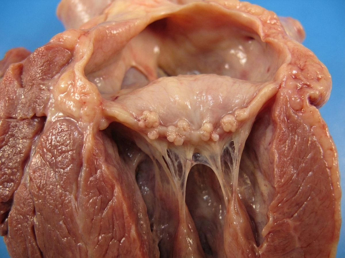
Gross findings of the heart from an 18-year-old female presented with a history of sore throat 3 months back and joint pains have been shown below. On auscultatory finding, a murmur was noted. What is your diagnosis?

What is the average time taken for a myocardial infarction to be completely healed?
A 36-year-old man with a history of long-standing rheumatic heart disease and mitral stenosis dies during cardiac surgery. Autopsy findings are consistent with mitral stenosis and reveal the presence of "heart failure cells." What process leads to the observation of "heart failure cells"?
At the time of autopsy of a 39-year-old female who died of complications of systemic lupus erythematosus, several medium-sized vegetations are found on both sides of the mitral valve and tricuspid valve. These cardiac vegetations are most likely the result of?
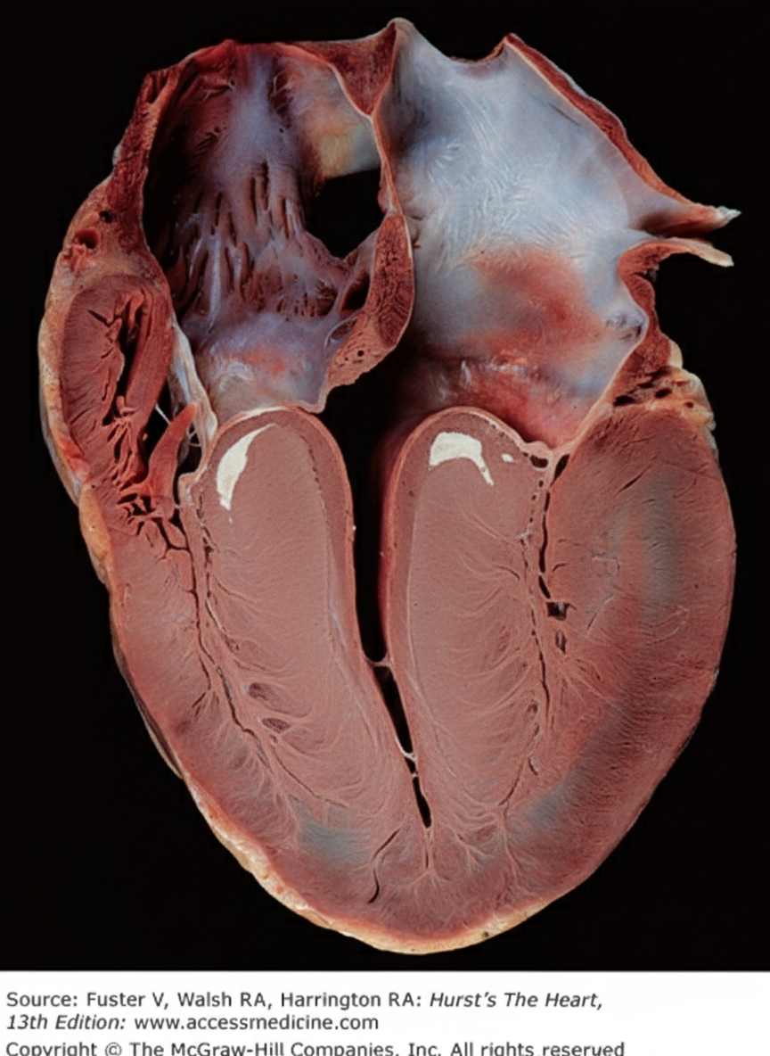
Explanation: **Explanation:** **1. Why Anterior Transmural MI is Correct:** Ventricular aneurysms are a late complication of myocardial infarction (MI), typically occurring weeks to months after the event. They result from the thinning and stretching of necrotic myocardium that has been replaced by fibrous scar tissue [1]. **Anterior transmural MIs** (usually involving the LAD artery) are the most common precursors because the anterior wall and apex are thinner and subjected to higher systolic wall stress compared to other regions [2]. The transmural nature ensures that the entire thickness of the wall is weakened, allowing the intraventricular pressure to cause outward bulging (paradoxical movement) during systole [1]. **2. Why the Other Options are Incorrect:** * **Posterior and Inferior Wall MI:** While these can lead to complications like papillary muscle rupture (leading to mitral regurgitation), they are statistically much less likely to result in true aneurms compared to anterior wall infarcts [1]. * **Subendocardial MI:** By definition, these involve only the inner 1/3 to 1/2 of the ventricular wall [2]. The remaining viable epicardial myocardium provides enough structural integrity to prevent the wall from bulging outward into an aneurysm [4]. **3. Clinical Pearls for NEET-PG:** * **Most Common Site:** The apex of the left ventricle is the most frequent site for post-MI aneurysms [1]. * **Complications:** Ventricular aneurysms do **not** typically rupture (due to dense fibrous scarring), but they predispose patients to **mural thrombus** (and subsequent embolism), refractory heart failure, and ventricular arrhythmias [1], [3]. * **ECG Finding:** Persistent ST-segment elevation in the same leads as the original MI is a classic sign of a ventricular aneurysm. * **True vs. False Aneurysm:** A true aneurysm involves all layers of the thinned heart wall; a pseudoaneurysm (false aneurysm) is a contained rupture by the pericardium and carries a high risk of sudden rupture. **References:** [1] Kumar V, Abbas AK, et al.. Robbins and Cotran Pathologic Basis of Disease. 9th ed. The Heart, pp. 556-557. [2] Cross SS. Underwood's Pathology: A Clinical Approach. 6th ed. Common Clinical Problems From Cardiovascular Disease, pp. 288-289. [3] Cross SS. Underwood's Pathology: A Clinical Approach. 6th ed. Common Clinical Problems From Cardiovascular Disease, pp. 289-290. [4] Kumar V, Abbas AK, et al.. Robbins and Cotran Pathologic Basis of Disease. 9th ed. The Heart, p. 550.
Explanation: **Explanation:** **1. Why Verrucous Endocarditis is correct:** Systemic Lupus Erythematosus (SLE) is classically associated with **Libman-Sacks Endocarditis**, also known as **Verrucous Endocarditis** [1]. The characteristic lesions are small, sterile, pinkish, "wart-like" vegetations (verrucae) that can occur on any heart valve [3]. * **Pathognomonic Feature:** Unlike infective endocarditis, these vegetations can be found on **both sides** of the valve leaflets (surface and undersurface), as well as on the chordae tendineae and endocardial surfaces [2]. * **Histology:** They consist of eosinophilic fibrinoid material with an inflammatory infiltrate, but notably **lack microorganisms** (sterile) [1]. **2. Why other options are incorrect:** * **A. Pericarditis:** While pericarditis is actually the **most common** clinical cardiac manifestation of SLE, it is not considered the "characteristic" or pathognomonic lesion often tested in pathology [3]. * **C. Myocardial fibrosis:** This is a non-specific finding that may occur due to small vessel vasculitis or secondary to hypertension in SLE patients with renal involvement, but it is not a hallmark of the disease. * **D. Valvular incompetence:** While chronic Libman-Sacks endocarditis can lead to scarring and subsequent valvular regurgitation (incompetence), the primary characteristic lesion is the formation of the verrucae themselves [3]. **High-Yield Clinical Pearls for NEET-PG:** * **Location:** Most common on the Mitral and Aortic valves [3]. * **Sterility:** Always remember these are **non-bacterial** (sterile) vegetations [1]. * **Association:** Strongly associated with **Antiphospholipid Antibody Syndrome (APS)** [4]. * **Hematoxylin Bodies:** These may be found within the vegetations, representing the cardiac equivalent of the LE cell. **References:** [1] Kumar V, Abbas AK, et al.. Robbins and Cotran Pathologic Basis of Disease. 9th ed. The Heart, p. 570. [2] Kumar V, Abbas AK, et al.. Robbins and Cotran Pathologic Basis of Disease. 9th ed. The Heart, p. 568. [3] Kumar V, Abbas AK, et al.. Robbins and Cotran Pathologic Basis of Disease. 9th ed. Diseases of the Immune System, pp. 232-233. [4] Kumar V, Abbas AK, et al.. Robbins and Cotran Pathologic Basis of Disease. 9th ed. Hemodynamic Disorders, Thromboembolic Disease, and Shock, pp. 134-135.
Explanation: **Explanation:** The pathognomonic lesion of Acute Rheumatic Heart Disease (RHD) is the **Aschoff body**, which is a form of granulomatous inflammation [1]. However, unlike the "caseating granulomas" seen in Tuberculosis, Aschoff bodies are **non-epithelioid granulomas**. 1. **Why Option B is correct:** **Epithelioid cells** (activated macrophages that resemble epithelial cells) are the hallmark of "true" granulomas seen in TB, Sarcoidosis, or Leprosy [2]. In Aschoff bodies, the macrophages transform into specialized **Anitschkow cells**, not classical epithelioid cells [1]. 2. **Why Options A, C, and D are incorrect:** * **Anitschkow cells (A):** These are specialized macrophages with abundant cytoplasm and "caterpillar-like" nuclei (due to condensed chromatin) [1]. They are the most characteristic feature of Aschoff bodies. * **Giant cells (C):** Aschoff bodies often contain multinucleated giant cells (Aschoff giant cells) formed by the fusion of Anitschkow cells. * **Fibrinoid necrosis (D):** The early (exudative) phase of an Aschoff body is characterized by a central focus of eosinophilic, proteinaceous material known as fibrinoid necrosis. **High-Yield Clinical Pearls for NEET-PG:** * **Phases of Aschoff Body:** 1. Exudative (Fibridoid necrosis) → 2. Proliferative (Anitschkow/Giant cells) → 3. Healing (Fibrosis/Scarring). * **Anitschkow Cells:** Also called "Caterpillar cells" in longitudinal section and "Owl-eye cells" in cross-section [1]. * **Location:** Aschoff bodies can be found in all three layers of the heart (**Pancarditis**), but are most commonly found in the **interstitial connective tissue of the myocardium** [1]. * **MacCallum Patch:** A subendocardial thickening, usually in the left atrium, caused by inflammatory regurgitant jets. **References:** [1] Kumar V, Abbas AK, et al.. Robbins and Cotran Pathologic Basis of Disease. 9th ed. The Heart, pp. 566-567. [2] Kumar V, Abbas AK, et al.. Robbins and Cotran Pathologic Basis of Disease. 9th ed. Inflammation and Repair, p. 109.
Explanation: **Explanation:** **Troponin T (cTnT)** is a component of the troponin complex (along with Troponin I and C) that regulates the calcium-dependent interaction between actin and myosin in cardiac muscle. [2] **Why Myocardial Infarction is correct:** When myocardial cells are damaged due to ischemia (Myocardial Infarction), the cell membrane integrity is lost, causing cardiac-specific proteins to leak into the bloodstream. [1] Cardiac Troponin T (cTnT) and Troponin I (cTnI) are the **gold standard biomarkers** for diagnosing MI because they are highly sensitive and specific to cardiac injury. [2] They typically rise within 3–12 hours of injury, peak at 24 hours, and cTnT can remain elevated for up to 10–14 days. [1] **Why other options are incorrect:** * **Renal disease:** While cTnT can be chronically elevated in end-stage renal disease (due to decreased clearance or silent micro-infarcts), it is not a diagnostic marker for renal disease itself. * **Muscular dystrophy:** This condition primarily involves skeletal muscle. While skeletal troponins exist, the clinical "Troponin T" test specifically targets the **cardiac isoform**, making it a poor marker for primary muscular dystrophies (where Creatine Kinase/CK is more relevant). * **Cirrhosis of the liver:** Liver damage is monitored via ALT, AST, and Bilirubin. Troponin has no physiological role in hepatic pathology. **High-Yield Clinical Pearls for NEET-PG:** * **Most Specific Marker:** Troponin I is considered slightly more cardio-specific than Troponin T. * **Earliest Marker:** Myoglobin (rises in 1–2 hours), but it lacks specificity. * **Marker for Re-infarction:** CK-MB is preferred because it returns to baseline within 48–72 hours, whereas Troponins stay elevated for over a week. * **Troponin C** is not used clinically for MI diagnosis because it is identical in both skeletal and cardiac muscle. **References:** [1] Kumar V, Abbas AK, et al.. Robbins and Cotran Pathologic Basis of Disease. 9th ed. The Heart, pp. 552-554. [2] Cross SS. Underwood's Pathology: A Clinical Approach. 6th ed. Common Clinical Problems From Cardiovascular Disease, pp. 288-289.
Explanation: **Explanation:** The correct answer is **Constrictive Pericarditis**. **Why it is correct:** Tuberculosis (TB) is the most common cause of constrictive pericarditis in developing countries. Pathologically, TB causes a chronic inflammatory response characterized by granulomatous inflammation. Over time, the healing process involves extensive **fibrosis and calcification** of the pericardium [1]. This results in a rigid, "shell-like" casing (concretio cordis) that prevents the heart from expanding during diastole, leading to the clinical features of constriction. **Why the other options are incorrect:** * **Hemorrhagic:** While TB can occasionally cause bloody effusions, hemorrhagic pericarditis is most classically associated with **malignancy** (metastatic spread) or post-cardiac surgery [1]. * **Fibrinous:** This is characterized by a "bread and butter" appearance and is most commonly seen in **Acute Myocardial Infarction** (Dressler syndrome) or Uremia. * **Serofibrinous:** This is the most common type of *acute* pericarditis overall, often seen in viral infections or Rheumatic Fever. While TB may start as a serofibrinous exudate, its hallmark and most definitive presentation in pathology exams is the progression to **constriction** [1]. **High-Yield Clinical Pearls for NEET-PG:** * **Imaging:** Chest X-ray may show a "calcified ring" around the heart. * **Clinical Sign:** **Kussmaul’s sign** (paradoxical rise in JVP during inspiration) and a **Pericardial Knock** (early diastolic sound) are classic findings. * **Treatment:** Surgical pericardiectomy is the definitive treatment for symptomatic constrictive pericarditis. * **Gold Standard:** Cardiac catheterization showing the "Square root sign" (dip-and-plateau pattern) in ventricular pressure tracings. **References:** [1] Kumar V, Abbas AK, et al.. Robbins and Cotran Pathologic Basis of Disease. 9th ed. The Heart, pp. 582-583.
Explanation: **Explanation:** **Concentric hypertrophy** is characterized by an increase in ventricular wall thickness with a decrease or no change in the internal chamber diameter. This occurs in response to **pressure overload** or genetic mutations affecting sarcomeric proteins [1]. 1. **Why Hypertrophic Cardiomyopathy (HCM) is correct:** HCM is a genetic disorder (most commonly mutations in the Beta-myosin heavy chain) that leads to significant **concentric hypertrophy**, often involving the interventricular septum disproportionately (Asymmetric Septal Hypertrophy) [2, 5]. Histologically, it is marked by **myocyte disarray**. 2. **Why other options are incorrect:** * **Systemic Hypertension:** While chronic hypertension *does* cause concentric hypertrophy, in the context of NEET-PG questions, if both HCM and Hypertension are listed, HCM is the classic pathological prototype for primary concentric changes [3]. However, note that many textbooks consider both A and C as causes; HCM is the intrinsic genetic cause, while Hypertension is the extrinsic hemodynamic cause. * **Congestive Heart Failure (CHF):** This is a clinical syndrome, not a specific morphological pattern. Most end-stage CHF cases involve **eccentric hypertrophy** (dilated chambers) due to systolic dysfunction. * **Mitral Regurgitation:** This causes **volume overload**, which leads to **eccentric hypertrophy** (chamber dilation with compensatory wall thinning) [1]. **High-Yield Clinical Pearls for NEET-PG:** * **Pressure Overload (Concentric):** Seen in Systemic Hypertension and Aortic Stenosis [1, 4]. New sarcomeres are added in **parallel**. * **Volume Overload (Eccentric):** Seen in Valvular Regurgitation (AR/MR) [1]. New sarcomeres are added in **series**. * **HCM Triad:** Sudden cardiac death in young athletes, systolic anterior motion (SAM) of the mitral valve, and myocyte disarray [2]. **References:** [1] Kumar V, Abbas AK, et al.. Robbins and Cotran Pathologic Basis of Disease. 9th ed. The Heart, p. 536. [2] Cross SS. Underwood's Pathology: A Clinical Approach. 6th ed. Common Clinical Problems From Cardiovascular Disease, pp. 303-304. [3] Kumar V, Abbas AK, et al.. Robbins and Cotran Pathologic Basis of Disease. 9th ed. The Heart, pp. 560-562. [4] Kumar V, Abbas AK, et al.. Robbins and Cotran Pathologic Basis of Disease. 9th ed. The Heart, pp. 576-577.
Explanation: ***Rheumatic carditis*** - History of **sore throat** 3 months prior followed by joint pains suggests **Group A Streptococcal** infection leading to **acute rheumatic fever** with carditis. - Gross pathology shows characteristic **small warty vegetations** along the **line of closure** of mitral valve leaflets, consistent with **rheumatic valvulitis**. *Libman-Sacks endocarditis* - Associated with **systemic lupus erythematosus (SLE)**, not preceded by streptococcal throat infection. - Vegetations occur on **both surfaces** of valve leaflets (atrial and ventricular), unlike the linear pattern in rheumatic carditis. *Infective endocarditis* - Characterized by **large, irregular, friable vegetations** that can embolize, not small warty ones. - Typically presents with **fever, positive blood cultures**, and constitutional symptoms, which are absent in this case. *Marantic carditis* - Also known as **non-bacterial thrombotic endocarditis (NBTE)**, associated with **debilitating illnesses** like cancer. - Occurs in patients with **hypercoagulable states** or **cachexia**, not in healthy young individuals following streptococcal infection.
Explanation: **Explanation:** The healing process of a Myocardial Infarction (MI) follows a predictable chronological sequence of inflammation, tissue repair, and remodeling. **1. Why 6 weeks is correct:** Healing of the myocardium occurs by **replacement fibrosis** (scarring), as cardiac myocytes are permanent cells and cannot regenerate [1]. * **0–2 weeks:** Characterized by acute inflammation (neutrophils) followed by macrophage infiltration to clear necrotic debris [1], [2]. * **2–4 weeks:** Granulation tissue is at its peak, characterized by neovascularization and collagen deposition [1]. * **4–8 weeks:** The granulation tissue is gradually replaced by a dense, white fibrous scar [1]. By the end of **6 weeks**, the necrotic area is usually completely replaced by a mature collagenous scar, marking the completion of the healing phase. **2. Why other options are incorrect:** * **A (3 weeks):** At this stage, the tissue is still in the "granulation tissue" phase. It is highly vascular and lacks the tensile strength of a mature scar [1]. * **C & D (12–15 weeks):** While the scar may continue to undergo subtle "remodeling" (contraction) for several months, the primary healing process and the formation of the fibrous bridge are completed much earlier, typically by the 6-to-8-week mark. **High-Yield NEET-PG Pearls:** * **Earliest Gross Change:** 4–12 hours (Occasional dark mottling) [2]. * **Earliest Microscopic Change:** 0.5–4 hours (Wavy fibers) [2]. * **Maximum Risk of Rupture:** 3–7 days (when the wall is softest due to macrophage-mediated lysis of necrotic tissue, known as *myomalacia cordis*) [1]. * **Staining:** Triphenyltetrazolium chloride (TTC) stains viable myocardium **red** (due to LDH enzyme); infarcted areas remain **pale/white** [1]. **References:** [1] Kumar V, Abbas AK, et al.. Robbins and Cotran Pathologic Basis of Disease. 9th ed. The Heart, pp. 552-554. [2] Kumar V, Abbas AK, et al.. Robbins and Cotran Pathologic Basis of Disease. 9th ed. The Heart, p. 552.
Explanation: **Explanation:** **1. Why Option B is Correct:** "Heart failure cells" are **hemosiderin-laden alveolar macrophages**. In the setting of mitral stenosis, there is an obstruction to left atrial outflow, leading to increased pressure in the left atrium and pulmonary veins. This results in **chronic passive congestion of the lungs** [1]. The increased hydrostatic pressure causes capillary rupture and intra-alveolar hemorrhage. Alveolar macrophages phagocytose the extravasated red blood cells and break down the hemoglobin into hemosiderin, a golden-brown pigment. The presence of these cells is a classic histological hallmark of chronic left-sided heart failure [1]. **2. Why Incorrect Options are Wrong:** * **Option A:** While the coagulation cascade is involved in thrombus formation (common in the left atrium in mitral stenosis), it does not directly produce pigmented macrophages in the alveoli. * **Option C:** Hypoxic myocardial injury refers to ischemia [1]. While heart failure can lead to hypoxia, the specific formation of "heart failure cells" is a pulmonary manifestation of back-pressure, not a direct result of myocardial cell death. * **Option D:** Hyperemia is an active process involving increased arterial inflow. Passive congestion (the cause here) is a passive process resulting from impaired venous outflow. **3. NEET-PG High-Yield Pearls:** * **Brown Induration of Lung:** This is the gross appearance of the lungs in chronic passive congestion, caused by the combination of hemosiderin deposition and compensatory alveolar septal fibrosis. * **Nutmeg Liver:** This refers to chronic passive congestion of the **liver**, typically seen in **right-sided** heart failure (centrilobular necrosis and congestion) [1]. * **Prussian Blue Stain:** This stain is used to confirm the presence of iron (hemosiderin) within heart failure cells, appearing as bright blue granules. **References:** [1] Kumar V, Abbas AK, et al.. Robbins and Cotran Pathologic Basis of Disease. 9th ed. The Heart, pp. 536-537.
Explanation: The clinical presentation describes **Libman-Sacks Endocarditis (LSE)**, also known as verrucous endocarditis, which is a classic cardiac manifestation of **Systemic Lupus Erythematosus (SLE)** [1]. ### **Explanation of the Correct Answer** Libman-Sacks endocarditis is characterized by small-to-medium-sized, sterile, pinkish vegetations [1]. Unlike most other forms of endocarditis, these vegetations can occur on **both sides of the valve leaflets** (undersurface and flow surface) and on the chordae tendineae [1], [3]. * **Pathogenesis:** The formation of these vegetations is strongly associated with **Antiphospholipid Syndrome (APS)**, specifically the presence of **anticardiolipin antibodies** and lupus anticoagulant [2]. These antibodies promote a prothrombotic state and endothelial injury, leading to the deposition of fibrin-platelet thrombi on the valves. ### **Analysis of Incorrect Options** * **Option B:** Turbulent flow through an incompetent valve is the mechanism for **Non-Bacterial Thrombotic Endocarditis (NBTE)**, typically seen in wasting diseases (Marantic endocarditis) [3]. While LSE is also non-bacterial, the specific association with SLE and "both sides of the valve" points to LSE. * **Option C:** Abnormal secretion of vasoactive amines (like serotonin) causes **Carcinoid Heart Disease**, which typically affects the right-sided valves (tricuspid/pulmonary) and presents as fibrous plaque-like thickenings rather than vegetations on both sides. * **Option D:** Bacterial colonization is the hallmark of **Infective Endocarditis**. These vegetations are usually large, friable, and occur only on the flow surface of the valve [3]. ### **NEET-PG High-Yield Pearls** * **LSE Location:** Mitral valve is most common; uniquely occurs on **both surfaces** of the valve [3]. * **Histology:** Vegetations consist of fibrin, inflammatory cells, and "hematoxylin bodies" (LE cells). * **Sterility:** Vegetations are **sterile** (non-infective) [1]. * **Association:** Highly associated with **Antiphospholipid Antibody Syndrome (APS)** [2]. **References:** [1] Kumar V, Abbas AK, et al.. Robbins and Cotran Pathologic Basis of Disease. 9th ed. The Heart, p. 570. [2] Kumar V, Abbas AK, et al.. Robbins and Cotran Pathologic Basis of Disease. 9th ed. Hemodynamic Disorders, Thromboembolic Disease, and Shock, pp. 134-135. [3] Kumar V, Abbas AK, et al.. Robbins and Cotran Pathologic Basis of Disease. 9th ed. The Heart, p. 568.
Congenital Heart Disease
Practice Questions
Ischemic Heart Disease
Practice Questions
Hypertensive Heart Disease
Practice Questions
Valvular Heart Disease
Practice Questions
Myocarditis and Cardiomyopathies
Practice Questions
Pericardial Disease
Practice Questions
Cardiac Tumors
Practice Questions
Heart Failure Pathophysiology
Practice Questions
Cardiac Transplantation Pathology
Practice Questions
Endocarditis
Practice Questions
Get full access to all questions, explanations, and performance tracking.
Start For Free