Biphasic histopathology is seen in which of the following conditions?
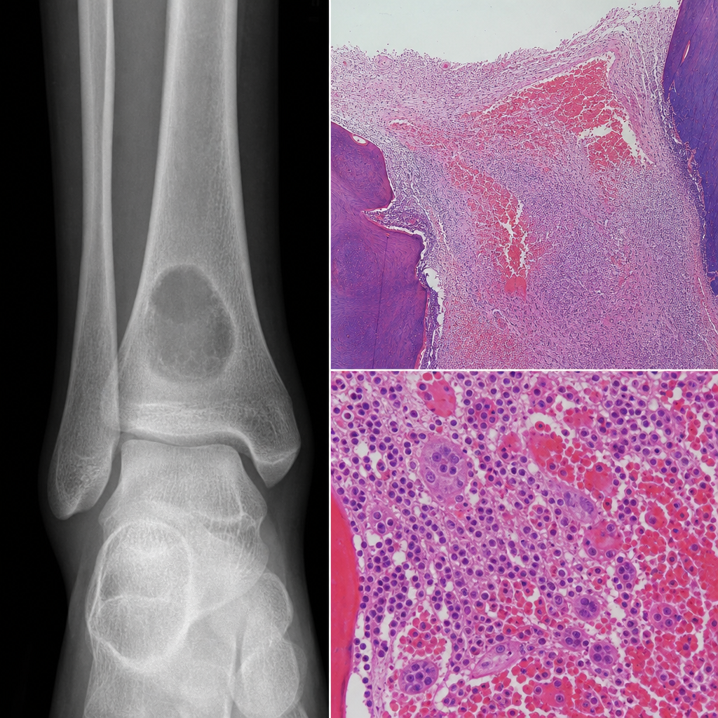
A patient presented with pain in his ankle. X-ray shows an osteolytic lesion with a sclerotic rim. Histopathological findings are available. What is your diagnosis?
All are congenital myopathies EXCEPT?
A 70-year-old male patient presents with a single, well-defined lytic lesion of the skull. The patient has no other complaints, and urine examination shows no abnormalities. What is the most likely diagnosis?
A 50-year-old patient presents with a midline lesion involving the sacrum, which is found to be sclerotic. What is the most probable diagnosis?
A 70-year-old male has a pathologic fracture of the femur. X-rays show a lytic lesion with a circumscribed, punched-out appearance. Curettage from the fracture site is most likely to show which of the following?
Which of the following is false regarding Cherubism?
Precancerous potential in Plummer-Vinson syndrome may be due to which change in the epithelium?
All of the following are examples of round cell tumors except?
Which of the following is the diagnostic characteristic of peripheral giant cell granuloma?
Explanation: **Explanation:** The term **"Biphasic"** in histopathology refers to the presence of two distinct cell populations within a single tumor—typically an **epithelial component** (forming glands or nests) and a **mesenchymal/spindle cell component** (stromal elements). **1. Why Mesothelioma is correct:** Malignant Mesothelioma classically presents in three histological patterns: Epithelioid (most common), Sarcomatoid (spindle cells), and **Biphasic** [3]. The biphasic type contains both cuboidal/epithelial-like cells and malignant spindle cells. It is a classic "high-yield" example of a biphasic tumor in pathology exams. **2. Analysis of other options:** * **Synovial Sarcoma:** This is a **controversial** point. Synovial sarcoma is indeed a biphasic tumor (containing epithelial and spindle cells) [1]. However, in the context of this specific question (often a repeat from older AIIMS/NEET patterns), **Mesothelioma** is frequently prioritized as the "most classic" answer unless "Both" is an option. *Note: If this were a multiple-choice question where more than one could be right, Synovial Sarcoma would also be correct.* * **Rhabdomyoma:** This is a benign skeletal muscle tumor characterized by "spider cells" (vacuolated cells with glycogen). It is monophasic. * **Neurofibromatosis:** This is a genetic condition (NF1/NF2). The associated tumors (Neurofibromas) consist of a mixture of Schwann cells, fibroblasts, and perineural cells, but they are not classified as "biphasic" in the traditional epithelial-mesenchymal sense. **Clinical Pearls for NEET-PG:** * **Other Biphasic Tumors:** Fibroadenoma (breast), Phyllodes tumor, Pleomorphic adenoma (salivary gland) [2], and Wilms tumor (which is actually *triphasic*: blastemal, stromal, and epithelial). * **Mesothelioma Marker:** Calretinin is the most specific immunohistochemical marker. * **Asbestos Link:** While asbestos is the primary risk factor, the most common histological type is Epithelioid, not Biphasic. **References:** [1] Kumar V, Abbas AK, et al.. Robbins and Cotran Pathologic Basis of Disease. 9th ed. Bones, Joints, and Soft Tissue Tumors, pp. 1225-1226. [2] Kumar V, Abbas AK, et al.. Robbins and Cotran Pathologic Basis of Disease. 9th ed. Diseases of the Immune System, pp. 274-276. [3] Cross SS. Underwood's Pathology: A Clinical Approach. 6th ed. Common Clinical Problems From Respiratory Tract Disease, pp. 339-340.
Explanation: **Explanation:** **Hemophilic Pseudotumor** is a rare but serious complication of severe hemophilia (Factor VIII or IX deficiency). It results from repeated, chronic subperiosteal or intraosseous hemorrhages. The pressure from the expanding hematoma leads to pressure necrosis of the bone, appearing on X-ray as a well-defined **osteolytic lesion** often surrounded by a **sclerotic rim** (representing the bone's attempt at repair). Histologically, it reveals organized blood clots, hemosiderin-laden macrophages, and fibrous tissue. **Analysis of Incorrect Options:** * **Eumycosis (Madura foot):** Typically presents with soft tissue swelling, multiple draining sinuses, and "sulfur granules." X-rays show "moth-eaten" bone destruction rather than a clean sclerotic rim. * **Ochronosis (Alkaptonuria):** Characterized by the deposition of homogentisic acid in connective tissues. It typically involves the spine (disc calcification) and large joints, presenting with joint space narrowing and subchondral sclerosis, not isolated lytic lesions. * **Pigmented Villonodular Synovitis (PVNS):** This is a proliferative lesion of the synovium. While it can cause "apple-core" erosions at the joint margins, it is primarily a soft tissue process characterized by brown, frond-like synovial projections and massive hemosiderin deposition on MRI (blooming effect). **NEET-PG High-Yield Pearls:** * **Location:** Most common in the femur, pelvis, and small bones of the hands/feet. * **Radiology:** Can mimic a malignant bone tumor (like Osteosarcoma) due to its size, but the sclerotic border and clinical history of a bleeding disorder are diagnostic clues. * **Management:** Factor replacement is the first line; surgery is high-risk due to potential uncontrollable hemorrhage.
Explanation: **Explanation:** Congenital myopathies are a group of genetically determined primary muscle disorders characterized by specific structural abnormalities in muscle fibers, early-onset hypotonia ("floppy infant syndrome"), and a non-progressive or slowly progressive clinical course [1]. **Why Option C is the Correct Answer:** **"Z band myopathy"** is not a recognized clinical entity or a specific category of congenital myopathy. While many congenital myopathies involve abnormalities of the Z-disk (such as Nemaline myopathy), there is no specific disease named "Z band myopathy." Therefore, it is the "except" in this list. **Analysis of Incorrect Options:** * **Central Core Myopathy (Option A):** Characterized by pale, "core-like" areas in the center of Type 1 muscle fibers that lack oxidative enzyme activity. It is strongly associated with mutations in the **RYR1 gene** and carries a high risk of **malignant hyperthermia**. * **Nemaline Myopathy (Option B):** Defined by the presence of thread-like structures called **"Nemaline rods"** (derived from Z-band material/alpha-actinin) within muscle fibers. It is one of the most common congenital myopathies. * **Centronuclear Myopathy (Option D):** Characterized by the presence of nuclei located in the center of the muscle fiber (resembling fetal myotubes) rather than the normal peripheral location. The X-linked form is known as **Myotubular Myopathy** (MTM1 mutation). **High-Yield NEET-PG Pearls:** 1. **Gomori Trichrome Stain:** This is the gold standard for identifying **Nemaline rods** (they appear red). 2. **Malignant Hyperthermia:** Always associate **Central Core Disease** with anesthesia-induced malignant hyperthermia due to RYR1 mutations. 3. **NADH-TR Stain:** Used to visualize the "cores" in Central Core Disease (cores appear as pale zones). **References:** [1] Kumar V, Abbas AK, et al.. Robbins and Cotran Pathologic Basis of Disease. 9th ed. Peripheral Nerves and Skeletal Muscles, pp. 1247-1248.
Explanation: ### Explanation **Correct Answer: A. Langerhans cell histiocytosis (LCH)** The clinical presentation of a **single, well-defined lytic lesion** in the skull (often described as a "punched-out" lesion) in an otherwise asymptomatic patient is a classic description of **Eosinophilic Granuloma**, the most common and localized form of Langerhans Cell Histiocytosis (LCH). While LCH is more frequent in children, it can occur in adults [1]. The skull is the most common site of involvement. The absence of urinary abnormalities (like Bence-Jones proteins) helps rule out Multiple Myeloma, which would be a primary differential for lytic lesions in a 70-year-old [2]. **Why other options are incorrect:** * **B & C (Myeloproliferative Disorders):** These typically present with splenomegaly, hypercellular bone marrow, and systemic symptoms (fever, weight loss). They do not characteristically present as solitary, well-defined lytic bone lesions. * **D (Tumor of osteoblasts):** Osteoblastoma or Osteoid osteoma usually present with painful lesions (often relieved by NSAIDs) and typically show sclerotic margins or a central nidus, rather than a purely lytic "punched-out" appearance. **NEET-PG High-Yield Pearls for LCH:** * **Pathognomonic feature:** **Birbeck granules** on Electron Microscopy (tennis-racket shaped) [1]. * **Immunohistochemistry (IHC):** Positive for **CD1a**, **S100**, and **Langerin (CD207)** [1]. * **Radiology:** "Punched-out" lytic lesions without a sclerotic rim. * **Hand-Schüller-Christian triad:** Bone lesions, exophthalmos, and diabetes insipidus (seen in multifocal chronic LCH). * **Letterer-Siwe disease:** The aggressive, multisystem form seen in infants (<2 years). **References:** [1] Kumar V, Abbas AK, et al.. Robbins and Cotran Pathologic Basis of Disease. 9th ed. Diseases of White Blood Cells, Lymph Nodes, Spleen, and Thymus, pp. 629-630. [2] Cross SS. Underwood's Pathology: A Clinical Approach. 6th ed. Common Clinical Problems From Blood And Bone Marrow Disease, pp. 616-617.
Explanation: ### Explanation **1. Why Chordoma is Correct:** Chordoma is a rare, slow-growing malignant tumor derived from **notochordal remnants**. It has a strong predilection for the axial skeleton, specifically the **midline**. The two most common sites are the **sacrococcygeal region** (50%) and the **spheno-occipital (clivus) region** (35%). While chordomas are typically described as osteolytic or destructive on imaging, they can frequently induce reactive sclerosis or present as mixed lesions, especially in the sacrum. The combination of a **midline location** and **sacral involvement** in a middle-aged adult is the classic "textbook" presentation for Chordoma. **2. Why Other Options are Incorrect:** * **Metastasis:** While common in the sacrum, metastases are usually multiple and rarely strictly confined to the midline [2]. Prostatic metastasis is typically sclerotic, but Chordoma is a more specific diagnosis for a primary midline sacral mass. * **Osteosarcoma:** This typically affects the metaphysis of long bones (like the distal femur) in adolescents. When it occurs in adults, it is usually secondary to Paget’s disease or radiation and is rarely localized specifically to the midline sacrum [3]. * **Chondrosarcoma:** Although it can involve the pelvis, it usually presents as a large, lobulated mass with "popcorn" calcifications [1]. It does not have the same specific affinity for the midline notochordal axis as Chordoma [3]. **3. High-Yield Clinical Pearls for NEET-PG:** * **Histology:** Characterized by **Physaliphorous cells** (large, vacuolated cells with "soap-bubble" appearance) in a myxoid stroma. * **Immunohistochemistry (IHC):** Positive for **Brachyury** (most specific), S-100, and Cytokeratin (CK). * **Age Group:** Typically occurs in the 40–60 year age range [1]. * **Prognosis:** Locally aggressive with a high rate of recurrence; surgical resection is the primary treatment. **References:** [1] Kumar V, Abbas AK, et al.. Robbins and Cotran Pathologic Basis of Disease. 9th ed. Bones, Joints, and Soft Tissue Tumors, p. 1204. [2] Cross SS. Underwood's Pathology: A Clinical Approach. 6th ed. Common Clinical Problems From Osteoarticular And Connective Tissue Disease, pp. 671-672. [3] Cross SS. Underwood's Pathology: A Clinical Approach. 6th ed. Common Clinical Problems From Osteoarticular And Connective Tissue Disease, pp. 673-674.
Explanation: ### Explanation **Correct Answer: B. Sheets of atypical plasma cells** The clinical presentation of a **70-year-old male** [3] with a **pathologic fracture** and classic **"punched-out" lytic lesions** on X-ray [1] is highly suggestive of **Multiple Myeloma**. Multiple myeloma is a plasma cell dyscrasia characterized by the neoplastic proliferation of a single clone of plasma cells [4]. These cells secrete cytokines (like IL-6) and RANKL, which activate osteoclasts, leading to bone resorption and the characteristic radiolucent lesions [1]. Histologically, curettage of such a lesion reveals dense sheets of atypical plasma cells (large cells with eccentric nuclei, "clock-face" chromatin, and a prominent perinuclear Golgi zone/hof) [2]. **Analysis of Incorrect Options:** * **A. Osteopenia/Osteoporosis:** While this causes thinned trabeculae and fractures in the elderly, it presents with generalized bone loss rather than focal, circumscribed "punched-out" lytic lesions [2]. * **C. Metastatic prostatic adenocarcinoma:** Prostate cancer typically produces **osteoblastic (sclerotic)** lesions, which appear dense and white on X-ray, rather than lytic lesions. * **D. Malignant cells forming osteoid:** This describes **Osteosarcoma**. While it causes bone destruction, it is characterized by the production of mineralized or unmineralized bone (osteoid) by malignant cells and typically follows a bimodal age distribution (teens or elderly with Paget’s disease), usually presenting with a "sunburst" appearance or Codman’s triangle. **High-Yield Clinical Pearls for NEET-PG:** * **CRAB Criteria for Myeloma:** **C**alcium (high), **R**enal insufficiency, **A**nemia, **B**one lesions [5]. * **Radiology:** "Raindrop skull" (multiple lytic spots on the cranium) [1]. * **Diagnosis:** Serum/Urinary Protein Electrophoresis (M-spike) and Bone Marrow Biopsy (>10% plasma cells) [2], [3]. * **Bence-Jones Proteins:** Free light chains in urine that precipitate at 40-60°C and redissolve at 100°C [3]. **References:** [1] Kumar V, Abbas AK, et al.. Robbins and Cotran Pathologic Basis of Disease. 9th ed. Diseases of White Blood Cells, Lymph Nodes, Spleen, and Thymus, p. 608. [2] Cross SS. Underwood's Pathology: A Clinical Approach. 6th ed. Common Clinical Problems From Blood And Bone Marrow Disease, pp. 617-618. [3] Cross SS. Underwood's Pathology: A Clinical Approach. 6th ed. Common Clinical Problems From Blood And Bone Marrow Disease, pp. 616-617. [4] Kumar V, Abbas AK, et al.. Robbins and Cotran Pathologic Basis of Disease. 9th ed. Diseases of White Blood Cells, Lymph Nodes, Spleen, and Thymus, pp. 606-607. [5] Cross SS. Underwood's Pathology: A Clinical Approach. 6th ed. Common Clinical Problems From Blood And Bone Marrow Disease, pp. 618-619.
Explanation: **Explanation:** **Cherubism** is a rare, autosomal dominant fibro-osseous disorder characterized by symmetrical, non-neoplastic enlargement of the jaws. It is caused by mutations in the **SH3BP2 gene** on chromosome 4p16. 1. **Why Option A is the Correct Answer (False Statement):** Radiographically, Cherubism presents as **multilocular**, expansive radiolucencies (often described as a "soap-bubble" appearance). These lesions are characteristically **bilateral** and involve the mandible (angle and ramus) and/or the maxilla. A unilocular presentation is inconsistent with the classic pathology of this condition. 2. **Analysis of Other Options:** * **Option B (Bilateral):** This is a hallmark feature. The symmetrical involvement of the lower face gives the child a characteristic "cherubic" (angel-like) appearance with upward-looking eyes due to maxillary involvement stretching the skin. * **Option C (Presence of Giant cells):** Histologically, the lesions consist of vascularized fibrous tissue containing numerous **multinucleated giant cells**, resembling a Central Giant Cell Granuloma (CGCG). * **Option D (Delayed eruption):** The massive expansion of the alveolar bone often leads to the displacement of tooth buds, failure of permanent teeth to erupt, or premature loss of primary teeth. **High-Yield Clinical Pearls for NEET-PG:** * **Inheritance:** Autosomal Dominant (High penetrance in males, lower in females). * **Age of Onset:** Typically appears between ages 2–5 years. * **Natural History:** The lesions usually undergo **spontaneous regression** or remodeling after puberty; hence, surgery is often delayed. * **Histology Tip:** Look for **perivascular collagen cuffing** (eosinophilic cuffing around small capillaries), which helps differentiate Cherubism from other giant cell lesions.
Explanation: **Plummer-Vinson Syndrome (PVS)**, also known as Paterson-Brown-Kelly syndrome, is characterized by the triad of iron-deficiency anemia, dysphagia (due to esophageal webs), and glossitis [1]. ### **Mechanism of Malignancy** The correct answer is **Atrophy**. Iron is a critical cofactor for cytochrome oxidase and other enzymes necessary for the normal maturation and maintenance of rapidly dividing mucosal cells. Chronic iron deficiency leads to a depletion of these enzymes, resulting in **mucosal atrophy** of the upper aerodigestive tract (tongue, pharynx, and esophagus). Atrophic epithelium is thin, fragile, and has a diminished regenerative capacity. This compromised state makes the DNA of the basal cells more susceptible to carcinogens (like tobacco or alcohol) and spontaneous mutations, significantly increasing the risk of **Squamous Cell Carcinoma (SCC)** of the post-cricoid region and esophagus [1]. ### **Why Other Options are Incorrect** * **Hypertrophy:** This refers to an increase in cell size. In PVS, the lack of iron leads to a "wasting" or thinning of the tissue, the exact opposite of hypertrophy. * **Acanthosis:** This is a thickening of the stratum spinosum (prickle cell layer). While seen in some inflammatory skin conditions, PVS is defined by the loss of epithelial layers, not their thickening. ### **High-Yield Clinical Pearls for NEET-PG** * **The Triad:** Iron deficiency anemia + Dysphagia + Esophageal webs [1]. * **Demographics:** Most common in middle-aged Scandinavian women. * **Clinical Signs:** Koilonychia (spoon-shaped nails), glossitis (smooth red tongue), and cheilosis [1]. * **Cancer Risk:** It is a premalignant condition for **Post-cricoid Squamous Cell Carcinoma** [1]. * **Radiology:** Esophageal webs are best visualized using a **Barium Swallow** (seen as thin, eccentric membrane-like projections). **References:** [1] Cross SS. Underwood's Pathology: A Clinical Approach. 6th ed. Common Clinical Problems From Blood And Bone Marrow Disease, pp. 591-592.
Explanation: **Explanation:** Small round blue cell tumors (SRBCTs) are a group of malignant neoplasms characterized by small, primitive-appearing cells with high nuclear-to-cytoplasmic ratios, hyperchromatic nuclei, and scant cytoplasm. On H&E staining, they appear predominantly blue due to the dense chromatin. **Why Osteosarcoma is the Correct Answer:** Osteosarcoma is primarily a **spindle cell tumor**, not a round cell tumor [1]. Its hallmark histological feature is the production of **osteoid** (unmineralized bone) by malignant mesenchymal cells [1]. While some variants (like small cell osteosarcoma) may mimic round cell tumors, the classic presentation involves pleomorphic spindle cells with significant cellular atypia [1]. **Analysis of Incorrect Options:** * **Neuroblastoma:** A classic pediatric SRBCT derived from neural crest cells. It is characterized by **Homer-Wright rosettes** and elevated urinary catecholamines (VMA/HVA). * **Non-Hodgkin’s Lymphoma (NHL):** Specifically types like Burkitt lymphoma or lymphoblastic lymphoma, these consist of sheets of small, round lymphoid cells [3]. * **Ewing’s Sarcoma:** A prototypical round cell tumor of the bone. It is associated with the **t(11;22)** translocation and typically shows **Homer-Wright or Flexner-Wintersteiner-like rosettes** and PAS-positive cytoplasm (due to glycogen). **High-Yield Clinical Pearls for NEET-PG:** * **Mnemonic for Round Cell Tumors:** "**MR ENB**" (Medulloblastoma, Retinoblastoma, Ewing’s, NHL, Neuroblastoma). * **Ewing’s Sarcoma:** Look for "Onion-skin" periosteal reaction on X-ray and CD99 (MIC2) positivity. * **Osteosarcoma:** Look for "Sunburst appearance" and "Codman’s triangle" on X-ray [2]. It is the most common primary malignant bone tumor in adolescents. **References:** [1] Cross SS. Underwood's Pathology: A Clinical Approach. 6th ed. Common Clinical Problems From Osteoarticular And Connective Tissue Disease, pp. 673-674. [2] Kumar V, Abbas AK, et al.. Robbins and Cotran Pathologic Basis of Disease. 9th ed. Bones, Joints, and Soft Tissue Tumors, pp. 1200-1202. [3] Cross SS. Underwood's Pathology: A Clinical Approach. 6th ed. Common Clinical Problems From Diseases Of The Urinary And Male Genital Tracts, pp. 560-561.
Explanation: **Explanation:** **Peripheral Giant Cell Granuloma (PGCG)** is a common reactive exophytic lesion of the oral cavity, specifically occurring on the gingiva or alveolar ridge. **Why Option B is Correct:** The hallmark histological feature of PGCG is the presence of numerous **multinucleated giant cells** (resembling osteoclasts) scattered within a highly vascularized fibrocellular stroma. These giant cells are considered the diagnostic signature of the lesion. They are often separated by spindle-shaped mesenchymal cells and extravasated red blood cells, which frequently lead to **hemosiderin deposition**, giving the lesion its characteristic reddish-blue clinical appearance. **Analysis of Incorrect Options:** * **Option A:** While PGCG is highly vascular, it is distinct from "granulation tissue" (which is the hallmark of a Pyogenic Granuloma). PGCG is a specific reactive hyperplasia, not simple inflammatory repair tissue. * **Option C:** Keloid-like enlargement refers to excessive collagen deposition (hypertrophic scarring), which is not a feature of PGCG. PGCG is cellular and vascular, not densely collagenous. * **Option D:** While the overlying epithelium may be ulcerated due to trauma, atrophic epithelium is not a diagnostic or defining characteristic of this pathology. **NEET-PG High-Yield Pearls:** * **Origin:** It arises from the **periosteum** or periodontal ligament. * **Clinical Site:** Exclusively found on the **gingiva** (unlike Central Giant Cell Granuloma, which is intraosseous). * **Radiology:** May show "cupping" resorption of the underlying alveolar bone. * **Differential Diagnosis:** Must be clinically differentiated from Pyogenic Granuloma and Peripheral Ossifying Fibroma. * **Association:** Always rule out **Hyperparathyroidism** (Brown tumor) if multiple giant cell lesions are present.
Bone Development and Growth
Practice Questions
Fracture Healing
Practice Questions
Osteomyelitis and Infectious Diseases
Practice Questions
Metabolic Bone Diseases
Practice Questions
Bone Tumors and Tumor-like Lesions
Practice Questions
Joints and Rheumatologic Diseases
Practice Questions
Soft Tissue Tumors
Practice Questions
Muscular Dystrophies and Myopathies
Practice Questions
Diseases of Tendons and Fascia
Practice Questions
Pathology of Orthopedic Implants
Practice Questions
Get full access to all questions, explanations, and performance tracking.
Start For Free