What is the gold standard for the diagnosis of osteoporosis?
Which vitamin deficiency is most commonly associated with rickets in children?
A child presents with rachitic changes in the limbs that are not responding to Vitamin D supplementation. Investigations reveal the following results: - Calcium: $9.5 \mathrm{mg} / \mathrm{dl}$ - Phosphorus: $1.6 \mathrm{mg} / \mathrm{dl}$ - Alkaline phosphatase (ALP): 814 IU - Serum PTH: $24.2 \mathrm{pg} / \mathrm{ml}$ - Serum electrolytes, creatinine, and blood gases: Normal. What is the most likely diagnosis?
The most important regulator of serum 1,25(OH)2 vitamin D concentration is:
A 70 year old male, known case of chronic renal failure suffers from a pathological fracture of Rt femur, the diagnosis is -
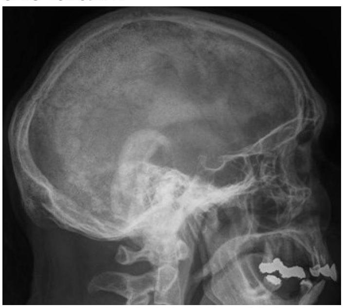
What type of lesions in the skull bones can be identified on this X-ray?

Select the type of bone disease which is most likely to be associated with genetically determined disorder in the structure or processing of type I collagen (SELECT 1 DISEASE)
All are causes of Osteoporosis, except:
Albers-Schönberg disease is:
Marble bone disease is:
Explanation: ***Dual energy X-ray absorptiometry*** - **DXA** is the current **gold standard** for diagnosing osteoporosis and assessing fracture risk due to its high precision and accuracy in measuring **bone mineral density (BMD)**. - It measures BMD at clinically relevant sites such as the **lumbar spine** and **hip**, providing T-scores and Z-scores for comparison. *Single energy X-ray absorptiometry* - **SXA** measures BMD at peripheral sites but is **less accurate** and comprehensive than DXA for diagnosing osteoporosis. - It has **limited utility** as a diagnostic tool for osteoporosis at the hip or spine, which are critical sites for fracture risk. *Ultrasound* - **Quantitative ultrasound (QUS)** can assess bone quality but is primarily used for **screening** and is not accurate enough for definitive diagnosis or treatment monitoring of osteoporosis. - It does **not provide direct bone mineral density measurements** comparable to DXA for diagnostic purposes. *Quantitative computed tomography* - **QCT** can measure volumetric bone density and is useful for assessing **trabecular bone**, but involves higher radiation exposure than DXA. - It is **more expensive** and less readily available than DXA, making it a secondary option for osteoporosis diagnosis.
Explanation: ***Vitamin D*** - **Vitamin D** plays a crucial role in the absorption of **calcium** and **phosphate**, which are essential for proper **bone mineralization**. - A deficiency leads to impaired mineralization of newly formed bone matrix, resulting in soft, weak bones characteristic of **rickets** in children and **osteomalacia** in adults. *Vitamin A* - **Vitamin A** is primarily involved in **vision**, immune function, and cell growth and differentiation. - While essential for health, its deficiency is not directly linked to the skeletal deformities seen in rickets. *Vitamin C* - **Vitamin C** is vital for **collagen synthesis**, a key component of connective tissues, skin, and bone matrix. - Its deficiency causes **scurvy**, characterized by bleeding gums, poor wound healing, and joint pain, not the bone deformities of rickets. *Vitamin K* - **Vitamin K** is essential for **blood coagulation** and plays a role in bone metabolism through the carboxylation of certain bone proteins like **osteocalcin**. - However, its deficiency primarily leads to bleeding disorders and does not cause rickets.
Explanation: ***Hypophosphatemic rickets*** - The combination of **rachitic changes** not responding to Vitamin D, **low serum phosphorus (1.6 mg/dl)**, and **normal calcium and PTH levels** strongly points to hypophosphatemic rickets, a condition characterized by impaired renal phosphate reabsorption. - The **elevated alkaline phosphatase** indicates increased bone turnover as the body tries to mineralize bone despite phosphate deficiency. *Vitamin D-dependent rickets type 2* - This condition involves resistance to **1,25-dihydroxyvitamin D**, leading to **hypocalcemia** and elevated PTH, none of which are present here. - It would also typically show an inadequate response to Vitamin D, but the primary biochemical derangement is different. *Vitamin D-dependent rickets type 1* - This type is caused by a defect in **1-alpha-hydroxylase**, leading to an inability to convert 25-hydroxyvitamin D to its active form, resulting in **hypocalcemia** and elevated PTH, which are not observed. - It would also show a poor response to standard Vitamin D supplementation. *Vitamin D deficiency rickets* - This is the most common form of rickets caused by inadequate Vitamin D intake or synthesis, presenting with **hypocalcemia**, **elevated PTH**, and **low phosphorus**. - However, it typically responds well to Vitamin D supplementation, unlike the presentation here, and would show elevated PTH levels. *Chronic renal failure* - Chronic renal failure would present with **elevated creatinine**, and typically leads to **secondary hyperparathyroidism** (elevated PTH), **hyperphosphatemia**, and metabolic acidosis, none of which are suggested by the provided lab results. - The serum electrolytes, creatinine, and blood gases are explicitly stated as normal.
Explanation: ***Parathyroid hormone*** - **Parathyroid hormone (PTH)** directly stimulates the **kidney's 1-alpha hydroxylase** enzyme, which converts **25(OH)D** to its active form, **1,25(OH)2D (calcitriol)**. - This regulation is critical for maintaining **calcium and phosphate homeostasis**, with PTH levels increasing when serum calcium is low, thereby boosting 1,25(OH)2D production. *Calcium levels in serum* - While **low serum calcium** indirectly stimulates **PTH** release, which then regulates 1,25(OH)2 vitamin D, calcium itself is not the direct or most important regulator. - The direct regulatory action on the conversion enzyme is mediated by PTH. *Magnesium levels in serum* - **Magnesium** plays a cofactor role in various enzymatic reactions, including those involving vitamin D metabolism, but it is not a direct or primary regulator of **1,25(OH)2 vitamin D concentration**. - Severe **hypomagnesemia** can sometimes impair PTH secretion and action, indirectly affecting vitamin D, but this is a secondary effect. *25-hydroxyvitamin D in serum* - **25-hydroxyvitamin D** is the precursor to **1,25(OH)2 vitamin D**, and its availability limits the maximum potential production of the active form. - However, the *rate* of conversion into the active form and thus the *concentration* of 1,25(OH)2D is primarily dictated by PTH, not the precursor itself.
Explanation: ***Secondary Hyperparathyroidism*** - **Chronic renal failure** causes **hyperphosphatemia** and **decreased production of calcitriol (active vitamin D)**. - This leads to hypocalcemia, which stimulates the parathyroid glands to produce excessive **parathyroid hormone (PTH)**, resulting in bone demineralization and **pathological fractures** [2]. *Scurvy* - Caused by **vitamin C deficiency**, leading to impaired collagen synthesis and fragility of blood vessels. - While it can cause bone pain and potential for fractures in severe cases, it is not directly associated with **chronic renal failure** as a primary cause of pathological fracture. *Vitamin D Resistant rickets* - This is a genetic disorder (e.g., X-linked hypophosphatemia) characterized by impaired renal phosphate reabsorption and normal or elevated PTH levels. - While it causes bone demineralization, it is typically a **childhood-onset condition** [1] and not directly linked to **acquired chronic renal failure** in a 70-year-old male. *Primary Hyperparathyroidism* - Characterized by autonomous **overproduction of PTH** due to parathyroid gland adenoma or hyperplasia, leading to **hypercalcemia** and hypophosphatemia. - Unlike secondary hyperparathyroidism, which is a compensatory response to hypocalcemia in the context of renal failure, primary hyperparathyroidism is a direct parathyroid gland pathology.
Explanation: ***Paget's disease*** - An X-ray of the skull in Paget's disease typically shows **thickening of the skull vault** and areas of both **osteolysis** and **osteosclerosis**, leading to a characteristic "cotton wool" appearance. - The disease involves abnormal bone remodeling, leading to enlarged and weakened bones susceptible to deformity and fracture. *Multiple myeloma* - On a skull X-ray, multiple myeloma usually presents as multiple, sharply-defined, **"punched-out" lytic lesions** without a sclerotic border. - These lesions reflect areas where malignant plasma cells have destroyed bone, which is distinct from the mixed lytic and sclerotic changes of Paget's disease. *Osteosarcoma* - Osteosarcoma is a **primary bone malignancy** that typically presents as a solitary lesion with a mixture of lytic and sclerotic areas, often with a **sunburst or Codman's triangle** periosteal reaction. - It most commonly affects long bones in younger individuals and is a much less common presentation in the skull compared to other bone conditions. *Osteomyelitis* - Osteomyelitis is an **infection of the bone** that would appear on an X-ray as areas of bone destruction (lysis) and new bone formation (sclerosis), often with **sequestrum** (dead bone) and **involucrum** (new bone formation around the infection). - While it can affect the skull, its imaging features would typically be localized signs of infection rather than the widespread, generalized changes seen in Paget's disease.
Explanation: ***Osteogenesis imperfecta*** - This condition is primarily caused by **genetic defects** in the production of **type I collagen**, leading to fragile bones. - Due to these defects, bones are prone to **fractures** with minimal trauma. *Osteopetrosis* - Characterized by abnormally **dense bones** due to a defect in **osteoclast function**, not collagen structure [1]. - This leads to bones that are brittle and prone to fracture, but the underlying cause is different from collagen abnormalities [1]. *Osteomalacia* - This refers to the **softening of bones** due to impaired **mineralization**, most commonly from **vitamin D deficiency** or phosphate imbalance. - It does not involve a primary defect in the genetic structure or processing of type I collagen. *Osteitis fibrosa cystica* - This is a bone lesion caused by **severe hyperparathyroidism**, leading to excessive bone resorption and replacement by fibrous tissue and cysts. - It is an endocrine disorder affecting **calcium metabolism**, not a primary collagenopathy. **References:** [1] Kumar V, Abbas AK, et al.. Robbins and Cotran Pathologic Basis of Disease. 9th ed. Bones, Joints, and Soft Tissue Tumors, p. 1188.
Explanation: Hypothyroidism - **Hypothyroidism** is generally associated with decreased bone turnover and can lead to **increased bone mineral density**, rather than osteoporosis. - In some cases, severe hypothyroidism might cause secondary osteoporosis due to associated **vitamin D deficiency** or other factors, but it is not a direct cause. Thyrotoxicosis - **Thyrotoxicosis** (hyperthyroidism) accelerates **bone remodeling**, leading to increased **bone resorption** and a net loss of bone mass. - This increases the risk of osteoporosis and fractures due to the catabolic effects of excess **thyroid hormone**. Old age - **Old age** is a major risk factor for osteoporosis due to a natural decline in **bone mineral density** and bone formation over time [2]. - Hormonal changes, such as **estrogen deficiency** in postmenopausal women and reduced **testosterone** in men [1], contribute significantly to age-related bone loss [2]. Chronic heparin therapy - **Chronic heparin therapy** (especially **unfractionated heparin**) can cause osteoporosis due to its effects on **osteoblast activity** and **collagen synthesis**. - It interferes with **bone formation** and can enhance **bone resorption**, leading to a decrease in bone density.
Explanation: ***Osteopetrosis*** - **Albers-Schönberg disease** is another name for **osteopetrosis**, also known as **marble bone disease** [1]. - It is a group of rare genetic disorders characterized by abnormally **dense bones** due to a defect in **osteoclast** function, leading to impaired bone resorption [1]. *Osteoporosis* - **Osteoporosis** is characterized by decreased bone density and structural deterioration of bone tissue, leading to an increased risk of fractures. - It results from an imbalance where **bone resorption outpaces bone formation**, the opposite of osteopetrosis. *Paget* (Paget's disease of bone) - **Paget's disease of bone** involves localized areas of increased bone turnover, leading to disorganized bone remodeling and weakened, enlarged bones. - It is distinct from osteopetrosis, which involves a generalized increase in bone density. *Osteogenesis imperfecta* - **Osteogenesis imperfecta** (OI), or brittle bone disease, is a genetic disorder causing extremely fragile bones prone to fractures, often due to defects in **collagen production** [1]. - This condition presents with bone fragility and often blue sclera, which is the opposite of the increased bone density seen in osteopetrosis. **References:** [1] Kumar V, Abbas AK, et al.. Robbins and Cotran Pathologic Basis of Disease. 9th ed. Bones, Joints, and Soft Tissue Tumors, pp. 1188-1189.
Explanation: ***Osteopetrosis*** - **Osteopetrosis**, also known as **marble bone disease**, is a rare genetic disorder characterized by abnormally dense bones due to a defect in **osteoclast function** [1]. - Impaired bone resorption leads to an accumulation of woven bone, causing bones to be fragile despite their density [1]. *Osteosclerosis* - **Osteosclerosis** is a general term for increased bone density and can be a feature of various conditions, including osteopetrosis. - However, it is a descriptive term rather than a specific disease diagnosis equivalent to marble bone disease. *Histiocytosis X* - **Histiocytosis X**, also known as **Langerhans cell histiocytosis**, is a rare disorder involving the proliferation of Langerhans cells. - It primarily affects bone but can also involve other organs, presenting with lytic lesions rather than increased bone density. *Osteomalacia* - **Osteomalacia** is a condition characterized by inadequate mineralization of bone tissue, leading to soft and weakened bones. - It is typically caused by **vitamin D deficiency** and is the opposite of increased bone density. **References:** [1] Kumar V, Abbas AK, et al.. Robbins and Cotran Pathologic Basis of Disease. 9th ed. Bones, Joints, and Soft Tissue Tumors, pp. 1188-1189.
Get full access to all questions, explanations, and performance tracking.
Start For Free