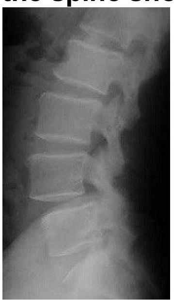
A 45-year-old patient presents with chronic lower back pain. X-ray shows anterior displacement of a vertebral body. What is the likely diagnosis?
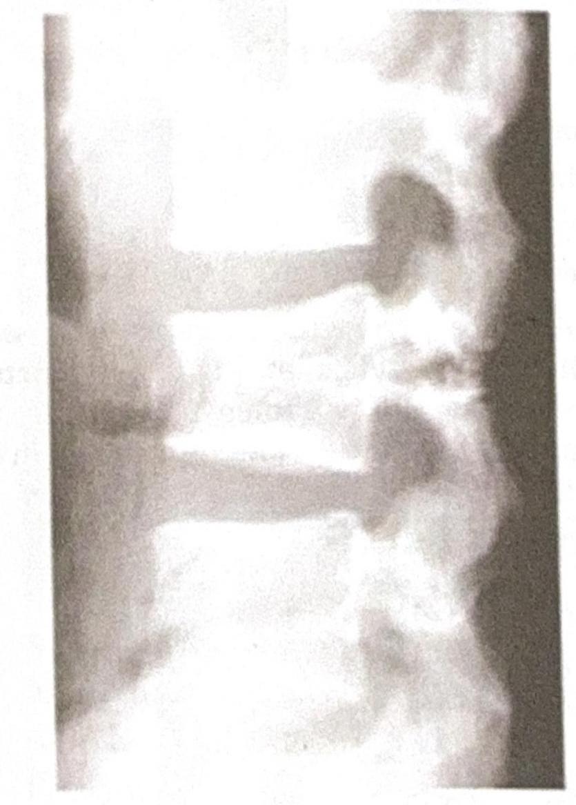
A 35-year-old male presents to the emergency department following a high-speed motor vehicle accident. He complains of severe lower back pain but denies any loss of consciousness or abdominal pain. A lateral X-ray of the lumbar spine is obtained, as shown in the image. The image reveals a horizontal fracture through the vertebral body, extending through the posterior elements. Based on the clinical presentation and imaging findings, what is the most likely diagnosis?

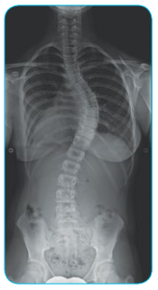
X-ray spine of a child is shown. What is the probable diagnosis?

Earliest investigation for diagnosis of Ankylosing spondylitis:
False about fracture of vertebrae
Burst fracture of spine is a type of:
Identify the condition shown in the image:

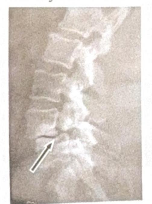
What should be the most likely diagnosis of this 65-year-old lady who presents with backache?

Condition in which there is anterior or posterior displacement of a vertebra in relation to the vertebrae below:
Hangman's fracture is
Explanation: ***Spondylolisthesis*** - This condition involves the **anterior displacement** (slipping forward) of one vertebral body over the one below it, which perfectly matches the X-ray finding. - It often causes **chronic lower back pain**, especially in active individuals or those with degenerative changes. *Spondylosis* - Refers to **degenerative changes** in the spine, including **osteophytes** and **disc space narrowing**, but typically does not involve anterior vertebral displacement. - While it can cause chronic back pain, the specific X-ray finding points away from isolated spondylosis. *Compression fracture* - Involves a **collapse of the vertebral body**, usually due to trauma or osteoporosis, leading to a **decreased vertebral height**. - It does not present as an anterior displacement of an entire vertebral body. *Osteoporosis* - This is a condition of **decreased bone density**, making bones fragile and prone to fractures (e.g., compression fractures), but it does not directly cause anterior vertebral displacement. - While osteoporosis can be an underlying factor for some spinal conditions, it is not the direct diagnosis for the described X-ray finding.
Explanation: ***Chance fracture*** - A **chance fracture** is characterized by a **horizontal fracture** through the entire vertebral body and posterior elements, including the neural arch, typically caused by a **flexion-distraction mechanism** in accidents like those involving seatbelts (lap belt only), consistent with the high-speed motor vehicle accident scenario. - The image distinctly shows a fracture line traversing the vertebral body and extending into the posterior elements, which is the hallmark of this type of injury. *Burst fracture* - A **burst fracture** involves a comminuted fracture of the vertebral body with **retropulsion of bone fragments** into the spinal canal due to axial loading, which is not clearly depicted here. - While it can result from high-impact trauma, the characteristic horizontal disruption of both anterior and posterior segments points away from a solely compressive mechanism. *Compression fracture* - A **compression fracture** primarily involves the **anterior wedging** or collapse of the vertebral body, resulting from only axial compression forces without significant involvement of the posterior elements. - The presented image shows a fracture extending through the posterior elements, which is not typical for a simple compression fracture. *Spondylolisthesis* - **Spondylolisthesis** is the **anterior slippage of one vertebral body over another**, often due to pars interarticularis defects (spondylolysis) or degenerative changes. - This condition involves vertebral displacement, not a fresh fracture line across the body and posterior elements as seen in the image.
Explanation: ***Rugger Jersey spine*** - This X-ray image shows alternating bands of **increased density (sclerosis) at the superior and inferior endplates** with **central lucency** in the vertebral bodies, creating the characteristic "Rugger Jersey spine" appearance. - This pattern is commonly associated with **secondary hyperparathyroidism**, most often seen in patients with **chronic renal failure**. - The alternating bands resemble the horizontal stripes on a rugby jersey, hence the name. *Bamboo spine* - **Bamboo spine** is a characteristic finding in advanced **ankylosing spondylitis**, where ossification of the annulus fibrosus and anterior longitudinal ligament leads to **syndesmophytes** bridging adjacent vertebral bodies. - This creates a smooth, continuous appearance resembling bamboo, which is not the pattern seen in this image. *Normal spine* - A **normal spine** would show **uniform bone density** throughout the vertebral bodies without the alternating lucent and sclerotic bands seen in this image. - Normal vertebral bodies have homogeneous trabecular bone density without endplate sclerosis. *Ivory spine* - An **ivory vertebra** refers to a **homogeneously dense and enlarged** vertebral body, typically observed in conditions like **metastatic prostate cancer** (blastic metastases), **Paget's disease**, or **lymphoma**. - This is typically a **localized finding** in a single or few vertebrae, distinct from the **generalized alternating band pattern** seen in Rugger Jersey spine.
Explanation: ***MRI STIR sequence*** - An **MRI STIR (Short Tau Inversion Recovery) sequence** is highly sensitive for detecting early inflammatory changes in the **sacroiliac joints** and spine, such as **bone marrow edema**, which is a hallmark of early ankylosing spondylitis. - It can identify disease activity and structural changes *before* they are visible on conventional X-rays, making it the earliest diagnostic tool. *CT scan* - While a **CT scan** provides excellent detailed images of bone, it is not as sensitive as MRI for detecting early inflammatory changes like **bone marrow edema** in the sacroiliac joints. - It involves significant **radiation exposure** and is typically used for more advanced structural assessment rather than early diagnosis. *Bone scan* - A **bone scan** (scintigraphy) shows areas of increased bone turnover but is **not specific** for ankylosing spondylitis and has lower spatial resolution compared to MRI. - It can indicate inflammation or increased metabolic activity but cannot differentiate specific causes or provide detailed anatomical information as effectively as MRI. *X-ray* - **X-rays** are often the initial imaging modality due to their accessibility, but they only show **structural changes** (like erosions, sclerosis, or fusion) in the sacroiliac joints and spine at a later stage of the disease. - Early inflammatory changes, such as **bone marrow edema**, are typically not visible on plain radiographs, leading to a delay in diagnosis compared to MRI.
Explanation: ***Anterior longitudinal ligament runs along the posterior surface of vertebral bodies*** - The **anterior longitudinal ligament (ALL)** runs along the **anterior aspect** of the vertebral bodies, preventing hyperextension. - The **posterior longitudinal ligament (PLL)** runs along the posterior surface of the vertebral bodies, within the vertebral canal. *Fracture dislocation is common in flexion rotation injury* - **Flexion-rotation injuries** are highly unstable and frequently lead to **fracture-dislocations** of the vertebral column. - The combined forces cause significant disruption of both bony and ligamentous structures, increasing the likelihood of displacement. *Chance fracture occurs due to flexion distraction injury* - A **Chance fracture** (or seatbelt fracture) is caused by a **flexion-distraction injury**, typically seen in individuals wearing lap belts during deceleration. - This mechanism results in a horizontal splitting of the vertebral body and posterior elements. *Wedge compression causes flexion injury* - A **wedge compression fracture** is the most common type of vertebral fracture and results from a **flexion injury** (hyperflexion). - The anterior portion of the vertebral body collapses, creating a wedge shape, while the posterior column remains intact.
Explanation: ***Compression injury*** - A **burst fracture** occurs due to a high-energy axial load or significant compression force impacting the spine. - This force causes the vertebral body to **shatter or "burst"** outwards, often into the spinal canal. *Extension injury* - **Extension injuries** typically result from hyperextension of the spine, such as in whiplash. - This mechanism often leads to **posterior element fractures** or disc injuries, not the bursting of the vertebral body. *Rotation injury* - **Rotational injuries** involve twisting forces on the spine, which generally result in **facet joint dislocations** or **fracture-dislocations**. - While they can cause instability, they do not primarily manifest as the compressive shattering seen in a burst fracture. *Flexion injury* - **Flexion injuries** are caused by forward bending forces, leading to **wedge fractures** or **flexion-distraction injuries**. - These typically spare the posterior vertebral wall from bursting into the spinal canal, unlike burst fractures.
Explanation: ***Spondylolysis*** * The image shows a **break in the pars interarticularis** of a vertebra, indicated by the arrow, which is characteristic of spondylolysis. * This condition is a **stress fracture** or defect in the pars interarticularis, a bony segment connecting the superior and inferior articular facets. *Renal osteodystrophy* * Renal osteodystrophy refers to a spectrum of **bone abnormalities** that occur in chronic kidney disease, not a specific vertebral fracture pattern. * It typically involves features such as **osteomalacia**, **osteitis fibrosa cystica**, or **osteoporosis**, which are not directly depicted as a fracture in this image. *Spondylolisthesis* * Spondylolisthesis is the **anterior slippage** of one vertebral body over another, which can be caused by bilateral spondylolysis but is not directly shown as a slip in this specific image. * The image distinctly highlights the **fracture line** itself, rather than the displacement of the vertebral body. *Tuberculosis (TB)* * Spinal tuberculosis (Pott's disease) typically presents with **destruction of vertebral bodies**, disc space narrowing, and often a **paravertebral abscess**. * The image does not show these features; instead, it demonstrates a clear **bony defect** in the pars interarticularis.
Explanation: ***Spondylolisthesis*** - The lateral X-ray image reveals an **anterior displacement of one vertebral body over the one below it**, which is characteristic of spondylolisthesis. - In a 65-year-old lady, degenerative spondylolisthesis due to **arthritic changes and instability** is a common cause of backache. *Osteoporotic fracture* - An osteoporotic fracture would typically show a **compression deformity** or a wedge-shaped vertebral body, which is not clearly depicted here. - While osteoporosis is common in this age group, the primary finding on this image is vertebral slippage, not fracture. *Spondylolysis* - Spondylolysis is a **defect in the pars interarticularis** (a thin segment of bone connecting the superior and inferior articular facets) and is best seen on oblique views or CT. - Although spondylolysis can *lead to* spondylolisthesis, the immediate and most striking finding on this lateral view is the slippage itself. *Discitis* - Discitis, an **inflammation or infection of the intervertebral disc and adjacent vertebrae**, would typically show **loss of disc height** and **endplate irregularities or erosions**. - These features are not the predominant finding on this image, which clearly demonstrates vertebral body displacement.
Explanation: ***Spondylolisthesis*** - This term specifically refers to the **slippage** (anterior or posterior displacement) of one vertebra relative to an adjacent one. - It often results from conditions like **spondylolysis** (a defect in the pars interarticularis) or degenerative changes. *Spondylitis* - This condition involves **inflammation of the vertebrae**, often seen in diseases like ankylosing spondylitis. - While it can cause pain and stiffness, it does not primarily describe the displacement of one vertebra over another. *Spondylolysis* - This refers to a **defect or stress fracture in the pars interarticularis** of a vertebra. - It is a common *cause* of spondylolisthesis, but not the slippage itself. *Spondylosis* - This is a general term for **degenerative changes** in the spine, including osteoarthritis of the spinal joints and disc degeneration. - It describes age-related wear and tear, not the direct displacement of a vertebra.
Explanation: ***traumatic spondylolisthesis of axis vertebra (C2)*** - A Hangman's fracture is a specific type of **traumatic spondylolisthesis** involving the **C2 (axis) vertebra**. - It results from bilateral fractures of the **pars interarticularis** of C2, often due to hyperextension and distractive forces. *traumatic spondylolisthesis of atlas vertebra (C1)* - A fracture of the atlas (C1) is typically called a **Jefferson fracture**, not a Hangman's fracture. - A Jefferson fracture usually involves ruptures of facets or arches, often caused by **axial loading**. *traumatic spondylolisthesis of lumbar vertebra (L1)* - **Spondylolisthesis** can occur in the lumbar spine, but it most commonly affects L5-S1 or L4-L5, and is usually a **fatigue fracture** not a traumatic spondylolisthesis. - Fractures in the lumbar region have different causes and clinical implications than cervical fractures, and are not termed a Hangman's fracture. *traumatic spondylolisthesis of thoracic vertebra (T1)* - While traumatic spine fractures can occur in the **thoracic spine**, spondylolisthesis is much less common due to the rib cage's stabilizing effect. - Fractures in this region are distinctly different from the characteristic C2 pars interarticularis fracture of a Hangman's fracture.
Get full access to all questions, explanations, and performance tracking.
Start For Free