Which of the following is not a typical symptom of a lumbar disc herniation?
All of the following contribute to the intervertebral disc EXCEPT:
A patient while lifting a heavy weight presents with sudden onset pain in the lower back radiating along the postero-lateral thigh and lateral leg to the big toe with numbness. The most likely diagnosis is:
A patient presented with Saddle anaesthesia with bladder and bowel involvement and muscle power is normal. The diagnosis is:
A 60-year-old woman with a history of chronic back pain presents with acute-onset sharp pain radiating down the right leg. She also reports numbness and tingling in the foot. What is the best next step in management?
A right-sided disc herniation at the L5-S1 level typically may cause:
Investigation of choice for lumbar prolapsed disc -
Tuberculosis of the spine commonly affects all of the following parts of the vertebra except:
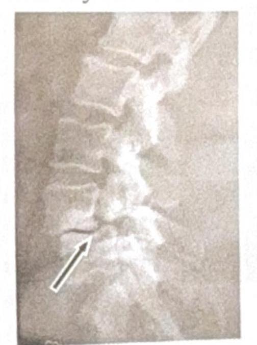
Identify the condition shown in the image:

Which of the following statements about Pott's spine is false?
Explanation: ***Saddle anesthesia*** - While a severe complication, **saddle anesthesia** is indicative of **cauda equina syndrome**, a medical emergency, and not a typical, isolated symptom of a simple lumbar disc herniation. - It suggests **compression of multiple nerve roots** in the lumbosacral region, beyond what is usually seen with a single disc herniation. *Positive straight leg raise test* - This is a common and reliable physical exam finding in patients with **lumbar disc herniation**, indicating nerve root irritation. - It elicits radiating pain down the leg when the affected leg is raised between 30 and 70 degrees. *Radicular leg pain* - **Radicular pain**, often described as sharp, shooting pain down the leg, is the hallmark symptom of a lumbar disc herniation as it signifies **nerve root compression**. - The pain typically follows a **dermatomal pattern**, corresponding to the specific nerve root involved. *Weakness in foot dorsiflexion* - Weakness in **foot dorsiflexion** (commonly affecting the **L4 or L5 nerve roots**) is a frequently observed neurological deficit in lumber disc herniation. - This can be assessed through manual muscle testing and is a sign of **motor nerve root compression**.
Explanation: ***Elastic cartilage*** - **Elastic cartilage** is characterized by the presence of **elastic fibers**, providing flexibility to structures like the ear and epiglottis. - It is **not found** within the intervertebral disc, which requires specific properties for weight-bearing and shock absorption. *Fibrocartilage* - **Fibrocartilage** is a primary component of the **annulus fibrosus** and plays a crucial role in providing tensile strength and resisting compressive forces [1]. - Its presence is essential for the structural integrity and function of the intervertebral disc. *Annulus fibrosus* - The **annulus fibrosus** is the **tough, outer fibrous ring** of the intervertebral disc, composed of concentric layers of fibrocartilage. - It encircles the nucleus pulposus, containing it and providing stability to the disc. *Nucleus pulposus* - The **nucleus pulposus** is the **gel-like core** of the intervertebral disc, rich in proteoglycans and water. - It acts as a shock absorber and allows for flexibility between vertebrae.
Explanation: ***L4 - L5 Disc prolapsed*** - A disc prolapse at the **L4-L5 level** typically compresses the **L5 nerve root**. - This compression leads to pain radiating along the **postero-lateral thigh** and **lateral leg**, reaching the **big toe**, often accompanied by numbness in the same distribution due to **L5 dermatome** involvement. *L3 - IA Disc prolapsed* - A prolapse at the **L3-L4 level** would compress the **L4 nerve root**, causing pain in the **anterior thigh** and medial leg, with potential numbness over the **medial calf** and ankle. - This presentation does not match the described symptoms of pain radiating to the big toe and lateral leg. *L5 fracture* - An **L5 fracture** would primarily manifest as localized lower back pain, often exacerbated by movement, and might not necessarily cause radiating pain or numbness in a dermatomal pattern to the big toe unless there's associated nerve root compression. - The sudden onset with radiating pain in a specific distribution points more towards nerve impingement from a disc prolapse rather than a fracture. *L5 - SI Disc prolapse* - A disc prolapse at the **L5-S1 level** compresses the **S1 nerve root**. - This typically results in pain radiating down the **posterior thigh**, **calf**, and to the **little toe** and lateral foot, along with numbness in the **S1 dermatome**, which is different from the big toe and lateral leg involvement described.
Explanation: ***Conus medullaris lesion*** - A **conus medullaris lesion** typically presents with **saddle anesthesia**, early and severe **bladder and bowel dysfunction**, and often **symmetrical neurological deficits** [1], [3]. - **Motor weakness in the legs** is usually minimal or absent, which aligns with the patient's normal muscle power in this case. *L4-L5 disc prolapsed* - A **L4-L5 disc prolapse** primarily causes **radicular pain** and weakness in the distribution of the L5 nerve root (e.g., foot drop, weakness of ankle dorsiflexion) [2]. - While it can cause some sensory changes, **saddle anesthesia** and severe bladder/bowel dysfunction are not typical features. *L3-L4 root involvement* - **L3-L4 root involvement**, often from a disc herniation, would typically present with **quadriceps weakness**, absent patellar reflex, and sensory loss over the medial thigh [2]. - It would not cause **saddle anesthesia** or significant bladder/bowel dysfunction as a primary symptom. *Cauda equina syndrome* - **Cauda equina syndrome** presents with **saddle anesthesia** and **bladder/bowel dysfunction**, but it is generally characterized by **significant motor weakness** in the lower extremities (e.g., severe leg weakness, foot drop), which is absent here. - The onset of bladder and bowel symptoms in cauda equina syndrome is often more gradual and less severe initially compared to conus medullaris lesions.
Explanation: ### MRI of the spine - The patient's symptoms of acute-onset sharp pain radiating down the right leg with numbness and tingling strongly suggest **radiculopathy**, likely due to **nerve root compression** from a herniated disc, stenosis, or other pathology [1]. - An **MRI of the spine** is the **most sensitive and specific imaging modality** to visualize soft tissue structures like intervertebral discs, nerve roots, and the spinal cord, allowing for accurate diagnosis and guiding further management. *Bed rest* - While historically recommended, **prolonged bed rest** is generally discouraged for acute low back pain and radiculopathy as it can lead to deconditioning and delayed recovery [2]. - **Modified activity** and early mobilization are often preferred over strict bed rest, even for severe pain [2]. *X-ray of the spine* - An **X-ray of the spine** can identify bony abnormalities like fractures, severe degenerative changes, or spondylolisthesis, but it **cannot visualize soft tissue structures** that are typically responsible for radicular symptoms, such as herniated discs or nerve root compression. - Therefore, it is not the best initial imaging choice for pinpointing the cause of the patient's neurological symptoms. *Physical therapy* - **Physical therapy** is an important component of long-term management for back pain and radiculopathy, focusing on exercises, stretching, and education to improve function and reduce pain. - However, in the setting of **acute, severe radicular symptoms** with numbness and tingling, it is crucial to first establish a definitive diagnosis through imaging to rule out more serious compression and guide appropriate therapeutic interventions.
Explanation: ***Low back pain and right-sided sciatica*** - A right-sided disc herniation at the L5-S1 level typically compresses the **S1 nerve root** on the right side. - This compression leads to **sciatica**, which presents as radiating pain along the S1 dermatome (back of the leg and sole of the foot), often accompanied by localized **low back pain**. *Weakness of dorsiflexion of the right foot* - **Dorsiflexion weakness** is primarily associated with compression of the **L4 or L5 nerve roots**, not S1. - The muscles responsible for dorsiflexion (e.g., tibialis anterior) are predominantly innervated by L4 and L5. *Normal ankle jerk reflex* - The **ankle jerk reflex** is mediated by the **S1 nerve root**. - Therefore, compression of the S1 nerve root would typically lead to a **diminished or absent ankle jerk reflex**, not a normal one. *Diminished sensation over the medial aspect of the foot* - **Diminished sensation** over the **medial aspect of the foot** corresponds to the **L4 dermatome**. - A herniation at L5-S1 primarily affects the S1 dermatome, which involves the **lateral aspect of the foot** and sole.
Explanation: ***MRI*** - An **MRI** provides the best visualization of **soft tissues**, including the intervertebral discs, spinal cord, and nerve roots, making it the **gold standard** for diagnosing lumbar prolapsed disc. - It can accurately show the **degree of disc herniation**, its impact on neural structures, and associated edema, which are crucial for treatment planning. *CT Scan* - While a **CT scan** provides good bony detail and can show disc herniation, its ability to visualize soft tissues is inferior to MRI for this specific condition. - It involves **ionizing radiation** and may miss subtle nerve root compression or spinal cord abnormalities apparent on MRI. *Myelogram* - A **myelogram** involves injecting contrast dye into the spinal canal and then performing X-rays or CT scans to outline the spinal cord and nerve roots. - Though effective in showing **nerve compression**, it is an **invasive procedure** with potential complications and has largely been replaced by MRI as a first-line diagnostic investigation. *X-ray* - **X-rays** primarily visualize **bony structures** and are useful for detecting fractures, spinal alignment issues, or severe degenerative changes. - They **cannot directly visualize intervertebral discs** or nerve compression, making them unsuitable for diagnosing a prolapsed disc.
Explanation: ***Spinous process*** - **Tuberculosis of the spine (Pott's disease)** typically affects the anterior columns of the vertebrae, primarily the vertebral bodies, due to their rich vascular supply. - The **spinous process** (posterior element) is rarely involved in tuberculosis because it has a relatively poor blood supply compared to the vertebral body. *Lamina* - The **lamina**, part of the vertebral arch (posterior element), is also less commonly affected by tuberculous spondylitis compared to the vertebral body. - While possible in advanced or disseminated disease, initial involvement is usually anterior. *Body* - The **vertebral body** is the most commonly affected part of the vertebra in tuberculosis of the spine. - This is due to its abundant blood supply, allowing for easy hematogenous spread of the *Mycobacterium tuberculosis* bacteria. *Pedicle* - The **pedicle** connects the vertebral body to the lamina and is considered an anterior element, albeit less frequently involved than the vertebral body itself. - Involvement of the pedicle tends to occur via direct extension from an affected vertebral body or disk space.
Explanation: ***Spondylolysis*** * The image shows a **break in the pars interarticularis** of a vertebra, indicated by the arrow, which is characteristic of spondylolysis. * This condition is a **stress fracture** or defect in the pars interarticularis, a bony segment connecting the superior and inferior articular facets. *Renal osteodystrophy* * Renal osteodystrophy refers to a spectrum of **bone abnormalities** that occur in chronic kidney disease, not a specific vertebral fracture pattern. * It typically involves features such as **osteomalacia**, **osteitis fibrosa cystica**, or **osteoporosis**, which are not directly depicted as a fracture in this image. *Spondylolisthesis* * Spondylolisthesis is the **anterior slippage** of one vertebral body over another, which can be caused by bilateral spondylolysis but is not directly shown as a slip in this specific image. * The image distinctly highlights the **fracture line** itself, rather than the displacement of the vertebral body. *Tuberculosis (TB)* * Spinal tuberculosis (Pott's disease) typically presents with **destruction of vertebral bodies**, disc space narrowing, and often a **paravertebral abscess**. * The image does not show these features; instead, it demonstrates a clear **bony defect** in the pars interarticularis.
Explanation: ***Always heals by chemotherapy*** - This statement is false because while **chemotherapy** (anti-tubercular drugs) is the primary treatment for **Pott's disease** (tuberculosis of the spine), healing is not always guaranteed and can sometimes require **surgical intervention** in cases of severe neurological deficit or instability. - The success of treatment depends on early diagnosis, patient compliance, and the severity of the disease, and not all cases resolve completely without residual issues. *Commonest at dorsolumbar junction* - **Pott's spine**, or **vertebral tuberculosis**, most frequently affects the **thoracic** and **lumbar regions**, particularly the **dorsolumbar junction** (T9-L1). - This predilection is attributed to the rich vascular supply and increased mechanical stress in this area. *Back pain is an early symptom* - **Back pain** is often one of the **earliest and most common symptoms** of Pott's spine, due to inflammation and destruction of vertebral bodies. - The pain is typically **localized**, progressive, and may worsen with movement. *There is disc space narrowing on x-ray* - **X-rays** of Pott's spine often show **disc space narrowing** along with vertebral destruction and collapse, differentiating it from pyogenic osteomyelitis where disc spaces might be initially preserved. - This narrowing is a consequence of the tuberculous infection spreading from the vertebral body to the adjacent **intervertebral disc**.
Get full access to all questions, explanations, and performance tracking.
Start For Free