An 8-year-old boy with a history of a fall from a 10-foot height complains of right ankle pain. Initial X-rays were normal and showed no fracture line. However, after 2 years, he developed a calcaneovalgus deformity. What is the most likely diagnosis?
What is the typical duration of delayed onset of muscle soreness (DOMS) in an 8-month-old child?
Sprengel's shoulder is a deformity of which bone?
All are true about Achondroplasia except?
What type of cast is typically used after closed reduction for a 4-month-old child diagnosed with developmental dysplasia of the hip (DDH)?
A child is spinned around by holding his hand by his father. While doing this, the child started crying and does not allow his father to touch his elbow. What is the most likely diagnosis?
A 12-year-old boy presents with chronic knee pain for 2 years. The X-ray shows significant pathology. What is the probable diagnosis?

Congenital Pseudoarthrosis is:
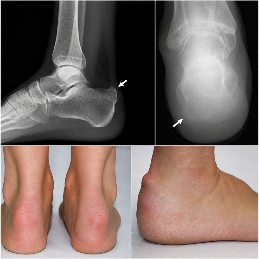
Which of the following statements about Haglund's deformity is false?
Which of the following represents an appropriate management of Legg-Calve-Perthes disease in a young patient?
Explanation: ### Explanation **Correct Answer: C. Tibial epiphyseal injury** The clinical scenario describes a classic presentation of a **Salter-Harris Type V (crush) injury** or a focal physeal arrest. In pediatric trauma, the growth plate (physis) is the weakest link. 1. **The "Hidden" Injury:** Initial X-rays are often normal in Type V injuries because there is no displaced fracture line; the damage is a microscopic compression of the germinal cells of the physis. 2. **The Deformity:** Over time, the damaged portion of the growth plate stops growing (physeal arrest), while the undamaged portion continues to grow. If the lateral aspect of the distal tibial physis is arrested while the medial side or the fibula continues to grow, it results in a progressive **calcaneovalgus deformity**. The 2-year delay is the typical "incubation period" for a growth disturbance to become clinically evident. --- ### Why the other options are incorrect: * **A. Undiagnosed malunited fracture:** A malunion would typically show an obvious fracture or callus formation on initial or early follow-up X-rays. It would cause immediate deformity rather than one that progresses over two years. * **B. Avascular necrosis (AVN) of the talus:** While a fall from height can cause talar neck fractures (Hawkins sign), AVN leads to joint collapse and pain, not a progressive angular growth deformity like calcaneovalgus. * **D. Ligamentous injury:** In children, the ligaments are stronger than the physis. A force significant enough to cause a long-term deformity would almost certainly result in a physeal fracture rather than a simple sprain. --- ### NEET-PG High-Yield Pearls: * **Salter-Harris Type V:** Highest risk of growth arrest; most difficult to diagnose acutely on X-ray. * **Heuter-Volkmann Law:** Increased pressure on a physis inhibits growth, while decreased pressure accelerates it (the mechanism behind progressive deformities). * **Management:** If <50% of the physis is involved, **Langenskiöld procedure** (resection of the bony bridge and fat graft interposition) may be attempted.
Explanation: **Explanation:** The correct answer is **D. 0-2 days**. **Underlying Medical Concept:** Delayed Onset Muscle Soreness (DOMS) refers to the pain and stiffness felt in muscles several hours to days after unaccustomed or strenuous eccentric exercise. In the context of pediatric orthopaedics—specifically during the treatment of **Congenital Talipes Equinovarus (CTEV)** using the **Ponseti method**—the term "delayed onset" is often discussed regarding the physiological response to serial casting and manipulation. In infants, the metabolic rate and tissue turnover are significantly higher than in adults. While DOMS typically peaks at 24–72 hours in adults, in an 8-month-old child, any localized muscle soreness or inflammatory response due to therapeutic manipulation or increased physical activity typically resolves rapidly, usually within **0 to 2 days**. **Analysis of Incorrect Options:** * **A (4-5 days):** This is longer than the typical recovery window for minor muscle soreness in infants, whose tissues possess high remodeling potential. * **B & C (21-30 days / 6-7 weeks):** These timeframes are characteristic of bone healing (callus formation) or ligamentous remodeling, not muscle soreness. For example, 6 weeks is the standard duration for a neonatal femoral fracture to achieve solid union. **High-Yield Clinical Pearls for NEET-PG:** * **Ponseti Technique:** The gold standard for CTEV. It involves weekly serial casting. The soreness associated with the manipulation is transient (0-2 days). * **Order of Correction in CTEV (CAVE):** **C**avus, **A**dduction, **V**arus, **E**quinus. * **Tissue Elasticity:** Infants have a higher proportion of collagen and water in their musculoskeletal tissues, leading to faster recovery from mechanical strain compared to adults. * **DOMS Mechanism:** Primarily caused by microscopic eccentric muscle fiber tears; it is *not* caused by lactic acid buildup.
Explanation: **Explanation:** **Sprengel’s deformity** is the most common congenital anomaly of the shoulder girdle. It is characterized by a **high-sitting, undescended scapula**. 1. **Why Scapula is correct:** During the 5th week of gestation, the scapula normally develops at the level of the C4–C5 vertebrae and descends to its permanent thoracic position (T2–T7) by the 8th week. Sprengel’s deformity occurs due to an **interruption in this caudal migration**, resulting in a hypoplastic, abnormally high, and medially rotated scapula. 2. **Why other options are incorrect:** * **Humerus:** While shoulder abduction is limited in this condition, the primary pathology is scapular, not humeral. * **Clavicle:** The clavicle may be shorter or straighter in these patients, but it is a secondary finding, not the primary site of deformity. * **Vertebra:** Although associated with vertebral anomalies (like Klippel-Feil syndrome), the "Sprengel's shoulder" specifically refers to the scapular malposition. **High-Yield Clinical Pearls for NEET-PG:** * **Omovertebral Bone:** A pathognomonic finding (seen in ~30% of cases) where a fibrous, cartilaginous, or osseous bridge connects the superior angle of the scapula to the cervical spine. * **Associated Conditions:** Frequently associated with **Klippel-Feil Syndrome** (triad of low hairline, short neck, and restricted neck ROM), scoliosis, and rib anomalies. * **Clinical Presentation:** Restricted shoulder abduction and cosmetic deformity (webbing of the neck). * **Treatment:** Surgical correction (e.g., **Woodward procedure** or Green’s procedure) is ideally performed between ages 3 and 8 to improve function and appearance.
Explanation: **Explanation:** Achondroplasia is the most common form of disproportionate short-limbed dwarfism. It is caused by a gain-of-function mutation in the **FGFR3 gene** (Fibroblast Growth Factor Receptor 3) on chromosome 4p, which leads to abnormal endochondral ossification. **Why Option B is the correct answer:** **Acral osteolysis** refers to the resorption of the distal phalanges of the fingers and toes. This is **not** a feature of Achondroplasia. It is classically associated with conditions like **Hadju-Cheney syndrome**, Pycnodysostosis, Hyperparathyroidism, and Scleroderma. **Analysis of other options:** * **Option A (Autosomal dominant):** Achondroplasia follows an autosomal dominant inheritance pattern. However, approximately 80% of cases are due to *de novo* mutations, often associated with advanced paternal age. * **Option C (Trident hand):** This is a classic clinical sign where there is an increased space between the third and fourth fingers, giving the hand a "three-pronged" appearance. * **Option D (Champagne glass pelvis):** Radiographically, the pelvis in achondroplasia is characterized by a short, broad ilium and a flat acetabular roof, resulting in a pelvic inlet that resembles a champagne glass. **High-Yield Clinical Pearls for NEET-PG:** * **Rhizomelic shortening:** The proximal segments (humerus/femur) are more affected than distal segments. * **Radiological signs:** "Bullet-shaped" vertebrae, narrowing of the interpedicular distance (caudally), and a "squared-off" iliac wing. * **Neurological complication:** Foramen magnum stenosis is a critical risk, potentially leading to hydrocephalus or sudden infant death. * **Intelligence:** Patients typically have normal intelligence and a normal lifespan.
Explanation: **Explanation:** The management of Developmental Dysplasia of the Hip (DDH) is age-dependent. For an infant aged **0 to 6 months**, the **Pavlik harness** is the gold standard and the first line of treatment. **Why Pavlik Harness is Correct:** The Pavlik harness is a dynamic functional brace. It maintains the hip in the "human position" (flexion >90° and moderate abduction). This position centers the femoral head into the acetabulum, promoting normal joint development while allowing active movement. It is highly effective for both reducible and stable hips in this age group. **Analysis of Incorrect Options:** * **Hip Spica:** This is typically used for children aged **6 months to 2 years** or after a surgical (open) reduction. While it provides rigid immobilization, it is not the primary choice for a 4-month-old unless the Pavlik harness fails. * **Minerva Cast:** This is used for cervical and upper thoracic spine immobilization (e.g., cervical fractures), not for hip pathology. * **Riser’s Cast:** This is a specialized body cast used in the treatment of **Scoliosis** to apply corrective forces before surgery or as part of serial casting. **Clinical Pearls for NEET-PG:** * **Age Gold Standard:** 0–6 months = Pavlik Harness; 6 months–2 years = Closed/Open Reduction + Hip Spica. * **Complication:** The most serious complication of the Pavlik harness is **Avascular Necrosis (AVN)** of the femoral head, usually due to excessive abduction (forced vascular compromise). * **Pavlik Paralysis:** Excessive flexion in the harness can lead to femoral nerve palsy. * **Barlow & Ortolani Tests:** These clinical maneuvers are most reliable only up to 2–3 months of age.
Explanation: **Explanation:** **1. Why the correct answer is right:** The clinical scenario describes a classic case of **Pulled Elbow** (also known as **Nursemaid’s Elbow** or Subluxation of the Radial Head). It typically occurs in children aged 1–4 years when sudden longitudinal traction is applied to an extended, pronated arm (e.g., swinging a child or pulling them up a curb). * **Pathophysiology:** In young children, the radial head is relatively small and the **annular ligament** is lax. Sudden traction causes the annular ligament to slip over the radial head and become trapped in the radio-capitellar joint. * **Clinical Presentation:** The child holds the arm in a fixed position of **flexion and pronation**, refusing to move it. There is no significant swelling or deformity. **2. Why the incorrect options are wrong:** * **B. Radial head dislocation:** This is usually associated with high-energy trauma or a Monteggia fracture-dislocation (ulnar fracture). Pulled elbow is a *subluxation*, not a complete dislocation. * **C. Annular ligament tear:** While the ligament is displaced and trapped, it is rarely actually torn in this mechanism. The pathology is displacement, not a rupture. * **D. Fracture of the olecranon process:** This would present with significant localized swelling, ecchymosis, and a palpable gap or deformity, which are absent in a pulled elbow. **3. NEET-PG High-Yield Pearls:** * **Management:** Reduction is performed by **Supination followed by Flexion** (or the Hyperpronation technique). A "click" is often felt, and the child usually resumes normal arm use within minutes. * **X-ray Findings:** Usually normal; X-rays are only indicated if a fracture is suspected (e.g., history of a fall or focal bony tenderness). * **Age Group:** Most common between **1–4 years**; rare after age 5 as the annular ligament becomes thicker and the radial head grows.
Explanation: ***Osteosarcoma*** - Most common **primary bone malignancy** in children and adolescents, typically affecting the **metaphyseal region** around the knee (distal femur, proximal tibia). - Classic X-ray features include **Codman's triangle**, **sunburst pattern**, and **mixed lytic-sclerotic lesions** in the metaphysis, which would explain the "significant pathology" mentioned. *Chronic osteomyelitis* - Usually presents with **systemic symptoms** like fever and elevated inflammatory markers, which are not mentioned in this case. - X-ray typically shows **sequestrum formation** and **involucrum** rather than the aggressive bone destruction pattern seen in malignancy. *Rickets* - Primarily affects **growth plates** and causes **bone deformities** like bowing of legs, not localized knee pain. - X-ray shows **osteopenia**, **coxa vara**, and **delayed ossification** rather than aggressive bone lesions. *Hyperparathyroidism* - Causes **generalized bone demineralization** and **brown tumors**, not localized aggressive lesions around the knee. - More commonly presents with **hypercalcemia symptoms** and affects multiple bones, particularly the **skull** and **long bones** diffusely.
Explanation: **Explanation:** **Congenital Pseudoarthrosis of the Tibia (CPT)** is a rare but challenging condition characterized by a spontaneous fracture or bowing of the bone that fails to heal, leading to a "false joint" (pseudoarthrosis). **1. Why Option B is Correct:** There is a strong clinical association between CPT and **Neurofibromatosis Type 1 (NF-1)**. Approximately 50% of children with CPT have NF-1, and conversely, about 5% of NF-1 patients develop CPT. The underlying pathology involves a defect in the periosteum where fibrovascular tissue replaces normal bone-forming cells, preventing union. **2. Why Other Options are Incorrect:** * **Option A:** CPT is most commonly seen in the **tibia** (specifically the distal third), not the femur. * **Option C:** It is a **congenital/developmental** dysplasia of the bone and periosteum. While osteomyelitis can cause non-union, it is not the etiology of "Congenital Pseudoarthrosis." * **Option D:** It is called a "pseudoarthrosis" because it mimics a joint's mobility, but it lacks the anatomical structures of a **true joint** (synovium, articular cartilage, and capsule). **High-Yield Clinical Pearls for NEET-PG:** * **Most Common Site:** Distal third of the Tibia. * **Clinical Presentation:** Anterolateral bowing of the tibia is often the first sign, usually present at birth or within the first two years. * **Radiological Sign:** "Tea-cup and saucer" appearance or "pencil-point" tapering of the bone ends. * **Treatment:** Extremely difficult; often requires intramedullary nailing (Williams rod), Ilizarov fixation, or vascularized fibular grafts. Amputation is a last resort for recalcitrant cases.
Explanation: **Explanation:** The question appears to be based on a clinical scenario or image describing **Madelung’s Deformity**, but the options mistakenly label it as **Haglund’s Deformity**. In the context of pediatric orthopaedics and competitive exams like NEET-PG, this is a common point of differentiation. 1. **Why Option A is the Correct Answer (False Statement):** The condition described by the clinical features (bilateral involvement, association with Turner’s syndrome, and wrist deformity) is **Madelung’s Deformity**, not Haglund’s deformity. Haglund’s deformity is a bony enlargement on the back of the heel (retrocalcaneal exostosis). Therefore, stating "It is Haglund's deformity" is factually incorrect. 2. **Analysis of Other Options (True for Madelung’s Deformity):** * **Option B:** Madelung’s deformity is indeed **bilateral in approximately 50%** of cases. * **Option C:** It is strongly associated with **Turner’s Syndrome (45, XO)** and Leri-Weill dyschondrosteosis (SHOX gene mutation). * **Option D:** Despite the visible deformity (prominent ulnar head, volar bowing of the radius), patients typically maintain **excellent function** and are often asymptomatic until late adolescence when pain or decreased range of motion (especially supination) may occur. **Clinical Pearls for NEET-PG:** * **Madelung’s Deformity:** Caused by premature closure of the **volar-ulnar** part of the distal radial physis. * **Radiological Sign:** Increased volar tilt and ulnar tilt of the distal radius; the carpus "sinks" between the radius and ulna (V-shaped proximal carpal row). * **Haglund’s Deformity:** Known as "Pump Bump," it involves the posterosuperior aspect of the calcaneus and is associated with retrocalcaneal bursitis.
Explanation: **Explanation:** **Legg-Calvé-Perthes Disease (LCPD)** is an idiopathic avascular necrosis of the femoral head occurring in children (typically aged 4–8 years). The primary goal of management is to maintain the spherical shape of the femoral head as it re-ossifies to prevent early-onset osteoarthritis. **Why Option D is Correct:** The fundamental principle of LCPD management is **"Containment."** By ensuring the femoral head remains deep within the acetabulum, the acetabulum acts as a biological mold. This ensures that as the necrotic bone is resorbed and replaced (creeping substitution), the new femoral head develops a spherical shape rather than becoming flattened (coxa plana). Containment can be achieved via non-operative methods (Abduction braces like the Atlanta/Newington brace) or surgical methods (Varus derotation osteotomy). **Why Other Options are Incorrect:** * **A. Total Hip Replacement:** This is contraindicated in children. It is a salvage procedure reserved for end-stage arthritis in adults. * **B. Exercise:** While range-of-motion exercises are helpful to prevent stiffness, "exercise" alone is not a definitive management strategy and does not address the structural deformity. * **C. High dose calcium:** LCPD is a vascular/ischemic issue, not a nutritional deficiency. Calcium supplementation has no role in the revascularization of the femoral head. **Clinical Pearls for NEET-PG:** * **Classic Presentation:** A painless limp or hip/knee pain in a young boy (Male:Female = 4:1). * **Early Sign:** Limitation of **Abduction** and **Internal Rotation**. * **Radiology:** Look for the "Crescent sign" (subchondral fracture) and "Gage’s sign" (V-shaped lucency at the lateral epiphysis). * **Prognosis:** The most important prognostic factor is the **age at onset** (younger children <6 years have a better prognosis due to higher remodeling potential).
Developmental Dysplasia of Hip
Practice Questions
Clubfoot
Practice Questions
Pediatric Fractures
Practice Questions
Growth Plate Injuries
Practice Questions
Legg-Calvé-Perthes Disease
Practice Questions
Slipped Capital Femoral Epiphysis
Practice Questions
Pediatric Spine Deformities
Practice Questions
Cerebral Palsy: Orthopaedic Aspects
Practice Questions
Neuromuscular Disorders in Children
Practice Questions
Pediatric Bone and Joint Infections
Practice Questions
Limb Length Discrepancies
Practice Questions
Orthopedic Manifestations of Genetic Disorders
Practice Questions
Get full access to all questions, explanations, and performance tracking.
Start For Free