Which of the following statements regarding genu varum (bowleg) is true?
In treating a fractured clavicle in a 14-month-old child, what is the preferred management?
A 10-year-old girl presents with a fall from a tree. Radiographic and physical examinations reveal Osgood-Schlatter disease. Which bony structure is chiefly affected in this condition?
Irregular thigh folds are seen in?
What are the components of talipes equinovarus?
Which of the following is/are X-ray features of Legg-Calve-Perthes disease?
Which statement is NOT true regarding CTEV shoes?
What is a common consequence of impacted canines?
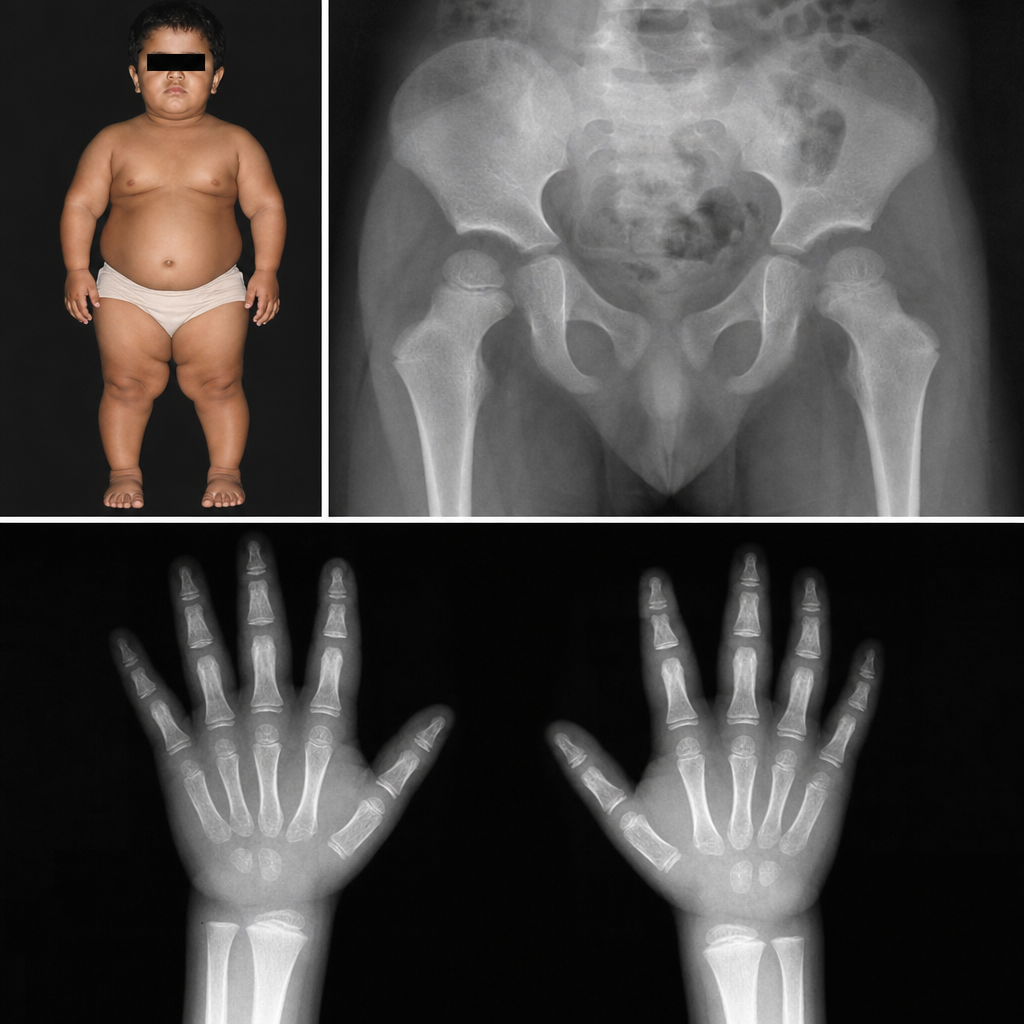
A child presents with short stature and abnormalities of the hand and pelvis. What is your diagnosis?
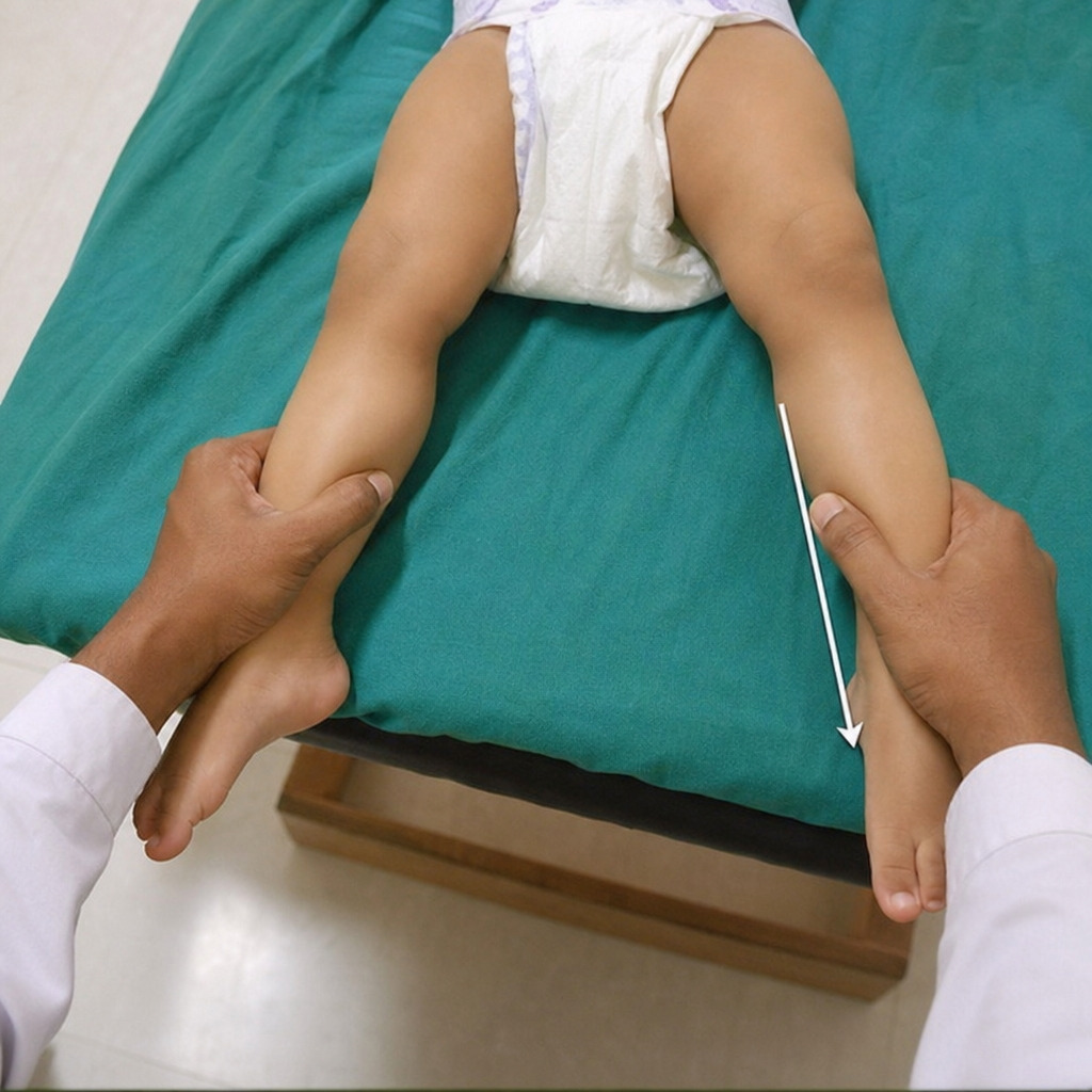
All the following are radiological signs that correlate positively with the outcome of Perthes' disease except?
Explanation: **Explanation:** Genu varum (bowleg) is a common clinical presentation in pediatric orthopaedics characterized by an outward bowing of the legs. **Why Option B is Correct:** Pathological genu varum is frequently associated with skeletal dysplasias, specifically **epiphyseal or metaphyseal dysplasias** (e.g., Achondroplasia, Multiple Epiphyseal Dysplasia). In these conditions, abnormal development or premature ossification of the growth plates leads to asymmetric growth, resulting in progressive angular deformities. Other pathological causes include Rickets and Blount’s disease (Tibia Vara). **Analysis of Incorrect Options:** * **Option A:** While "Physiological Genu Varum" is normal in infants up to 18–24 months, the question asks for a definitive statement regarding the pathology. In the context of standard orthopaedic examinations, pathological bowing is distinguished from the self-limiting physiological version. * **Option C:** Genu varum is **frequently** associated with tibial angulation. In fact, in Blount’s disease, the primary pathology is localized to the posteromedial aspect of the proximal tibial physis. * **Option D:** The deformity can involve both the **distal femur** and the **proximal tibia**. For example, in Rickets, softening of the bones leads to bowing across multiple segments of the lower limb. **NEET-PG High-Yield Pearls:** * **Developmental Timeline:** Remember the sequence: Genu Varum (Birth to 2 yrs) → Straight (2 yrs) → Genu Valgum (3 to 4 yrs) → Straight (6 to 7 yrs). * **Blount’s Disease:** Characterized by an abnormal **Metaphyseal-Diaphyseal Angle (>16°)** on X-ray. * **Rickets:** Look for "cupping, splaying, and fraying" of the metaphysis. * **Clinical Measurement:** Genu varum is assessed by measuring the **intercondylar distance** (distance between the medial femoral condyles).
Explanation: **Explanation:** Fractures of the clavicle are common in the pediatric population due to birth trauma or falls. In a 14-month-old child, the management is almost exclusively **conservative** because of the immense remodeling potential of the bone and the thick periosteal sleeve that stabilizes the fracture. **Why Figure-of-eight bandage is correct:** The **Figure-of-eight bandage** (or a simple broad arm sling) is the treatment of choice. It functions by pulling the shoulders back, which helps in aligning the fragments and providing symptomatic relief by limiting movement. In infants and toddlers, even significant displacement or malalignment (overlap) will remodel completely over time without functional deficit. **Why other options are incorrect:** * **Open reduction:** Surgical intervention is contraindicated in young children due to the risk of infection, scarring, and non-union. It is only considered in rare cases of open fractures or neurovascular compromise. * **Shoulder cast:** A full shoulder cast (like a shoulder spica) is unnecessarily restrictive, heavy, and uncomfortable for a toddler. It provides no additional benefit over a simple bandage. * **Kirschner pin:** Internal fixation with K-wires is avoided in children because of the risk of migration and damage to the growth plates or underlying vital structures. **NEET-PG High-Yield Pearls:** * **Most common site:** The middle third (junction of medial 2/3 and lateral 1/3) is the most common site of clavicle fractures. * **Remodeling:** The younger the child, the greater the remodeling potential. Clinical "lumps" (callus) usually disappear within 6–9 months. * **Birth Injury:** Clavicle fracture is the most common bone fractured during labor (often associated with shoulder dystocia). * **Adult Management:** While children are treated with slings/bandages, adults with significant shortening (>2cm) or comminution may require ORIF with plates and screws.
Explanation: **Explanation:** **Osgood-Schlatter disease** is a common cause of knee pain in active adolescents (typically aged 10–15 years). It is characterized as a **traction apophysitis** of the **tibial tuberosity**. 1. **Why the Correct Answer is Right:** The condition results from repetitive strain and microtrauma caused by the powerful pull of the **quadriceps femoris** muscle through the patellar tendon onto the immature tibial tuberosity. During the adolescent growth spurt, this area is a weak link (apophysis). Repeated stress leads to inflammation, micro-avulsion fractures, and subsequent bony prominence at the **tibial tuberosity (Option D)**. 2. **Why the Incorrect Options are Wrong:** * **Medial condyle of tibia (Option A):** This is the site involved in *Blount’s disease* (tibia vara), not traction apophysitis. * **Posterior intercondylar area (Option B):** This is the attachment site for the Posterior Cruciate Ligament (PCL); it is not involved in Osgood-Schlatter disease. * **Intercondylar eminence (Option C):** This is the attachment site for the Anterior Cruciate Ligament (ACL). Fractures here are common in children (ACL avulsion), but it is not the site of Osgood-Schlatter disease. **High-Yield Clinical Pearls for NEET-PG:** * **Clinical Presentation:** Pain, swelling, and tenderness localized over the tibial tuberosity, exacerbated by jumping, running, or kneeling. * **Radiology:** May show fragmentation or heterotopic ossification of the tibial tuberosity. * **Management:** Primarily conservative (Rest, Ice, NSAIDs, and activity modification). It is self-limiting and resolves once the physis closes. * **Differential Diagnosis:** Sinding-Larsen-Johansson syndrome (traction apophysitis at the *inferior pole of the patella*).
Explanation: **Explanation:** **Developmental Dysplasia of the Hip (DDH)** is the correct answer because **asymmetric skin folds** (thigh, gluteal, or inguinal) are one of the earliest clinical signs of a dislocated or subluxated hip in an infant. This occurs due to the proximal migration of the femur on the affected side, which causes the soft tissues to bunch up, resulting in an increased number or uneven positioning of the skin folds compared to the normal side. **Analysis of Incorrect Options:** * **Perthes Disease:** This is an avascular necrosis of the femoral head typically seen in children aged 4–8 years. Clinical features include a painless limp and restricted abduction/internal rotation, but not thigh fold asymmetry, as the hip is not dislocated. * **Slipped Capital Femoral Epiphysis (SCFE):** This occurs in obese adolescents (10–15 years). It presents with an external rotation deformity and "obligate external rotation" during hip flexion. Thigh folds are not a diagnostic feature here. * **Congenital Coxa Vara:** This involves a decreased neck-shaft angle (less than 120°). While it causes limb shortening and a painless limp, it does not typically present with the classic irregular thigh folds seen in the neonatal/infant screening for DDH. **Clinical Pearls for NEET-PG:** * **Galeazzi Sign:** Apparent shortening of the femur (uneven knee heights when hips/knees are flexed) is a classic sign of unilateral DDH. * **Screening Tests:** **Barlow’s maneuver** (dislocatable hip - "provocative") and **Ortolani’s maneuver** (reducible hip - "reductive"). * **Imaging:** Ultrasound is the investigation of choice for infants <6 months; X-ray (looking for Shenton’s line disruption and Hilgenreiner/Perkin lines) is used for infants >6 months. * **Treatment:** **Pavlik harness** is the gold standard for infants under 6 months of age.
Explanation: **Explanation:** Talipes Equinovarus (TEV), commonly known as **Clubfoot**, is a congenital deformity involving the foot and ankle. The correct sequence of deformities is remembered by the classic mnemonic **CAVE**, which describes the components in the order they are typically corrected using the Ponseti method. 1. **C – Cavus:** Increased longitudinal arch of the foot (due to forefoot pronation). 2. **A – Adduction:** The forefoot is deviated medially at the midtarsal joints. 3. **V – Varus:** The heel (calcaneum) is tilted toward the midline (inversion). 4. **E – Equinus:** Plantarflexion at the ankle joint (tight Tendo-Achilles). **Why Option B is correct:** The core components of TEV are **Equinus** (ankle), **Inversion/Varus** (subtalar/hindfoot), and **Adduction** (forefoot). These represent the inward and downward deviation characteristic of the "club" shape. **Why other options are incorrect:** * **Options A & D (Abduction):** Abduction is the opposite of the deformity found in TEV. Abduction is actually the corrective maneuver used during casting. * **Options C & D (Eversion):** Eversion is the opposite of Varus. In TEV, the sole of the foot faces inward (Inversion/Varus), not outward (Eversion/Valgus). **NEET-PG High-Yield Pearls:** * **Ponseti Method:** The gold standard treatment involving serial casting. The order of correction is **C-A-V-E**. * **Pirani Score:** Used to assess the severity of clubfoot (0 to 6 scale). * **Radiology:** On X-ray, the **Kite’s Angle** (talocalcaneal angle) is **decreased** (<20°) in both AP and lateral views. * **Last deformity to be corrected:** Equinus (often requires a percutaneous Tendo-Achilles Tenotomy). * **Maintenance:** After casting, a **Denis Browne Splint** (foot abduction brace) is used to prevent recurrence.
Explanation: **Explanation:** Legg-Calvé-Perthes Disease (LCPD) is an idiopathic avascular necrosis of the femoral head in children (typically aged 4–8 years). The radiological features evolve through stages (Waldenström classification), reflecting the process of necrosis, fragmentation, and eventual remodeling. **Why "All the Above" is correct:** * **Increased medial joint space (Waldenström’s Sign):** This is one of the earliest radiological signs. It occurs due to hypertrophy of the acetabular cartilage and the presence of joint effusion/synovitis. * **Metaphyseal cysts and rarefaction:** During the fragmentation stage, the metaphysis often shows radiolucent "cysts" and generalized rarefaction. This is thought to be due to increased vascularity and osteoclastic activity at the growth plate. * **Lateral extrusion of the femoral head:** As the femoral head softens and flattens (coxa plana), it may be displaced laterally out of the acetabulum. This is a critical "Head at Risk" sign, indicating a poorer prognosis. **High-Yield Clinical Pearls for NEET-PG:** * **Gage’s Sign:** A V-shaped radiolucency on the lateral side of the epiphysis (a "Head at Risk" sign). * **Crescent Sign:** A subchondral fracture line seen on the lateral X-ray, indicating the early fragmentation stage. * **Sagging Rope Sign:** A radiopaque line across the metaphysis seen in late stages, representing a deformed femoral neck. * **Prognosis:** The most important prognostic factor is the **age at onset** (children <6 years have a better prognosis). * **Management:** The primary goal is "containment" of the femoral head within the acetabulum to ensure it remains spherical during remodeling.
Explanation: In the management of **Congenital Talipes Equinovarus (CTEV)**, specialized shoes are prescribed once the child begins weight-bearing to maintain the correction achieved by the Ponseti technique (casting and tenotomy). ### Why "Straight outer border" is the correct answer: The statement is **NOT true** because CTEV shoes must have a **lateral (outer) convexity** or an **out-flare**. In CTEV, the forefoot is adducted (curved inward). To counteract this deformity and prevent recurrence, the shoe is designed with an **abducted (outward-curved) forefoot** rather than a straight one. A straight border would not provide the necessary corrective force to keep the forefoot in abduction. ### Explanation of other options: * **Used only from the age the child starts walking (True):** Before walking, correction is maintained using the **Steenbeek or Denis Browne splint** (foot abduction braces). CTEV shoes are functional only during ambulation. * **No heel (True):** CTEV shoes are "flat-soled" with no heel elevation. This prevents the development of **equinus** (plantarflexion) by ensuring the Achilles tendon remains stretched during walking. * **Raised outer portion (True):** These shoes feature a **lateral wedge (outer border elevation)**. This helps to counteract the **varus** (inward tilting) of the heel and the **supination** of the foot by forcing the foot into eversion. ### High-Yield Clinical Pearls for NEET-PG: * **Order of Correction (PONSETI):** **C**aveus $\rightarrow$ **A**dduction $\rightarrow$ **V**arus $\rightarrow$ **E**quinus. * **The "Gold Standard":** The Ponseti method is the treatment of choice. * **Bracing Protocol:** After casting, a foot abduction brace is worn 23 hours/day for 3 months, then at nap/night-time until age 4. * **Relapse:** The most common cause of deformity recurrence is poor compliance with the bracing protocol.
Explanation: **Explanation:** Impacted maxillary canines are a common clinical challenge in pediatric dentistry and orthodontics, second only to third molars in frequency of impaction. **Why Option B is correct:** The most significant and common complication of an impacted canine is **root resorption of adjacent teeth**, particularly the lateral and central incisors. As the canine attempts to erupt along an abnormal path, the pressure and proximity of its follicle can lead to the destruction of the cementum and dentin of neighboring roots. Studies suggest that up to 50% of cases involving impacted canines show some degree of root resorption on CBCT imaging. **Analysis of Incorrect Options:** * **Option A:** While impacted teeth *can* become ankylosed (fused to the bone), it is not the most common consequence. Ankylosis is more frequently associated with primary molars or teeth that have undergone trauma. * **Option C:** While canines are vital for "canine-guided occlusion" and aesthetic lip support, "good function" is not impossible without them. Premolars can often be orthodontically moved or "camouflaged" to take over their functional role. * **Option D:** Physiologically, teeth usually erupt when **two-thirds to three-quarters** of their root formation is complete. Waiting for complete root formation often leads to the tooth losing its eruptive force, making the impaction permanent. **NEET-PG High-Yield Pearls:** * **Incidence:** Maxillary canines are impacted 20 times more frequently than mandibular canines. * **Palatal vs. Buccal:** Palatal impactions are more common (85%) than buccal impactions (15%). * **Guidance Theory:** The lateral incisor root acts as a guide for canine eruption; if the lateral incisor is peg-shaped or absent, the risk of canine impaction increases. * **Management:** Surgical exposure followed by orthodontic traction is the gold standard treatment.
Explanation: ***Achondroplasia*** - Characterized by **rhizomelic shortening** (proximal limb shortening) and classic **trident hand** deformity with hand and pelvic abnormalities. - X-ray features include **champagne-glass pelvis**, **small squared iliac wings**, **horizontal acetabular roof**, and **narrow sacroiliac notch**. *Rickets* - Primarily causes **metaphyseal** changes with **widening**, **cupping**, and **fraying** of bone ends, not specific hand deformities. - Characterized by **bowing deformities** of weight-bearing bones and **delayed epiphyseal ossification**, but lacks trident hand pattern. *Osteogenesis imperfecta* - Presents with **multiple fractures** with minimal trauma and **blue sclerae** as hallmark features. - Bone deformities result from **recurrent fractures** rather than specific developmental abnormalities of hands and pelvis. *Cushing syndrome* - Causes **centripetal obesity**, **moon facies**, and **purple striae** rather than skeletal deformities. - Growth retardation occurs due to **cortisol excess** affecting growth hormone, not structural bone abnormalities.
Explanation: In Perthes' disease (Legg-Calvé-Perthes), the prognosis depends heavily on the degree of femoral head involvement and its containment within the acetabulum. To predict outcomes, Catterall described specific radiological features known as **"Head-at-Risk" signs**. ### Why "Shallow Acetabulum" is the Correct Answer A **shallow acetabulum** is not a "Head-at-Risk" sign. In fact, Perthes' disease often leads to a secondary *deepening* or remodeling of the acetabulum to accommodate a flattened femoral head (coxa plana). While acetabular dysplasia is a primary feature of Developmental Dysplasia of the Hip (DDH), it is not a prognostic marker for Perthes'. ### Explanation of "Head-at-Risk" Signs (Incorrect Options) The following signs correlate **positively with a poor outcome** (increased risk of femoral head collapse and permanent deformity): * **Lateral subluxation of the femoral head (Option A):** Indicates the head is moving out of the protective cover of the acetabulum, leading to eccentric loading. * **Speckled calcification lateral to the epiphysis (Option B):** Represents calcification in the cartilaginous part of the head outside the acetabulum, signifying severe extrusion. * **Gage sign (Option D):** A V-shaped radiolucency on the lateral side of the epiphysis/metaphysis. It indicates osteolysis and impending collapse of the lateral pillar. * *Other signs include:* Horizontal growth plate and diffuse metaphyseal reactions. ### NEET-PG High-Yield Pearls * **Age of Onset:** Most common in boys aged 4–8 years. * **Pathology:** Avascular necrosis of the proximal femoral epiphysis. * **Classification Systems:** * **Catterall:** Based on the extent of necrosis. * **Herring (Lateral Pillar):** Most widely used for prognosis (Group A, B, and C based on the height of the lateral 1/3rd of the epiphysis). * **Management Goal:** "Containment" of the femoral head within the acetabulum using braces or osteotomy.
Developmental Dysplasia of Hip
Practice Questions
Clubfoot
Practice Questions
Pediatric Fractures
Practice Questions
Growth Plate Injuries
Practice Questions
Legg-Calvé-Perthes Disease
Practice Questions
Slipped Capital Femoral Epiphysis
Practice Questions
Pediatric Spine Deformities
Practice Questions
Cerebral Palsy: Orthopaedic Aspects
Practice Questions
Neuromuscular Disorders in Children
Practice Questions
Pediatric Bone and Joint Infections
Practice Questions
Limb Length Discrepancies
Practice Questions
Orthopedic Manifestations of Genetic Disorders
Practice Questions
Get full access to all questions, explanations, and performance tracking.
Start For Free