
Pediatric Orthopaedics — MCQs

On this page
What is a Pulled Elbow?
What is the most common complication of lateral condyle humerus fracture?
In elbow, osteochondritis usually involves
Tardy ulnar nerve palsy is specifically associated with which type of fracture?
Which of the following statements is true regarding supracondylar fractures of the humerus?
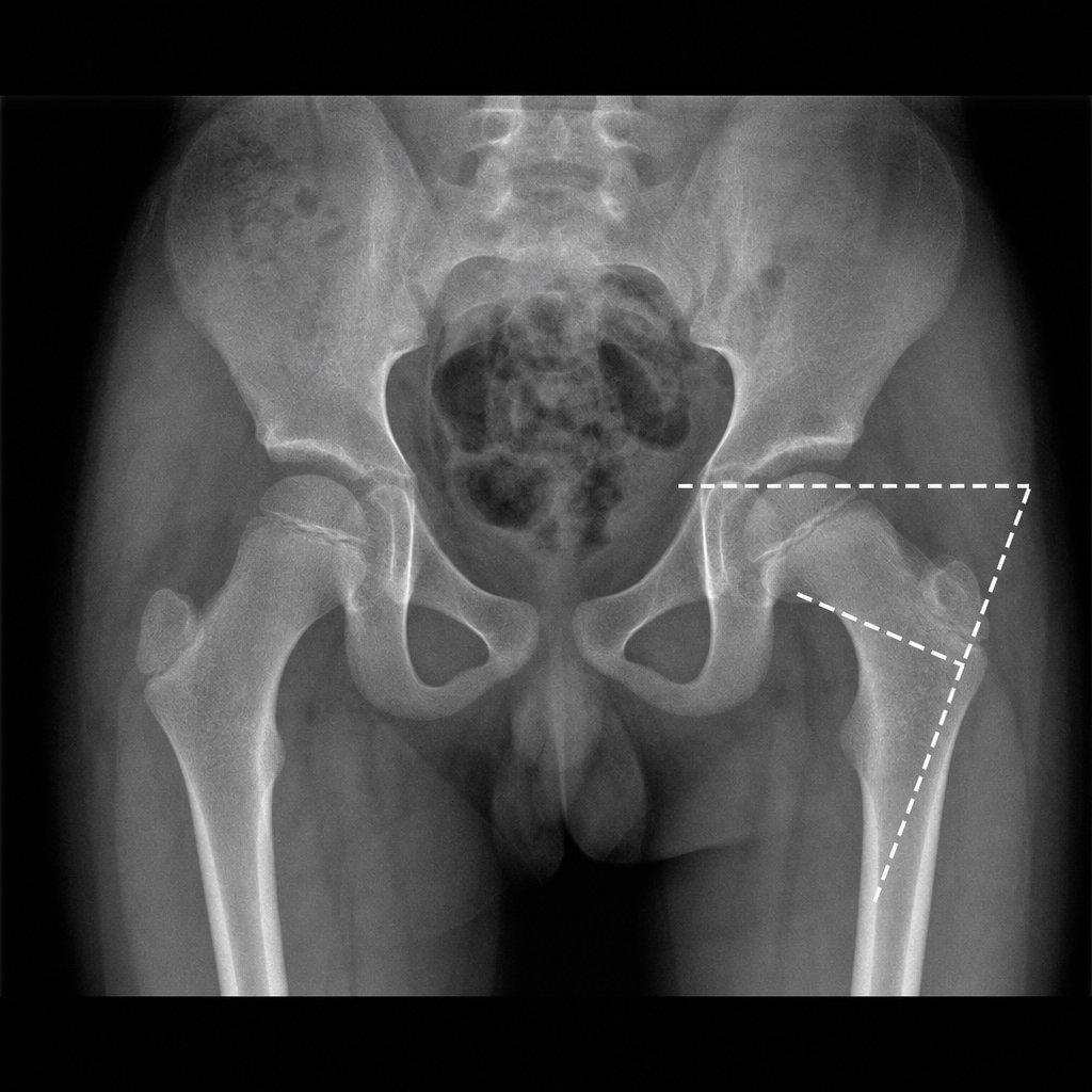
Fairbank triangle is seen in
Congenital pseudoarthrosis is seen in which of the following?
Which is the most common elbow fracture in children?
What is the most common bone fractured in children?
Blount's disease is associated with all of the following, except:
Practice by Chapter
Developmental Dysplasia of Hip
Practice Questions
Clubfoot
Practice Questions
Pediatric Fractures
Practice Questions
Growth Plate Injuries
Practice Questions
Legg-Calvé-Perthes Disease
Practice Questions
Slipped Capital Femoral Epiphysis
Practice Questions
Pediatric Spine Deformities
Practice Questions
Cerebral Palsy: Orthopaedic Aspects
Practice Questions
Neuromuscular Disorders in Children
Practice Questions
Pediatric Bone and Joint Infections
Practice Questions
Limb Length Discrepancies
Practice Questions
Orthopedic Manifestations of Genetic Disorders
Practice Questions
Want unlimited practice?
Get full access to all questions, explanations, and performance tracking.
Scan to download app