
Pediatric Orthopaedics — MCQs

On this page
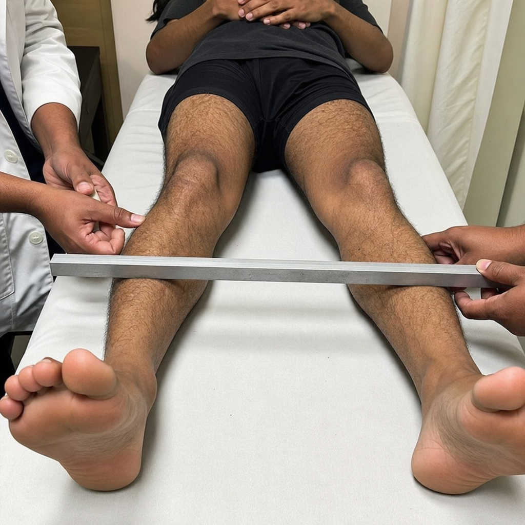
Which test is being performed?
Which of the following complications is possible following excision of the head of the radius in children?
What is the most common cause of acute compartment syndrome in children?
Pyle's disease is characterized by which of the following?
A 7-year-old boy presents with abrupt onset of hip pain and the hip is held in abduction. Hemogram is normal, but ESR is raised. What is the next line of management?
A 4-year-old child complains of a painful limb. What is the most likely cause?
What is the most common complication of a supracondylar fracture?
Posterior iliac horns are seen in which of the following conditions?
Which of the following are associated with supracondylar fracture of humerus?
All are true about supracondylar fracture of humerus except?
Practice by Chapter
Developmental Dysplasia of Hip
Practice Questions
Clubfoot
Practice Questions
Pediatric Fractures
Practice Questions
Growth Plate Injuries
Practice Questions
Legg-Calvé-Perthes Disease
Practice Questions
Slipped Capital Femoral Epiphysis
Practice Questions
Pediatric Spine Deformities
Practice Questions
Cerebral Palsy: Orthopaedic Aspects
Practice Questions
Neuromuscular Disorders in Children
Practice Questions
Pediatric Bone and Joint Infections
Practice Questions
Limb Length Discrepancies
Practice Questions
Orthopedic Manifestations of Genetic Disorders
Practice Questions
Want unlimited practice?
Get full access to all questions, explanations, and performance tracking.
Scan to download app