
Pediatric Orthopaedics — MCQs

On this page
Which splint is used in the management of a fracture of the shaft of the femur in the age group of 2 to 10 years?
A 24-year-old male presents for a routine checkup. Radiographs reveal supernumerary teeth. General examination shows hypermobility of the shoulders, and the Gorlin sign is absent. What is the most probable diagnosis?
Fairbank's triangle is seen in which of the following conditions?
What is the most common elbow injury in adolescents?
When should treatment for Congenital Talipes Equinovarus (CTEV) start?
What is the recommended conservative treatment for neurogenic dislocation?
Open reduction and internal fixation is definitely required in a child with which of the following fractures?
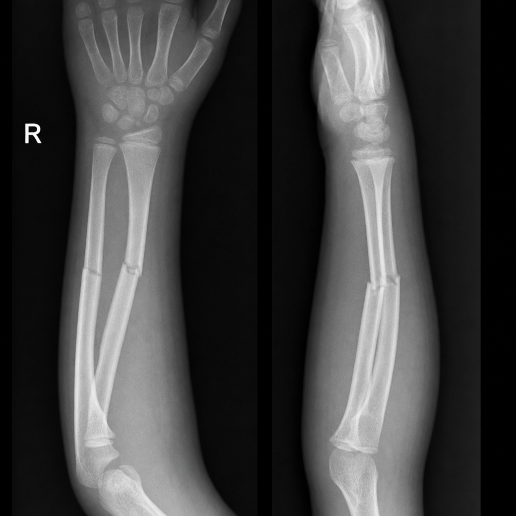
A 10-year-old child presents with pain and restricted movement in the right upper limb after falling from a wall with an outstretched hand. Radiographic findings are shown below. What is the management?
What imaging is required for a suspected medial epicondylar fracture of the humerus in a 4-year-old child?
Which of the following is NOT a feature of Achondroplasia?
Practice by Chapter
Developmental Dysplasia of Hip
Practice Questions
Clubfoot
Practice Questions
Pediatric Fractures
Practice Questions
Growth Plate Injuries
Practice Questions
Legg-Calvé-Perthes Disease
Practice Questions
Slipped Capital Femoral Epiphysis
Practice Questions
Pediatric Spine Deformities
Practice Questions
Cerebral Palsy: Orthopaedic Aspects
Practice Questions
Neuromuscular Disorders in Children
Practice Questions
Pediatric Bone and Joint Infections
Practice Questions
Limb Length Discrepancies
Practice Questions
Orthopedic Manifestations of Genetic Disorders
Practice Questions
Want unlimited practice?
Get full access to all questions, explanations, and performance tracking.
Scan to download app