
Pediatric Orthopaedics — MCQs

On this page
All the following are features of achondroplasia EXCEPT:
A 12-year-old child presents with a rapid increase in weight and height over the past year. The child experiences difficulty sitting cross-legged and squatting, with the knees going into the axilla with every hip and knee flexion. What is the most likely diagnosis?
The classical flexion and rotation deformities at hip and knee joints, as a sequela of poliomyelitis, are due to the contracture of which muscle?
What is the most likely diagnosis in a 23-year-old, mentally alert dwarf with disproportionate arm and leg to body growth, prominent forehead, and retruded maxilla?
An 8-year-old boy has a history of a fall from 10 feet with pain in the right ankle. The initial X-ray was normal. Two years later, he developed a calcaneovalgus deformity. What is the likely diagnosis?
What is the preferred treatment for a shaft femur fracture in a 3-year-old boy?
In congenital dislocation of the hip, which clinical sign shows that the affected thigh is at a lower level when the knees and hips are flexed to 90 degrees?
Apert syndrome is a disease of:
Ortolani's test is done in which condition?
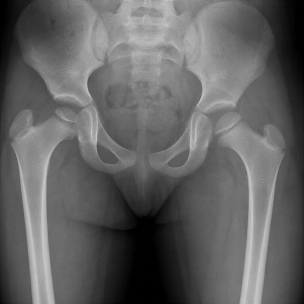
A child presents with a limp and limb shortening. An X-ray is provided. What is the diagnosis?
Practice by Chapter
Developmental Dysplasia of Hip
Practice Questions
Clubfoot
Practice Questions
Pediatric Fractures
Practice Questions
Growth Plate Injuries
Practice Questions
Legg-Calvé-Perthes Disease
Practice Questions
Slipped Capital Femoral Epiphysis
Practice Questions
Pediatric Spine Deformities
Practice Questions
Cerebral Palsy: Orthopaedic Aspects
Practice Questions
Neuromuscular Disorders in Children
Practice Questions
Pediatric Bone and Joint Infections
Practice Questions
Limb Length Discrepancies
Practice Questions
Orthopedic Manifestations of Genetic Disorders
Practice Questions
Want unlimited practice?
Get full access to all questions, explanations, and performance tracking.
Scan to download app