In which condition is the presence of an extra pair of ribs sometimes observed?
Which of the following is least commonly associated with Down's syndrome?
A 10-year-old boy presents with frequent fractures and blue sclerae. This patient most likely carries a mutation in a gene that encodes which of the following proteins?
A 10-year-old girl presents with severe joint laxity, scoliosis, and a history of easy bruising. Which of the following conditions is most likely?
Mutations in type I collagen fibres results in:
An intrauterine scan at the 13th week of pregnancy showed a fetus with multiple long bone fractures. What is commonly associated with this finding?
The most commonly involved nerve in lunate dislocation is -
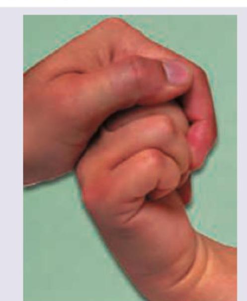
Identify the true statement regarding the clinical examination given in the image:

False about fracture of vertebrae
Slipped capital femoral epiphysis is seen most commonly in which age group?
Explanation: ***Turner syndrome*** - **Turner syndrome** (45,X) is often associated with skeletal abnormalities, including an extra pair of ribs (cervical ribs) in some cases. - Other common skeletal features include **short stature**, a **shield chest**, and **cubitus valgus**. *Down syndrome* - **Down syndrome** (trisomy 21) is characterized by specific facial features, intellectual disability, and congenital heart defects. - While it can manifest with various skeletal anomalies, an extra pair of ribs is not a characteristic feature. *Klinefelter syndrome* - **Klinefelter syndrome** (47,XXY) is characterized by hypogonadism, tall stature, gynecomastia, and learning difficulties. - Skeletal features may include long limbs and decreased bone density, but cervical ribs are not typically associated with this condition. *Holt-Oram syndrome* - **Holt-Oram syndrome** is a genetic disorder affecting heart and limb development, specifically the upper limbs (thumb abnormalities, phocomelia). - It does not typically involve the presence of an extra pair of ribs. *Fibrous dysplasia* - **Fibrous dysplasia** is a bone disorder where normal bone is replaced by fibrous tissue, leading to weakened bone and fractures. - It is a localized bone condition and does not involve the presence of supernumerary ribs.
Explanation: ***Premature loss of deciduous teeth*** - **Premature loss of deciduous teeth** is the **least commonly associated** feature with Down syndrome among the given options. - While individuals with Down syndrome have increased periodontal disease that can lead to tooth loss, **premature exfoliation of deciduous teeth as a primary developmental feature is uncommon**. - Tooth loss, when it occurs, is typically a **secondary consequence** of severe periodontal destruction rather than an intrinsic developmental anomaly causing premature shedding. - Unlike the delayed eruption pattern which is a consistent finding, premature loss is not a characteristic feature of Down syndrome itself. *Delayed eruption of deciduous teeth* - This is a **common characteristic** of Down syndrome, not the least common. - Infants with Down syndrome frequently experience **delayed eruption of both deciduous and permanent teeth**, reflecting the generalized slower maturation seen in this condition. - Dental developmental delay is well-documented and consistently observed. *Periodontal disease* - This is **very common** in Down syndrome, with prevalence rates of 58-96%. - Individuals have significantly **increased prevalence and severity of periodontal disease** due to compromised immune function, poor oral hygiene, and specific anatomical factors. - One of the most consistent oral manifestations of Down syndrome. *Retrognathia* - **Retrognathia** (recessed lower jaw) is a **common craniofacial feature** in Down syndrome. - Contributes to difficulties in feeding, speech, and can impact airway patency. - Part of the characteristic facial phenotype.
Explanation: ***Collagen*** - Collagen mutations commonly result in various **connective tissue disorders** and defects in structural integrity [1]. - It plays a critical role in **skin, bones, and cartilage**, making it a significant candidate for mutations affecting these areas [1]. *Keratin* - Keratin is a protein primarily found in **skin, hair, and nails**, and mutations typically cause **epidermal dysplasia or disorders** like keratoderma. - While important for structural integrity, keratin is less associated with broader connective tissue disorders. *Fibrillin* - Fibrillin is essential for maintaining the **elasticity and structure of connective tissues**, predominantly in **Marfan syndrome** [2]. - Mutations lead to issues with connective tissue but are not as broadly relevant as collagen in this context [2]. *Myosin* - Myosin is primarily involved in **muscle contraction** and is not typically associated with **connective tissue** disorders. - Mutations in myosin often relate to **muscle diseases**, which are not relevant to the context of connective tissue mutations. **References:** [1] Kumar V, Abbas AK, et al.. Robbins and Cotran Pathologic Basis of Disease. 9th ed. Genetic Disorders, pp. 154-155. [2] Kumar V, Abbas AK, et al.. Robbins and Cotran Pathologic Basis of Disease. 9th ed. Genetic Disorders, pp. 153-154.
Explanation: ***Ehlers-Danlos syndrome*** - This syndrome is characterized by defects in **collagen synthesis** and structure, leading to **joint hypermobility** (laxity) [1], skin hyperextensibility, and fragility, which explains the easy bruising [1]. - **Scoliosis** is a common musculoskeletal manifestation due to weakened connective tissue support [1]. *Marfan's syndrome* - While Marfan's syndrome also presents with **joint laxity** and **scoliosis**, its defining features include distinct skeletal abnormalities (e.g., **arachnodactyly**, sternal deformities) and **cardiovascular abnormalities** (e.g., aortic root dilatation), which are not mentioned here. - **Easy bruising** is not a prominent feature of Marfan's syndrome. *Rheumatoid arthritis* - This is an **autoimmune inflammatory arthritis** primarily affecting synovial joints, causing pain, swelling, and stiffness, often symmetrically. - It does not typically present with severe **joint laxity** throughout the body, **scoliosis**, or **easy bruising** as primary features in a 10-year-old. *Osteogenesis imperfecta* - This condition is characterized by **brittle bones** due to defective collagen, leading to recurrent **fractures** with minimal trauma. - While patients can have some **joint laxity** and **scoliosis**, the most prominent symptom is bone fragility, often accompanied by **blue sclerae**, which is not mentioned, and easy bruising is less indicative than in EDS.
Explanation: ***Osteogenesis imperfecta*** - This condition is primarily caused by **genetic defects** in the genes encoding **Type I collagen**, particularly *COL1A1* and *COL1A2*. - It leads to **fragile bones** that fracture easily, **blue sclera**, **hearing loss**, and **dentinogenesis imperfecta**, due to the impaired formation of collagen, a major component of bone and connective tissues. *Osteosclerosis* - This refers to a general term for **increased bone density** and hardening of bone, which can be a symptom of various conditions. - It is not caused by a specific mutation in Type I collagen but rather points to an **imbalance in bone remodeling** where bone formation outpaces resorption. *Osteopetrosis* - Also known as **Albers-Schönberg disease** or **marble bone disease**, this condition is characterized by **abnormally dense bones** due to a defect in **osteoclast function**, which impairs bone resorption [1]. - It is primarily caused by mutations in genes involved in osteoclast development and acidification, such as *CLCN7*, not Type I collagen genes [1]. *Marfan's syndrome* - This is a **connective tissue disorder** caused by a mutation in the *FBN1 gene* encoding **fibrillin-1**, a protein essential for the formation of elastic fibers. - It affects the **skeleton, eyes, heart, and blood vessels**, leading to features like tall stature, long limbs, and cardiovascular abnormalities, distinct from collagen defects causing bone fragility. **References:** [1] Kumar V, Abbas AK, et al.. Robbins and Cotran Pathologic Basis of Disease. 9th ed. Bones, Joints, and Soft Tissue Tumors, p. 1188.
Explanation: ***Osteogenesis imperfecta*** - **Multiple long bone fractures** detected early in pregnancy are a classic presentation of **osteogenesis imperfecta (OI)**, a genetic disorder characterized by **bone fragility**. - OI is primarily caused by mutations in genes encoding **type I collagen**, leading to defective bone formation. *Achondroplasia* - This condition is a form of **dwarfism** characterized by disproportionately short limbs and a normal-sized trunk, resulting from a mutation in the **FGFR3 gene**. - While it affects bone growth, it typically does not cause **multiple fractures** prenatally. *Marfan syndrome* - This is a connective tissue disorder affecting the skeletal, ocular, and cardiovascular systems, characterized by **tall stature**, **long limbs and fingers**, and **aortic root dilation**. - It results from a mutation in the **fibrillin-1 gene** and is not primarily associated with prenatal long bone fractures. *Cretinism* - This is a historical term for **congenital hypothyroidism**, which results from severely deficient thyroid hormone production in a newborn. - It leads to developmental delays, growth retardation, and intellectual disability, but not to **multiple bone fractures**.
Explanation: ***Median nerve*** - In a **lunate dislocation**, the lunate bone dislocates anteriorly and rotates. This displaced lunate can directly compress the **median nerve** within the carpal tunnel, which lies just anterior to it. - Compression of the median nerve leads to symptoms of **carpal tunnel syndrome**, including numbness and tingling in the thumb, index, middle, and radial half of the ring finger. *Ulnar nerve* - The **ulnar nerve** passes through Guyon's canal, which is located more medially and is generally not directly compressed by an isolated lunate dislocation. - While other wrist injuries can affect the ulnar nerve, it is not the most common nerve involved in lunate dislocation. *Posterior interosseous* - The **posterior interosseous nerve** is a branch of the radial nerve and supplies muscles in the posterior compartment of the forearm; it is located away from the carpal bones and is very rarely affected by lunate dislocation. - Injury to this nerve typically results in wrist drop or issues with finger extension. *Anterior interosseous* - The **anterior interosseous nerve** is a branch of the median nerve that supplies deep flexor muscles in the forearm; it also lies away from the direct path of a dislocated lunate. - Injury to this nerve leads to an inability to make the "ok" sign due to paralysis of the flexor pollicis longus and flexor digitorum profundus to the index finger.
Explanation: ***Wrist is held in forced flexion for 60 sec eliciting pain*** - The image depicts **Phalen's test**, used to diagnose **carpal tunnel syndrome**. In this test, the patient's wrists are held in maximal sustained **flexion** for 30-60 seconds. - The reproduction of **tingling or pain** in the median nerve distribution (thumb, index, middle, and radial half of the ring finger) within this time frame indicates a positive test. *Wrist is held in forced extension for 60 sec* - Holding the wrist in **forced extension** for 60 seconds describes **reverse Phalen's test**, not the standard Phalen's test shown. - While reverse Phalen's test also assesses for **carpal tunnel syndrome**, it typically involves holding the wrists in **extension**. *Wrist is held in forced flexion for 45 sec eliciting pain* - While **flexion** is correct for Phalen's test, the standard duration is up to **60 seconds**, not specifically 45 seconds to determine a positive result. - Pain should be elicited within this timeframe, but the 45-second duration is not the most accurate statement regarding the full range of the test's timing. *Wrist is held in forced extension for 45 sec* - This option incorrectly states **forced extension** rather than flexion for Phalen's test, and the specific duration of 45 seconds is not universally cited as the definitive endpoint for a positive result. - **Forced extension** is part of the reverse Phalen's maneuver, not the test shown.
Explanation: ***Anterior longitudinal ligament runs along the posterior surface of vertebral bodies*** - The **anterior longitudinal ligament (ALL)** runs along the **anterior aspect** of the vertebral bodies, preventing hyperextension. - The **posterior longitudinal ligament (PLL)** runs along the posterior surface of the vertebral bodies, within the vertebral canal. *Fracture dislocation is common in flexion rotation injury* - **Flexion-rotation injuries** are highly unstable and frequently lead to **fracture-dislocations** of the vertebral column. - The combined forces cause significant disruption of both bony and ligamentous structures, increasing the likelihood of displacement. *Chance fracture occurs due to flexion distraction injury* - A **Chance fracture** (or seatbelt fracture) is caused by a **flexion-distraction injury**, typically seen in individuals wearing lap belts during deceleration. - This mechanism results in a horizontal splitting of the vertebral body and posterior elements. *Wedge compression causes flexion injury* - A **wedge compression fracture** is the most common type of vertebral fracture and results from a **flexion injury** (hyperflexion). - The anterior portion of the vertebral body collapses, creating a wedge shape, while the posterior column remains intact.
Explanation: **Explanation:** **Slipped Capital Femoral Epiphysis (SCFE)** is a classic hip disorder characterized by the displacement of the capital femoral epiphysis from the femoral neck through the physeal plate. 1. **Why Adolescents is correct:** The condition occurs most commonly during the **adolescent growth spurt** (typically ages 10–16 years). During this period, the growth plate (physis) thickens and weakens under the influence of growth hormones, while the periosteum thins. This mechanical instability, often combined with **obesity**, leads to a "slip" where the epiphysis remains in the acetabulum while the neck moves anteriorly and superiorly. 2. **Why other options are incorrect:** * **Infants:** Hip pathology in this group is usually Developmental Dysplasia of the Hip (DDH) or Septic Arthritis. * **Childhood:** The most common hip pathology in younger children (ages 4–8) is **Legg-Calvé-Perthes disease** (avascular necrosis). * **Old age:** Hip issues in the elderly are typically related to Osteoarthritis or Neck of Femur fractures. **High-Yield Clinical Pearls for NEET-PG:** * **Typical Profile:** An obese adolescent male presenting with a limp and hip or **referred knee pain**. * **Clinical Sign:** **Drehmann Sign** (obligate external rotation of the hip during passive flexion). * **Radiology:** Look for **Trethowan’s Sign** (Klein’s line fails to intersect the femoral head). * **Associated Conditions:** If seen in younger children, suspect endocrine disorders like **Hypothyroidism** or Growth Hormone deficiency. * **Management:** The definitive treatment is **In-situ pinning** (Internal fixation with a single cannulated screw).
Get full access to all questions, explanations, and performance tracking.
Start For Free