All are characteristic features of cerebral palsy except
Appropriate treatment for mild congenital ptosis?
Unilateral high stepping gait is seen in
Match the following drugs in Column A with their contraindications in Column B. | Column A | Column B | | :-- | :-- | | 1. Morphine | 1. QT prolongation | | 2. Amiodarone | 2. Thromboembolism | | 3. Vigabatrin | 3. Pregnancy | | 4. Estrogen preparations | 4. Head injury |
Which of the following is a FALSE statement regarding Cerebral Palsy?
What is the significance of the persistence of the asymmetric tonic neck reflex in a 9-month-old infant?
Scissor gait is seen in which of the following conditions:
Combination of appearance in CTEV
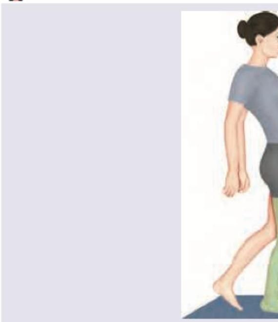
The following gait is seen due to weakness of:

Open reduction (OR) is not required in which fracture?
Explanation: ***Erb's palsy*** - **Erb's palsy** is a form of brachial plexus palsy, characterized by injury to the **upper brachial plexus** (C5-C6 nerve roots), typically occurring during birth. - It results in a characteristic **"waiter's tip" position** of the arm and is a distinct peripheral nerve injury, not a characteristic feature of **cerebral palsy**, which is a central neurological disorder. *Hypotonia* - While many forms of cerebral palsy present with **spasticity**, some individuals, particularly those with **ataxic cerebral palsy** or specific types of dyskinetic cerebral palsy, can exhibit **hypotonia** (low muscle tone). - Hypotonia can also be an early manifestation before the development of more prominent hypertonia or spasticity, making it an associated feature. *Epilepsy* - **Epilepsy** and seizure disorders are common co-morbidities seen in children with **cerebral palsy**, particularly in those with severe brain damage or certain types of CP. - The underlying brain injury that causes cerebral palsy can also disrupt normal electrical activity in the brain, leading to seizures. *Spasticity* - **Spasticity** is the most common motor type of **cerebral palsy**, affecting approximately 80% of individuals. - It is characterized by **increased muscle tone** and **hyperreflexia**, resulting in stiff, tight muscles and exaggerated reflexes, due to damage to the motor cortex or pyramidal tracts.
Explanation: ***LPS Resection*** - **LPS (levator palpebrae superioris) resection/advancement** is the most common surgical treatment for congenital ptosis, especially in mild to moderate cases. - This procedure strengthens the levator muscle, improving eyelid position and is appropriate when the **levator function is good** (typically greater than 4mm). *Frontalis sling procedure* - The **frontalis sling procedure** is generally reserved for severe congenital ptosis with poor levator function (<4mm) or in cases where the levator muscle is absent or highly dysfunctional. - It uses the frontalis muscle to lift the eyelid indirectly, which is less ideal for mild ptosis. *Antibiotics and hot compression* - **Antibiotics and hot compression** are treatments for infectious or inflammatory conditions of the eyelid, such as a **hordeolum** (stye) or **chalazion**. - They are not effective treatments for anatomical defects like congenital ptosis, which requires surgical intervention. *Wedge resection of conjunctiva* - **Wedge resection of the conjunctiva** might be used in some cases of conjunctival prolapse or for correction of specific conjunctival lesions or abnormalities. - It is not a standard or appropriate treatment for congenital ptosis.
Explanation: ***L5 radiculopathy*** - Damage to the **L5 nerve root** can cause weakness in the **tibialis anterior muscle**, leading to **foot drop**. [1] - To compensate for the foot drop and prevent tripping, the patient develops a **high stepping gait** (steppage gait) on the affected side. [1] *Distal radiculopathy* - This term is too general; **radiculopathy** refers to nerve root compression but does not specify which root or its precise distal effects. - While a radiculopathy can cause weakness, "distal" does not specifically localize to L5 or unilateral foot drop. *Cauda equina syndrome* - This is a serious condition involving **compression of multiple nerve roots** below the conus medullaris. - It typically causes bilateral symptoms, including **saddle anesthesia**, bowel/bladder dysfunction, and often bilateral leg weakness, not isolated unilateral foot drop. *None of the options* - This option is incorrect because **L5 radiculopathy** directly explains unilateral high stepping gait due to foot drop.
Explanation: ***A-4, B-1, C-3, D-2*** - **Morphine** is contraindicated in **head injury** as it can increase intracranial pressure and mask neurological symptoms. - **Amiodarone** is contraindicated in patients with **QT prolongation** due to its risk of inducing more severe arrhythmias like Torsades de Pointes. - **Vigabatrin** is contraindicated during **pregnancy** due to its potential for teratogenicity and adverse effects on fetal development. - **Estrogen preparations** are contraindicated in patients with a history of **thromboembolism** due to their increased risk of blood clot formation. *A-1, B-3, C-2, D-4* - This option incorrectly matches **Morphine** with QT prolongation and **Estrogen preparations** with head injury, which are not their primary contraindications. - It also incorrectly links **Vigabatrin** with thromboembolism and **Amiodarone** with pregnancy. *A-3, B-2, C-4, D-1* - This choice incorrectly associates **Morphine** with pregnancy and **Vigabatrin** with head injury, which are not the most critical or direct contraindications. - It also misaligns **Amiodarone** with thromboembolism and **Estrogen preparations** with QT prolongation. *A-2, B-4, C-1, D-3* - This option incorrectly matches **Morphine** with thromboembolism and **Amiodarone** with head injury, which are not their most significant contraindications. - It also incorrectly links **Vigabatrin** with QT prolongation and **Estrogen preparations** with pregnancy.
Explanation: ***Birth trauma is the most common cause of cerebral palsy*** - While birth trauma can contribute to brain injury, **prematurity** and **intrauterine growth restriction** are actually more frequent risk factors for cerebral palsy. - The majority of cerebral palsy cases originate from prenatal or perinatal events, with **birth asphyxia** being a less common cause than historically believed. *Cerebral palsy occurs due to one time insult to developing fetal brain* - Cerebral palsy is defined by a **non-progressive disturbance** in the developing fetal or infant brain, which is indeed a "one time insult" rather than a degenerative process. - This insult can occur before, during, or shortly after birth, leading to permanent but **non-worsening** motor impairments. *Periventricular leucomalacia causes spastic diplegia* - **Periventricular leucomalacia (PVL)**, a type of white matter brain injury, is strongly associated with **spastic diplegia**, particularly in premature infants. - PVL damages the periventricular white matter that contains descending motor tracts to the lower limbs, hence causing a **diplegic** (legs more affected than arms) presentation. *Persistent cortical thumb after 3 months of age is seen in spastic cerebral palsy* - A **cortical thumb**, where the thumb is held adducted and flexed across the palm, can be a sign of **upper motor neuron lesion** or spasticity. - Its persistence beyond 3 months of age is an indicator of neurological dysfunction and is often observed in infants who develop **spastic cerebral palsy**.
Explanation: ***Increased muscle tone*** - The **asymmetric tonic neck reflex (ATNR)** should integrate by **6 months of age**, and its persistence beyond this period is a sign of **neurological dysfunction**. - Persistent primitive reflexes, including ATNR, are often associated with **upper motor neuron lesions** and can manifest as increased muscle tone or **spasticity**. *Decreased muscle tone* - **Decreased muscle tone**, or **hypotonia**, is typically associated with **lower motor neuron lesions** or certain genetic conditions, not the persistence of primitive reflexes. - While some neurological conditions can cause hypotonia, persistent ATNR is a hallmark of problems leading to **hypertonia**. *Normal phenomenon* - The persistence of the ATNR beyond **6 months of age** is considered abnormal and indicates a potential developmental delay or neurological issue. - In a **9-month-old**, the reflex should have fully integrated, and its presence warrants further investigation. *None of the options* - As the persistence of the ATNR is indeed a significant finding, associated with increased muscle tone, this option is incorrect.
Explanation: ***Cerebral palsy*** - **Scissor gait** is a characteristic presentation in individuals with **spastic cerebral palsy**, due to hyperactivity of adductor muscles, causing the legs to cross over each other. - This **spasticity** often results from damage to the brain's motor control centers during development. *Polio* - **Polio** primarily causes **flaccid paralysis** due to damage to anterior horn cells, leading to muscle weakness and atrophy, not spasticity. - The gait in polio is often characterized by muscle weakness, leading to a **waddling or steppage gait**, not scissoring. *Hyperbilirubinemia* - Severe **hyperbilirubinemia** in neonates can lead to **kernicterus**, causing **choreoathetosis**, dystonia, and hearing loss. - While it affects motor control, it typically results in involuntary movements and muscle rigidity (dystonia), but **scissor gait** is not a hallmark. *Hyponatremia* - **Hyponatremia** is an electrolyte imbalance that can cause neurological symptoms such as confusion, seizures, and coma. - It does not directly cause specific gait abnormalities like **scissor gait**; any gait disturbances would be secondary to altered mental status or seizures.
Explanation: ***Equinus, inversion, forefoot adduction, cavus*** - The classic presentation of **clubfoot** (CTEV) involves a characteristic combination of deformities: **equinus** (fixed plantarflexion of the ankle), **inversion** (tilting of the heel inward), **forefoot adduction** (inward turning of the front of the foot), and **cavus** (an abnormally high arch). - These four components are essential for the diagnosis and classification of CTEV. *Equinus, eversion, forefoot adduction, cavus* - This option incorrectly lists **eversion** instead of inversion. Eversion involves the outward tilting of the heel and is not a feature of CTEV. - While equinus, forefoot adduction, and cavus are typical, the presence of eversion rules out classic CTEV. *Equinus, inversion, forefoot adduction, planus* - This option incorrectly lists **planus** (pes planus or flatfoot) instead of cavus. Cavus (high arch) is a defining characteristic of CTEV, not a flatfoot. - While equinus, inversion, and forefoot adduction are correct, the presence of planus makes this option incorrect. *Equinus, eversion, forefoot abduction, cavus* - This option incorrectly lists both **eversion** and **forefoot abduction**. Eversion is the outward tilting of the heel, and forefoot abduction is the outward turning of the front of the foot. - Both eversion and forefoot abduction are opposite to the deformities seen in classical CTEV.
Explanation: ***Gluteus medius*** - Weakness of the **gluteus medius** leads to a **Trendelenburg gait**, where the pelvis drops on the unsupported side during the swing phase of gait. - The image suggests pelvic tilting, which is characteristic of the body attempting to compensate for the inability of the gluteus medius to stabilize the pelvis. *Gluteus maximus* - Weakness of the gluteus maximus causes difficulty in **hip extension**, resulting in a **lurching gait** where the trunk is thrown backward at heel strike. - This is commonly known as a **gluteus maximus lurch**, which is not depicted in an obvious manner here. *Psoas major* - Weakness of the psoas major would primarily affect **hip flexion**, making it difficult to lift the leg off the ground (e.g., during the swing phase). - This would result in compensatory movements such as circumduction or hiking the hip, rather than the characteristic pelvic drop. *Tibialis anterior* - Weakness of the tibialis anterior causes **foot drop**, leading to a **steppage gait** where the knee is lifted high to avoid dragging the foot. - The image does not show a foot drop or high stepping, thus ruling out tibialis anterior weakness.
Explanation: ***Fracture of the outer one-third of the radius*** - Fractures of the **outer one-third of the radius** (distal radius fractures) often can be managed with **closed reduction and casting** if stable and adequately reduced. - While some unstable distal radius fractures require OR, many stable patterns, especially those with minimal displacement or good alignment after closed manipulation, do not. *Fracture of the patella* - Many patellar fractures lead to significant **extensor mechanism disruption**, necessitating OR with **tension band wiring** or screw fixation to restore quadriceps function. - Displaced patellar fractures, especially transverse ones, require surgical fixation to prevent extensor lag and **nonunion**. *Displaced fracture of the olecranon* - Displaced olecranon fractures disrupt the **triceps mechanism** and compromise elbow stability, almost always requiring **open reduction and internal fixation (ORIF)**, typically with tension band wiring. - Without surgical repair, a displaced olecranon fracture can lead to significant loss of extension strength and **nonunion**. *Fracture of the condyle of the humerus* - Fractures of the humeral condyle, particularly in children, often require OR due to the risk of **avascular necrosis** (especially lateral condyle) and the need for **precise anatomical reduction** to prevent joint incongruity and cubitus varus/valgus deformities. - Intra-articular and displaced condylar fractures almost invariably require surgical intervention to ensure harmonious joint function and prevent long-term complications like **stiffness and deformity**.
Get full access to all questions, explanations, and performance tracking.
Start For Free