Slipped capital femoral epiphysis is seen most commonly in which age group?
A 4-year-old child sustained a fall on an outstretched hand. X-rays revealed a fracture line at the physis with a small metaphyseal fragment. There was no epiphyseal fracture. What type of injury, according to the Salter-Harris classification, is this?
What is the most common fracture in children?
Which provocative test is used for the detection of developmental dysplasia of the hip (CDH)?
In Cleidocranial dysostosis, what is the typical morphology of the roots of the permanent teeth?
Which of the following conditions is associated with congenital dislocations?
What is the spot diagnosis for a foot deformity described as a rocker bottom foot?
The windswept deformity is seen in which of the following conditions?
Pseudoarthrosis of Tibia is a classical feature of which condition?
Siffert-Katz sign is seen in which of the following conditions?
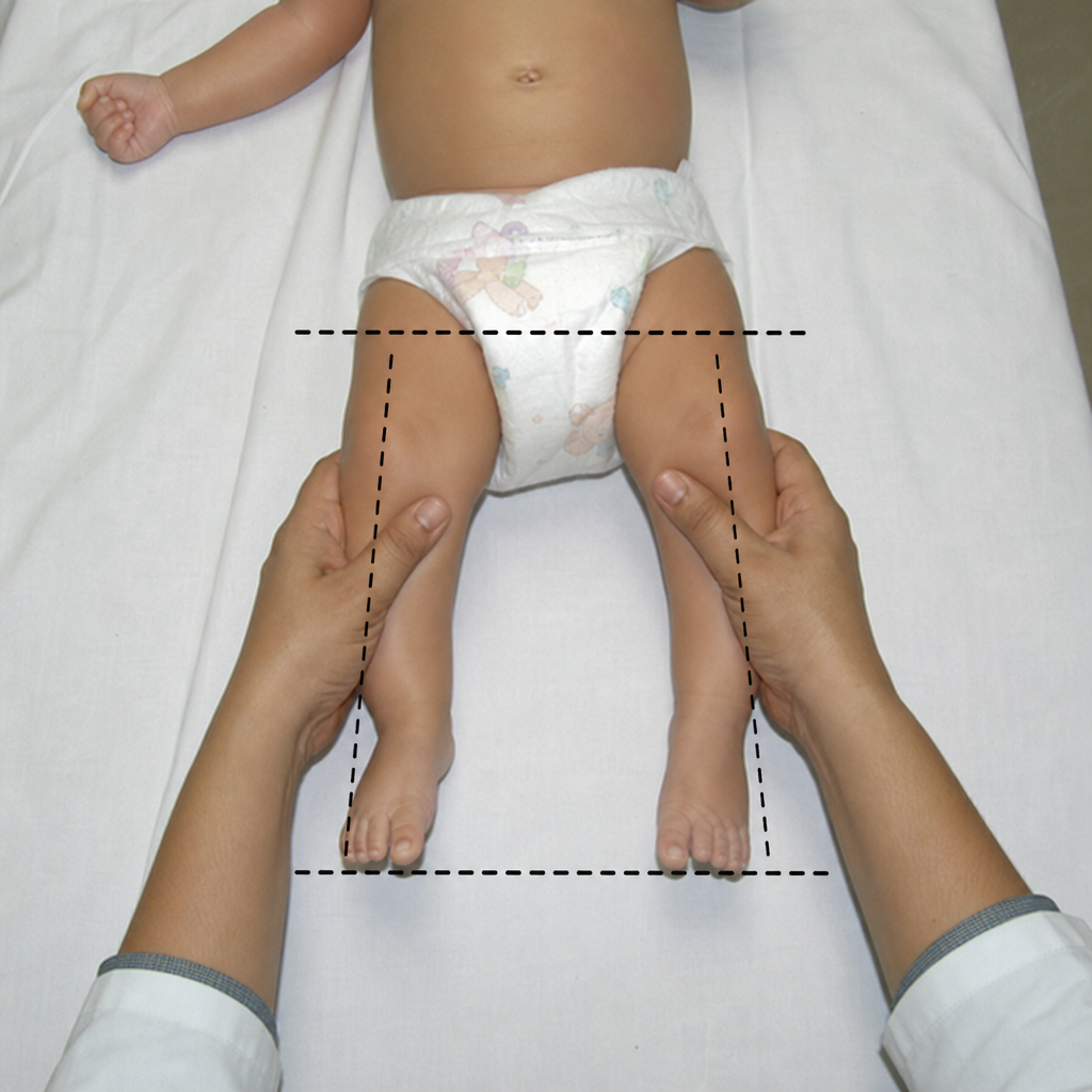
Explanation: **Explanation:** **Slipped Capital Femoral Epiphysis (SCFE)** is a classic hip disorder characterized by the displacement of the capital femoral epiphysis from the femoral neck through the physeal plate. 1. **Why Adolescents is correct:** The condition occurs most commonly during the **adolescent growth spurt** (typically ages 10–16 years). During this period, the growth plate (physis) thickens and weakens under the influence of growth hormones, while the periosteum thins. This mechanical instability, often combined with **obesity**, leads to a "slip" where the epiphysis remains in the acetabulum while the neck moves anteriorly and superiorly. 2. **Why other options are incorrect:** * **Infants:** Hip pathology in this group is usually Developmental Dysplasia of the Hip (DDH) or Septic Arthritis. * **Childhood:** The most common hip pathology in younger children (ages 4–8) is **Legg-Calvé-Perthes disease** (avascular necrosis). * **Old age:** Hip issues in the elderly are typically related to Osteoarthritis or Neck of Femur fractures. **High-Yield Clinical Pearls for NEET-PG:** * **Typical Profile:** An obese adolescent male presenting with a limp and hip or **referred knee pain**. * **Clinical Sign:** **Drehmann Sign** (obligate external rotation of the hip during passive flexion). * **Radiology:** Look for **Trethowan’s Sign** (Klein’s line fails to intersect the femoral head). * **Associated Conditions:** If seen in younger children, suspect endocrine disorders like **Hypothyroidism** or Growth Hormone deficiency. * **Management:** The definitive treatment is **In-situ pinning** (Internal fixation with a single cannulated screw).
Explanation: ### Explanation The **Salter-Harris classification** is the standard system used to describe physeal (growth plate) injuries in children. The key to identifying the correct type lies in observing which anatomical components (Physis, Metaphysis, or Epiphysis) are involved. **Why Type II is Correct:** In this case, the fracture line travels through the **physis** and exits through the **metaphysis**, creating a triangular metaphyseal fragment known as the **Thurston-Holland sign**. Crucially, the epiphysis remains uninvolved. Type II is the **most common** type of physeal injury, typically occurring in children over 10 years old, though it can occur at any age. **Analysis of Incorrect Options:** * **Type I (S):** The fracture occurs purely through the physis (Separation). There is no bony fragment from the metaphysis or epiphysis. It is often a clinical diagnosis as X-rays may appear normal. * **Type III (A):** The fracture runs through the physis and exits through the **epiphysis** (Above/Articular). This is an intra-articular fracture and requires anatomical reduction. * **Type IV (T):** The fracture line passes **Through** all three elements: the metaphysis, the physis, and the epiphysis. It carries a high risk of growth disturbance. **NEET-PG High-Yield Pearls:** * **Mnemonic (SALTER):** * **S** (Type I): **S**traight across/Separated. * **A** (Type II): **A**bove (Metaphysis). * **L** (Type III): **L**ower (Epiphysis). * **T** (Type IV): **T**hrough everything. * **ER** (Type V): **ER**asure/Crush of the growth plate (worst prognosis). * **Thurston-Holland Sign:** Pathognomonic for Salter-Harris Type II. * **Prognosis:** Generally, Types I and II have a good prognosis and can be managed closed, while Types III and IV often require ORIF (Open Reduction Internal Fixation) to prevent growth arrest and joint incongruity.
Explanation: **Explanation:** **Supracondylar fracture of the humerus** is the most common fracture in children, particularly in the age group of 5–8 years. The underlying anatomical reason is the relative thinning of the distal humerus at the olecranon and coronoid fossae. During a fall on an outstretched hand (FOOSH) with the elbow in extension, the olecranon process acts as a fulcrum against the weak supracondylar bone, leading to an extension-type fracture (95% of cases). **Analysis of Options:** * **A. Supracondylar humerus (Correct):** It accounts for nearly 60% of all pediatric elbow fractures and is the most frequent fracture requiring surgical intervention in children. * **B. Tibia fracture:** While common in toddlers (Toddler’s fracture) and older children due to sports, it is statistically less frequent than distal humerus injuries. * **C. Fracture shaft of the femur:** These usually occur due to high-energy trauma (e.g., RTA) or child abuse (in non-ambulatory infants) but are not the most common. * **D. Metatarsal fracture:** These are common in adolescents and adults but rare in young children compared to upper limb injuries. **High-Yield Clinical Pearls for NEET-PG:** * **Gartland Classification:** Used to grade supracondylar fractures (Type I: Undisplaced; Type II: Displaced with intact posterior cortex; Type III: Completely displaced). * **Most common nerve injured:** Anterior Interosseous Nerve (AIN)—a branch of the Median nerve. * **Most common vascular complication:** Brachial artery injury. * **Late complication:** Cubitus varus deformity (Gunstock deformity) due to malunion. * **Emergency:** Volkmann’s Ischemic Contracture (VIC) is the most dreaded complication of neglected compartment syndrome in these patients.
Explanation: **Explanation:** Developmental Dysplasia of the Hip (DDH), formerly known as Congenital Dislocation of the Hip (CDH), requires early clinical screening. The **Barlow test** is a provocative maneuver used to detect an unstable hip that is currently reduced but "dislocatable." 1. **Why Barlow test is correct:** The test is performed by adducting the hip while applying a posterior force through the femur. If the hip is unstable, the femoral head slips out of the acetabulum with a palpable "clunk." It essentially "pushes the hip out." 2. **Why other options are incorrect:** * **Peterson test:** This is not a standard orthopedic test for DDH; it is often confused with the Perthes disease classification or unrelated clinical signs. * **Perkin’s test:** This refers to **Perkin’s line**, a vertical line drawn downward from the lateral edge of the acetabulum on a pelvic X-ray. It is a radiological marker, not a provocative clinical test. * **Von Rosen test:** This refers to a specific **radiographic view** (AP view with legs abducted 45° and internally rotated) used to diagnose DDH in infants, rather than a clinical provocative maneuver. **High-Yield Clinical Pearls for NEET-PG:** * **Ortolani Test:** The counterpart to Barlow; it is a **reductive maneuver** used to "put the hip back in" by abducting the flexed hip. * **Mnemonic:** **B**arlow **B**acks it out (Dislocates); **O**rtolani **O**pens it (Reduces). * **Galeazzi Sign:** Look for apparent shortening of the femur (uneven knee heights) in older infants. * **Klisic Test:** Used to assess the position of the greater trochanter relative to the iliac spine. * **Ultrasound:** The gold standard for diagnosis in infants <6 months (before the femoral head ossifies).
Explanation: **Explanation:** Cleidocranial Dysostosis (CCD) is an autosomal dominant skeletal dysplasia caused by a mutation in the **RUNX2 (CBFA1) gene**, which is essential for osteoblast differentiation and dental follicle development. **Why "Thin and Short" is correct:** In CCD, there is a significant delay or failure in the eruption of permanent teeth. The underlying pathology involves a lack of cellular cementum on the roots. Because the teeth remain impacted or erupt very late, the roots do not develop fully, resulting in a characteristic **thin and short (stunted)** morphology. This is often accompanied by a lack of secondary alveolar bone resorption, preventing the teeth from emerging through the dense bone. **Analysis of Incorrect Options:** * **A. Thin and Long:** Roots in CCD are underdeveloped due to the lack of eruptive force and cementum defects; they do not reach normal or excessive lengths. * **C. Thick and Short:** While the roots are short, they are typically gracile/thin. "Thick" roots (hypercementosis) are seen in conditions like Paget’s disease, not CCD. * **D. Fused:** Fusion of roots (taurodontism or concrescence) is not a primary diagnostic feature of CCD, although general crowding is common. **NEET-PG High-Yield Pearls for CCD:** * **Triad:** Delayed closure of cranial sutures (wormian bones), absent/hypoplastic clavicles (ability to touch shoulders in midline), and multiple supernumerary teeth. * **Dental:** Retained deciduous teeth and a "third dentition" (supernumerary teeth). * **Stature:** Patients usually have mildly short stature but normal intelligence. * **Radiology:** Widened symphysis pubis and "drooping shoulder" appearance.
Explanation: **Explanation:** The correct answer is **Scurvy**. While the term "congenital dislocation" in this context refers to **pseudoparalysis** (the inability to move a limb due to intense pain), it is a classic clinical presentation in infantile scurvy (Barlow’s disease). **Why Scurvy is Correct:** Scurvy is caused by Vitamin C deficiency, which leads to defective collagen synthesis and fragile capillary walls. This results in **subperiosteal hemorrhages**, which are exquisitely painful. To avoid pain, the infant keeps the limb motionless, mimicking a dislocation or paralysis (Pseudoparalysis). Radiologically, this is associated with the "Pelkan spur" and "Wimberger’s ring sign." **Why Other Options are Incorrect:** * **Rickets:** Caused by Vitamin D deficiency, it leads to softening of bones and deformities like bowing of legs (Genu varum) or knock-knees (Genu valgum), but it does not typically present with pseudoparalysis or acute dislocations. * **Polio:** This is a viral infection affecting the anterior horn cells of the spinal cord. It causes **Lower Motor Neuron (LMN) type paralysis**, which is a true neurological paralysis, not a "congenital dislocation" or pain-induced pseudoparalysis. * **Osteomalacia:** This is the adult form of Vitamin D deficiency. It presents with generalized bone pain and "Looser’s zones" (pseudofractures), but not congenital dislocations. **NEET-PG High-Yield Pearls:** * **Pseudoparalysis Differential:** Scurvy, Osteomyelitis, Septic Arthritis, and Erb’s Palsy. * **Scurvy Radiology:** Look for the **White line of Frankel** (dense zone of calcification) and **Trummerfeld zone** (scurvy zone/radiolucent band). * **Key Distinction:** Unlike Rickets, Scurvy does *not* affect the spine; it primarily involves the long bones.
Explanation: ### **Explanation** **Correct Answer: B. Congenital Vertical Talus (CVT)** *(Note: While the provided key marks "Rocker bottom foot" as the answer, in clinical practice and NEET-PG exams, **Congenital Vertical Talus** is the pathological diagnosis synonymous with a "Rocker bottom foot" appearance.)* The "Rocker bottom foot" is a classic clinical description where the sole of the foot is convex, the heel is in equinus, and the forefoot is dorsiflexed at the midtarsal joint. The underlying pathology is a **fixed dorsal dislocation of the navicular on the head of the talus**. This forces the talus into a vertical position (plantarflexed), making its head the most prominent point on the sole, creating the "rocker" shape. #### **Why other options are incorrect:** * **A. Congenital Talipes Equinovarus (CTEV):** Characterized by the **CAVE** mnemonic (Cavus, Adduction, Varus, and Equinus). The foot is inverted and the sole is concave, the exact opposite of a rocker bottom foot. * **D. Pes Cavus:** Refers to an abnormally high medial longitudinal arch. It is often associated with neurological conditions (e.g., Friedreich's ataxia) but does not present with a convex sole. * **C. Rocker bottom foot:** This is a descriptive term for the deformity itself, while Congenital Vertical Talus is the specific orthopedic diagnosis. #### **High-Yield Clinical Pearls for NEET-PG:** * **Associations:** CVT is frequently associated with **Trisomy 18 (Edwards Syndrome)**, myelomeningocele, and arthrogryposis. * **Radiology:** The hallmark is a **non-reducible** vertical talus. On a lateral X-ray in maximal dorsiflexion, the talus remains vertical and does not align with the first metatarsal. * **Differential:** Must be distinguished from "Oblique Talus," which is a milder deformity that reduces on plantarflexion. * **Treatment:** Reverse Ponseti technique (serial casting) followed by surgical correction (e.g., pinning the talonavicular joint).
Explanation: **Explanation:** **Windswept deformity** is a classic clinical presentation of **Rickets**. It is characterized by a combination of **genu valgum (knock-knee)** in one leg and **genu varum (bow-leg)** in the other. This occurs because the softened, unmineralized osteoid in rickets cannot withstand the mechanical stresses of weight-bearing, leading to progressive bowing and angular deformities of the long bones. **Analysis of Options:** * **Rickets (Correct):** Nutritional or Vitamin D-resistant rickets leads to defective mineralization of the epiphyseal growth plate. Along with windswept deformity, other features include rachitic rosary, Harrison’s sulcus, and craniotabes. * **Achondroplasia:** This is a quantitative defect in cartilage formation (FGFR3 mutation) leading to rhizomelic dwarfism. While genu varum is common, the specific "windswept" combination is not characteristic. * **Ankylosing Spondylitis:** This is a chronic inflammatory arthritis primarily affecting the axial skeleton (SI joints and spine), leading to a "Bamboo spine." It does not cause pediatric long-bone bowing. * **Scurvy:** Caused by Vitamin C deficiency, it affects collagen synthesis. Clinical features include subperiosteal hemorrhages, "scorbutic rosary" (sharp), and gingival bleeding, but not windswept limbs. **High-Yield Clinical Pearls for NEET-PG:** * **Windswept Deformity:** Also seen in **Skeletal Dysplasias** and **Metabolic Bone Diseases** (like Osteomalacia in adults). * **Radiological Signs of Rickets:** Cupping, splaying, and fraying of the metaphysis (best seen at the lower end of the radius and ulna). * **Looser’s Zones (Pseudofractures):** A hallmark radiological finding of Osteomalacia/Rickets. * **Windswept Hip:** In Cerebral Palsy, this refers to one hip being adducted and the other abducted.
Explanation: **Explanation:** **Congenital Pseudoarthrosis of the Tibia (CPT)** is a rare but classic manifestation of **Neurofibromatosis Type 1 (NF1)**. It is characterized by spontaneous bowing of the tibia (usually anterolateral) followed by a fracture that fails to heal, resulting in a "false joint" (pseudoarthrosis). 1. **Why Neurofibromatosis Type 1 is correct:** Approximately **50%** of children with CPT have NF1. Conversely, about 3-5% of patients with NF1 will develop CPT. The underlying pathology involves a defect in the periosteum where fibrovascular tissue replaces normal bone-forming cells, leading to bone resorption and impaired healing. It is one of the "diagnostic criteria" for NF1 (specifically under the category of "distinctive bony lesions"). 2. **Why other options are incorrect:** * **Neurofibromatosis Type 2:** This is primarily characterized by bilateral acoustic neuromas (schwannomas) and does not typically involve the skeletal manifestations seen in NF1. * **Sturge-Weber Syndrome:** A neurocutaneous disorder characterized by port-wine stains (facial capillary malformation) and leptomeningeal angiomas; it is not associated with pseudoarthrosis. * **Tuberous Sclerosis:** Characterized by ash-leaf spots, facial angiofibromas, and cortical tubers. While it has skeletal findings (like periosteal new bone formation), CPT is not a feature. **Clinical Pearls for NEET-PG:** * **Direction of Bowing:** Anterolateral bowing of the tibia is highly suggestive of CPT/NF1. (Note: *Posteromedial* bowing is usually benign and resolves spontaneously). * **Boyd Classification:** Used to classify CPT; Type II (narrowed tibia with "hourglass" constriction) is the most common and most frequently associated with NF1. * **Treatment:** Extremely difficult; often requires intramedullary rodding (Williams rod) and vascularized fibular grafting. * **Other NF1 Bony Lesions:** Sphenoid wing dysplasia and thinning of long bone cortex.
Explanation: **Explanation:** **Blount’s Disease (Tibia Vara)** is the correct answer. The **Siffert-Katz sign** is a clinical/radiological finding specific to this condition. It refers to the **depression or "beaking" of the medial tibial metaphysis** along with an associated defect in the medial epiphysis. In Blount’s disease, disordered ossification of the medial aspect of the proximal tibial physis leads to progressive varus deformity. The Siffert-Katz sign specifically highlights the characteristic sloping and irregularity of the medial tibial plateau seen in advanced stages. **Why other options are incorrect:** * **Chondromalacia Patella:** This involves softening of the articular cartilage of the patella. Key clinical signs include the **Clarke’s test** (Patellar grind test) and Movie-goer’s sign (pain after prolonged sitting). * **Congenital Talipes Equinovarus (CTEV):** This is characterized by CAVE deformities (Cavus, Adduction, Varus, Equinus). High-yield signs include the **Pirani score** for clinical assessment and the **Kite’s angle** on X-ray. * **Rocker Bottom Foot:** This is a vertical talus deformity. It is often associated with Trisomy 18 (Edwards syndrome) and is characterized by a convex plantar surface, not the tibial changes seen in Blount’s. **High-Yield Clinical Pearls for NEET-PG:** * **Blount’s Disease:** Differentiated from physiological bowing by the **Drennan’s Metaphyseal-Diaphyseal Angle (>16° is diagnostic).** * **Langenskiöld Classification:** Used to stage the severity of Blount’s disease (Stages I-VI). * **Treatment:** Bracing (KAFO) is effective in early stages (Stage I-II), while osteotomy is required for advanced stages or older children.
Developmental Dysplasia of Hip
Practice Questions
Clubfoot
Practice Questions
Pediatric Fractures
Practice Questions
Growth Plate Injuries
Practice Questions
Legg-Calvé-Perthes Disease
Practice Questions
Slipped Capital Femoral Epiphysis
Practice Questions
Pediatric Spine Deformities
Practice Questions
Cerebral Palsy: Orthopaedic Aspects
Practice Questions
Neuromuscular Disorders in Children
Practice Questions
Pediatric Bone and Joint Infections
Practice Questions
Limb Length Discrepancies
Practice Questions
Orthopedic Manifestations of Genetic Disorders
Practice Questions
Get full access to all questions, explanations, and performance tracking.
Start For Free