Agnes hunt traction is used for which of the following conditions?
A cyst is 'deroofed' and the surrounding periosteum is sutured to the margins of the cyst wall. What is this procedure called?
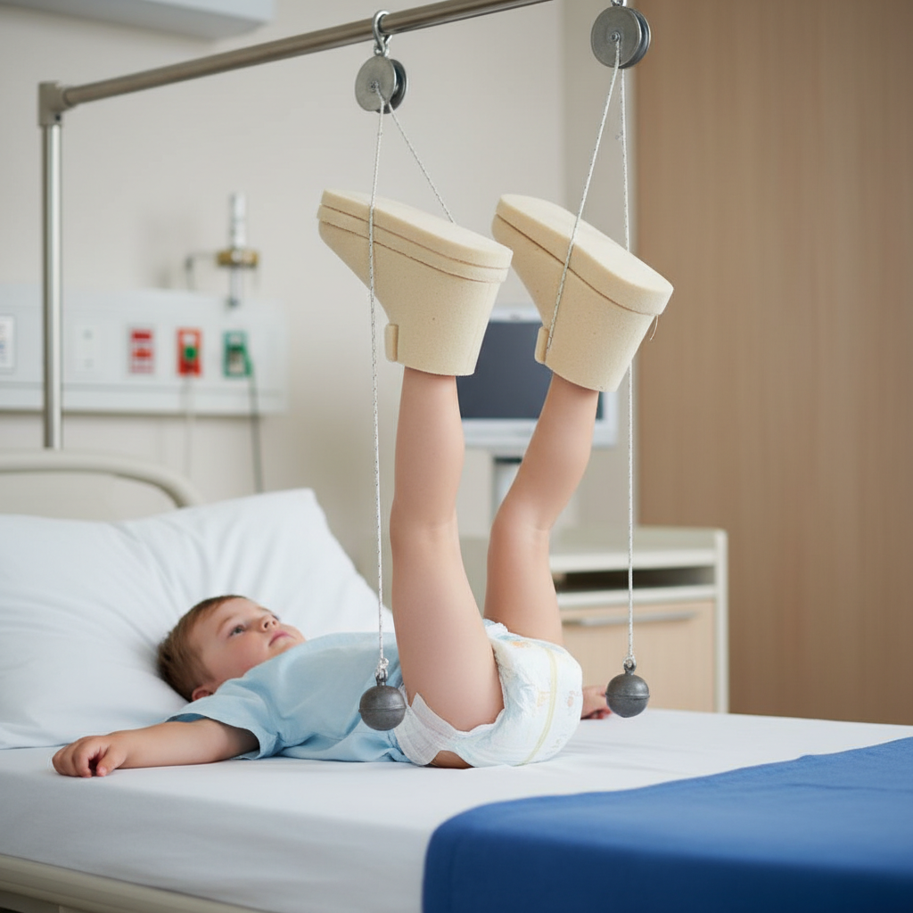
What is this mode of treatment called as?

Who is considered the father of distraction osteogenesis?
Triple arthrodesis involves which of the following joint combinations?
Perkins traction is used in which of the following conditions?
Watson Jones operation is done for:
A 55-year-old female presents with hip flexor contracture. What is the most likely test to be performed in this case?
Green extra-articular arthrodesis is done for which of the following conditions?
What is true about locking compression plates?
Explanation: **Explanation:** **Agnes Hunt Traction** is a specialized form of traction used specifically for the correction of **Flexion Deformity of the Hip**. The underlying medical concept involves applying traction to the affected limb while the contralateral (normal) limb is immobilized in a plaster cast in a position of maximum flexion. This stabilizes the pelvis and prevents compensatory lumbar lordosis, allowing the traction to act directly on the hip joint to gradually stretch the flexor contractures. **Analysis of Options:** * **A. Flexion deformity of the hip (Correct):** It is the classic indication. By neutralizing pelvic tilt, it effectively reduces fixed flexion deformities (FFD). * **B. Trochanteric fracture:** These are typically managed with skeletal traction (like Hamilton Russell traction) or, more commonly, surgical fixation (DHS or PFN). * **C. Fracture shaft of humerus:** This is managed using a U-slab, hanging cast, or skin traction like **Dunlop’s traction** (though Dunlop's is primarily for supracondylar fractures). * **D. Low backache:** This is usually managed with **Pelvic traction**, which helps in relieving muscle spasms and distracting the neural foramina. **High-Yield Clinical Pearls for NEET-PG:** * **Thomas Splint:** Used for immobilization of fractures of the shaft of the femur. * **Bryant’s Traction (Gallows):** Used for femur fractures in children below 2 years of age (weight <15-18kg). * **Russell’s Traction:** Used for trochanteric and subtrochanteric fractures; it uses a sling under the knee. * **Buck’s Traction:** A simple skin traction used for temporary immobilization of hip fractures or to reduce muscle spasms. * **90-90 Traction:** Commonly used in pediatric femoral shaft fractures to maintain the hip and knee at 90 degrees of flexion.
Explanation: **Explanation:** The procedure described is **Marsupialisation**. This technique involves opening the cyst (deroofing), evacuating its contents, and suturing the edges of the remaining cyst wall to the adjacent soft tissue or periosteum. This creates a permanent "pouch" or window, allowing the cavity to remain open to the exterior and heal by secondary intention. It is typically used for large cysts where complete removal (enucleation) might risk damaging vital structures or causing a pathological fracture. **Analysis of Options:** * **Decortication:** This involves the removal of the outer shell or "cortex" of a bone. In orthopaedics, it is often performed in cases of chronic osteomyelitis or non-union to improve vascularity and promote healing. * **Saucerization:** This is the surgical excavation of a bone cavity (usually in chronic osteomyelitis) to create a shallow, saucer-like depression. Unlike marsupialisation, it involves removing the overlying bone to allow the wound to heal from the base upwards, but it does not involve suturing the cyst wall to the periosteum. * **Enucleation:** This refers to the complete removal of a cyst in its entirety (shelling it out) without rupture. It is the treatment of choice for smaller, well-defined cystic lesions. **High-Yield Facts for NEET-PG:** * **Marsupialisation** is most commonly associated with the treatment of **Odontogenic Keratocysts (OKC)** in the jaw or large unicameral bone cysts. * **Brodie’s Abscess:** The surgical treatment of choice for this chronic localized bone abscess is **Saucerization** and curettage. * **Orr’s Technique:** A classic treatment for chronic osteomyelitis involving debridement, saucerization, and packing the wound open.
Explanation: ***Gallows traction*** - Also known as **overhead** or **Bryant's traction**, specifically designed for infants and children under 2 years weighing less than **12-15 kg**. - Both legs are suspended **vertically** with hips flexed at **90 degrees**, allowing the buttocks to just clear the bed surface for effective femoral fracture treatment. *Russell's traction* - Uses a **sling under the knee** with the leg supported horizontally, combining skin traction with **balanced suspension**. - Primarily indicated for **adult femoral shaft fractures** and hip fractures, not suitable for infants due to different anatomical requirements. *90-90 traction* - Involves hip and knee both flexed at **90 degrees** with the thigh vertical and lower leg horizontal using a **boot or pin**. - Used for **older children** and adolescents with femoral fractures, requiring more skeletal maturity than seen in infant applications. *Buck's traction* - **Straight skin traction** applied to the leg with the limb lying flat on the bed or slightly elevated. - Used for **hip fractures** in adults and **muscle spasms**, but lacks the bilateral vertical suspension characteristic of gallows traction.
Explanation: **Explanation:** **Gavriil Abramovich Ilizarov** is universally recognized as the **Father of Distraction Osteogenesis**. He pioneered the "Tension-Stress Effect," which describes how slow, steady traction on living tissues creates a metabolic stimulus that activates both proliferative and biosynthetic functions. This biological principle allows for the formation of new bone (callus) between two vascularized bone surfaces that are gradually pulled apart. Ilizarov developed the **Circular External Fixator** (Ilizarov Apparatus) to treat complex fractures, non-unions, and limb-length discrepancies. **Analysis of Incorrect Options:** * **Codivilla (A):** Alessandro Codivilla was the first to report a surgical technique for femoral lengthening in 1905. While he was a pioneer, his methods lacked the biological stability and gradual distraction principles perfected by Ilizarov. * **Snyder (B):** Snyder is associated with early experimental work in distraction, but he did not establish the clinical framework or the biological laws governing the process. * **Alexander (C):** Not a significant figure in the history of distraction osteogenesis; likely included as a distractor. **High-Yield Clinical Pearls for NEET-PG:** * **The Ilizarov Principle:** Distraction is typically performed at a rate of **1 mm per day**, divided into four increments (0.25 mm every 6 hours) to minimize soft tissue trauma and optimize osteogenesis. * **Latency Period:** The time between corticotomy and the start of distraction (usually **5–7 days**). * **Consolidation Phase:** The period where the newly formed bone (regenerate) matures and mineralizes; it usually takes twice as long as the distraction phase. * **Corticotomy:** Ilizarov emphasized a "low-energy" corticotomy to preserve the endosteal and periosteal blood supply, which is crucial for bone formation.
Explanation: **Explanation:** Triple arthrodesis is a surgical procedure aimed at stabilizing the hindfoot, correcting deformities, and relieving pain caused by arthritis or neuromuscular instability. The term "triple" refers to the fusion of the three primary joints of the hindfoot complex. **1. Why Option A is Correct:** The procedure involves the fusion of the following three joints: * **Talocalcaneal (Subtalar) joint:** Provides inversion and eversion. * **Talonavicular joint:** Part of the transverse tarsal joint; crucial for midfoot stability. * **Calcaneocuboid joint:** The lateral component of the transverse tarsal joint. By fusing these three joints, the hindfoot is locked into a neutral position, providing a stable base for weight-bearing. **2. Why Other Options are Incorrect:** * **Options B and C:** Both include the **Tibiotalar (Ankle) joint**. Triple arthrodesis specifically targets the hindfoot joints *below* the ankle. Fusing the ankle joint along with the hindfoot joints is termed a "pantalar arthrodesis," not a triple arthrodesis. The ankle joint must remain mobile in a standard triple arthrodesis to allow for dorsiflexion and plantarflexion. **Clinical Pearls for NEET-PG:** * **Indications:** Commonly used for Rigid Flatfoot (Pes Planus), Clubfoot (Talipes Equinovarus) deformities in older children/adults, and Rheumatoid Arthritis. * **Goal:** To provide a stable, plantigrade, and pain-free foot. * **Sequence of Fusion:** During surgery, the **Talonavicular joint** is considered the "key" to the reduction and is usually addressed first to set the alignment. * **Contraindication:** It is generally avoided in children under 10–12 years of age to prevent significant foot shortening due to interference with bone growth.
Explanation: **Explanation:** **Perkins Traction** is a specific type of **skeletal traction** used primarily for the management of **fractures of the shaft of the femur**. 1. **Why Option A is Correct:** Perkins traction is a "balanced" or "dynamic" traction system. Unlike fixed tractions, it does not use a Thomas splint. Instead, a Steinman pin is inserted through the proximal tibia, and the limb is placed on a specialized bed or a simple pillow. The key feature is that it allows for **early knee mobilization** while the traction is still being applied. This prevents knee stiffness (quadriceps fibrosis), which is a common complication of femoral shaft fractures. 2. **Why Other Options are Incorrect:** * **Foot drop:** This is a neurological deficit (often involving the common peroneal nerve). Management involves orthotic devices like an **AFO (Ankle Foot Orthosis)** or surgical tendon transfers, not traction. * **CTEV (Clubfoot):** The primary treatment follows the **Ponseti method**, which involves serial casting, Achilles tenotomy, and bracing (Denis Browne splint). * **Developmental Dysplasia of the Hip (DDH):** While traction (like Gallow’s or Bryant’s) was historically used to reduce the femoral head, Perkins traction is specifically designed for shaft fractures and knee movement, not hip reduction. **High-Yield Clinical Pearls for NEET-PG:** * **Bryant’s Traction (Gallow's):** Used for femur fractures in children <2 years old (weight <12-15kg). Both legs are suspended vertically. * **Russell’s Traction:** Used for trochanteric or femoral shaft fractures; it uses a sling under the knee to provide a resultant force along the femur. * **Hamilton Russell Traction:** Often used for hip fractures in the elderly. * **Thomas Splint:** Originally designed for TB spine, but most commonly used for the emergency immobilization of femur fractures.
Explanation: **Explanation:** **Watson-Jones Operation** is a classic reconstructive surgical procedure used for **Chronic Ankle Instability**, specifically to address lateral ligament laxity (the Anterior Talofibular Ligament - ATFL and Calcaneofibular Ligament - CFL). The underlying medical concept involves **tenodesis**. In this procedure, the **Peroneus Brevis tendon** is harvested (either partially or fully), passed through bone tunnels in the distal fibula and the neck of the talus, and then sutured back onto itself. This creates a "new" ligamentous structure that mechanically stabilizes the ankle joint and prevents recurrent inversion injuries. **Analysis of Incorrect Options:** * **Polio & Muscle Paralysis:** While various tendon transfers (like the Jones procedure for great toe deformity) are used in Polio or paralytic conditions to restore balance, the Watson-Jones operation specifically targets mechanical ligamentous stability rather than motor power restoration. * **Neglected Clubfoot:** This condition requires extensive soft tissue releases (e.g., Turco’s procedure) or bony corrections (e.g., Triple Arthrodesis or Ilizarov) rather than lateral ligament reconstruction. **High-Yield Clinical Pearls for NEET-PG:** * **Modified Broström Procedure:** Currently the "Gold Standard" for chronic ankle instability; it involves direct repair of the ligaments rather than using a tendon graft (unlike Watson-Jones). * **Evans and Chrisman-Snook:** Other historical tenodesis procedures for ankle instability using the Peroneus Brevis. * **Distinction:** Do not confuse the **Watson-Jones Operation** (Ankle) with the **Watson-Jones Approach** (Anterolateral approach to the Hip).
Explanation: ### Explanation **Correct Option: B. Thomas Test** The **Thomas test** is the clinical gold standard for assessing **fixed flexion deformity (FFD)** of the hip. In a normal individual, the lumbar spine has a natural lordosis that can be obliterated by flexing the contralateral hip. In a patient with a hip flexor contracture (most commonly involving the Iliopsoas muscle), the patient compensates for the deformity by increasing lumbar lordosis to keep the leg flat on the table. * **Mechanism:** When the clinician flexes the unaffected hip to flatten the lumbar spine against the examination table, the affected thigh spontaneously lifts off the table. The angle between the affected thigh and the table represents the degree of the flexion contracture. **Analysis of Incorrect Options:** * **A. Allis Test (Galeazzi Sign):** Used to assess **shortening of the femur or tibia**. It is performed by flexing both knees and hips with feet flat on the table; a difference in knee height indicates limb length discrepancy. * **C. Ober Test:** Used to identify contracture or tightness of the **Iliotibial (IT) band**. The patient lies on the unaffected side, and the clinician abducts and extends the affected hip; if the IT band is tight, the leg remains abducted and fails to adduct toward the table. * **D. Trendelenburg Test:** Assesses the stability of the hip and the strength of the **hip abductors (Gluteus medius and minimus)**. A positive sign occurs when the pelvis drops on the unsupported side during single-leg standing. **High-Yield Clinical Pearls for NEET-PG:** * **Thomas Test Pre-requisite:** Ensure the lumbar spine is flat (checked by placing a hand under the patient's lower back). * **Modified Thomas Test:** Can also assess tightness of the Rectus femoris (if the knee fails to flex to 90° while the hip is extended). * **Common Causes of Hip FFD:** Osteoarthritis, Rheumatoid Arthritis, and Psoas abscess.
Explanation: **Explanation:** **Green’s extra-articular arthrodesis** (specifically the Green-Grice procedure) is a surgical technique used to stabilize the subtalar joint without interfering with the growth of the tarsal bones. It is primarily indicated for the correction of severe paralytic flatfoot or **Congenital Vertical Talus (CVT)**. 1. **Why it is correct:** In Congenital Vertical Talus (Rocker-bottom foot), there is a rigid dislocation of the talonavicular joint. After initial reduction, maintaining the alignment of the talus and calcaneus is crucial. Green’s procedure involves placing a bone graft into the sinus tarsi (extra-articular) to fuse the subtalar joint in a neutral position. This provides stability and prevents the recurrence of the deformity while allowing the bones to continue growing in children. 2. **Why the other options are incorrect:** * **Genu Valgum (Knock-knees):** Managed by guided growth (hemiepiphyseodesis) using 8-plates or corrective osteotomies (e.g., Supracondylar femoral osteotomy). * **Coxa Vara:** Characterized by a decreased neck-shaft angle of the femur. It is treated with a **Pauwels’ Y-shaped osteotomy** or Valgus subtrochanteric osteotomy. * **Cubitus Varus (Gunstock deformity):** A common complication of malunited supracondylar fractures of the humerus. The gold standard treatment is a **French (Lateral closing wedge) osteotomy**. **High-Yield Pearls for NEET-PG:** * **Congenital Vertical Talus:** Also known as "Rocker-bottom foot." Radiologically, the talus is vertical, and the **first metatarsal-axis-talus-line (Meary’s angle)** is broken. * **Grice-Green Procedure:** It is an **extra-articular** subtalar arthrodesis, distinguishing it from intra-articular fusions like the Triple Arthrodesis (used in adults). * **Reverse Ponseti Technique:** The initial conservative management for CVT involves serial casting in a direction opposite to that used for Clubfoot (CTEV).
Explanation: ### Explanation **Locking Compression Plates (LCP)** represent a significant advancement in internal fixation, acting as an **"internal-external" fixator**. **Why Option C is correct:** LCPs are mechanically superior because the screw heads lock into the plate, creating a **fixed-angle construct**. Unlike conventional plates, which rely on friction between the plate and bone (requiring high torque), LCPs do not need to be pressed against the bone to achieve stability. This provides superior pull-out strength and stability, especially under axial loading. **Analysis of Incorrect Options:** * **Option A:** LCPs are actually the **implant of choice for osteoporotic bone**. In poor-quality bone, conventional screws often strip the threads; however, the fixed-angle stability of LCPs prevents "toggle" and screw back-out. * **Option B:** Conventional plates require tight bone-to-plate contact, which can compress and damage the periosteal blood supply. LCPs sit slightly off the bone, **preserving periosteal perfusion** and reducing the risk of necrosis. * **Option C:** LCPs feature **"Combi-holes"** (an oblong hole and a threaded hole). This allows them to be used for both dynamic compression (using cortical screws) and locking fixation, making them highly versatile. **High-Yield Clinical Pearls for NEET-PG:** * **Point Contact:** LCPs follow the "Limited Contact" principle to minimize vascular interference. * **Primary vs. Secondary Healing:** When used as a bridge plate (locking mode), LCPs allow for micromotion leading to **callus formation (secondary healing)**. When used in compression mode, they lead to **primary healing**. * **Hybrid Fixation:** Surgeons often use a combination of conventional screws (to reduce the fracture) and locking screws (to maintain the reduction).
Principles of Internal Fixation
Practice Questions
External Fixation
Practice Questions
Intramedullary Nailing
Practice Questions
Plate Osteosynthesis
Practice Questions
Tension Band Wiring
Practice Questions
Minimally Invasive Orthopaedic Surgery
Practice Questions
Arthroscopic Techniques
Practice Questions
Suture Techniques in Orthopaedics
Practice Questions
Navigation and Robotics
Practice Questions
3D Printing Applications
Practice Questions
Bone Grafting Techniques
Practice Questions
Local Flaps and Soft Tissue Coverage
Practice Questions
Get full access to all questions, explanations, and performance tracking.
Start For Free