Most common site of myositis ossificans ?
Osteitis fibrosa cystica is associated with which of the following conditions?
Most common benign tumor of bone?
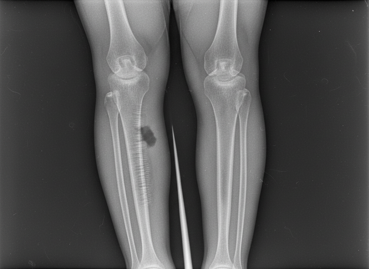
Following are seen in fibrous dysplasia EXCEPT
All of the following are features of myositis ossificans except -
A 33-year-old man presented with a slowly progressive swelling in the middle third of his right tibia. X-ray examination revealed multiple sharply demarcated radiolucent lesions separated by areas of dense and sclerotic bone. Microscopic examination of a biopsy specimen revealed islands of epithelial cells in a fibrous stroma. Which of the following is the most probable diagnosis?
Inspiratory stridor is found in what kind of lesions:
What is the most appropriate treatment for a soap bubble appearance at the lower end of the radius?
Most common site of osteogenic sarcoma is:
A 45 yrs male presented with an expansile lesion in the centre of femoral metaphysis. The lesion shows Endosteal scalloping and punctuate calcifications. Most likely diagnosis is:
Explanation: ***Quadriceps/Thigh*** - The **quadriceps and thigh** muscles are frequently affected due to their common involvement in sports injuries and trauma. - This region is prone to **hematoma formation** after contusions, which can predispose to ectopic bone formation. *Shoulder* - While the shoulder can be affected by myositis ossificans, it is **less common** than the quadriceps. - Traumatic myositis ossificans in the shoulder typically involves the **deltoid muscle**. *Wrist* - Myositis ossificans of the **wrist is rare** and usually occurs after severe trauma or crush injuries. - The small muscle mass and limited direct trauma to the wrist muscles make it an **unlikely primary site**. *Elbow* - Myositis ossificans can occur around the elbow, particularly in the **brachialis muscle**, often following dislocations or fractures. - However, the elbow is still **less commonly affected overall** compared to the large muscle groups of the thigh.
Explanation: ***Hyperparathyroidism*** - **Osteitis fibrosa cystica** is a classic bone manifestation of severe, chronic **hyperparathyroidism**, characterized by increased osteoclastic activity. - This condition results in the replacement of normal bone with **fibrous tissue** and **cysts**, often leading to bone pain, fractures, and deformities. *Hypoparathyroidism* - **Hypoparathyroidism** is characterized by low parathyroid hormone (PTH) levels and **hypocalcemia**, which typically leads to increased bone density, not bone degradation. - Its manifestations include muscle cramps, seizures, and tetany, without the bone lesions seen in osteitis fibrosa cystica. *Hypothyroidism* - **Hypothyroidism** is a state of thyroid hormone deficiency, affecting metabolism and growth but not directly causing osteitis fibrosa cystica. - It can lead to slowed bone turnover and delayed growth in children, but not the specific bone lesions associated with parathyroid dysfunction. *Hyperthyroidism* - **Hyperthyroidism** can cause increased bone turnover and **osteoporosis** due to accelerated bone resorption, but it does not lead to the distinct fibrous and cystic bone changes of osteitis fibrosa cystica. - Its effects on bone are generally a diffuse reduction in bone mineral density rather than specific lytic lesions or brown tumors.
Explanation: ***Osteochondroma*** - This is the **most common benign bone tumor**, characterized by a bony spur with a cartilaginous cap. - It typically arises from the **metaphysis of long bones**, especially around the knee. *Osteoma* - Osteomas are **benign, slow-growing tumors** composed of mature compact or cancellous bone. - They are most commonly found in the **skull and facial bones**, not typically in long bones. *Simple bone cyst* - This is a **fluid-filled lesion** of bone, not a true neoplasm, frequently found in the metaphysis of long bones in children. - It is often discovered incidentally or after a **pathological fracture**. *Osteoid osteoma* - Characterized by a **small, benign bone tumor** with a central nidus of osteoid and trabecular bone, surrounded by reactive sclerotic bone. - It classically causes **nocturnal pain** that is relieved by NSAIDs.
Explanation: ***No premalignant change*** - Fibrous dysplasia is a **benign condition** that does NOT undergo premalignant change [1]. - However, there is a small but definite risk of **malignant transformation** (not premalignant change), particularly to **osteosarcoma**, in about 0.5% of cases [1]. - This risk is higher in patients who have received **radiation therapy** for the condition. - **Key distinction**: Malignant transformation is different from premalignant change—fibrous dysplasia remains benign but can rarely transform directly to malignancy [1]. *Ground glass appearance on X-ray* - This is a **classic radiographic feature** of fibrous dysplasia, resulting from immature woven bone and fibrous tissue within the lesion [2]. - It describes the hazy, ill-defined radiodensity due to the lack of organized trabeculae. *Expanding rib lesions* - Fibrous dysplasia frequently affects the **ribs**, leading to their expansion and potential for pathological fractures [1]. - Rib involvement is particularly common in the **monostotic form** of the disease [1]. *Expanding lesions of maxilla* - The **maxilla** is a common site for fibrous dysplasia, especially in the **craniofacial form**. - Maxillary lesions can cause expansion, facial asymmetry, and dental abnormalities. **References:** [1] Kumar V, Abbas AK, et al.. Robbins and Cotran Pathologic Basis of Disease. 9th ed. Bones, Joints, and Soft Tissue Tumors, p. 1208. [2] Kumar V, Abbas AK, et al.. Robbins and Cotran Pathologic Basis of Disease. 9th ed. Bones, Joints, and Soft Tissue Tumors, pp. 1208-1209.
Explanation: ***It matures from inside out*** - This statement is incorrect; **myositis ossificans** characteristically matures from the **outside in**, meaning the periphery ossifies first, creating a distinct radiologic appearance. - The mature bone is found at the periphery of the lesion, while the more immature, cellular components are centrally located. *Commonly occurs around the elbow* - Myositis ossificans is frequently observed around joints, with the **elbow** being one of the most common sites due to its vulnerability to trauma. - Other common locations include the **thigh** and **shoulder**. *Can be post traumatic or occur without trauma also* - While most commonly **post-traumatic**, myositis ossificans can also occur atraumatically, sometimes referred to as **myositis ossificans progressiva** or fibrodysplasia ossificans progressiva, a rare genetic disorder. - The traumatic form is often preceded by a significant contusion or muscle injury. *Massaging is a known associated factor* - **Aggressive massage** or manipulation following muscle trauma can exacerbate local tissue injury and enhance the conditions conducive to heterotopic ossification, including myositis ossificans. - This is why gentle management of muscle contusions is crucial to prevent its development.
Explanation: ***Adamantinoma*** - This diagnosis is strongly supported by **multiple sharply demarcated radiolucent lesions** with areas of **dense and sclerotic bone** on X-ray, and islands of **epithelial cells in a fibrous stroma** on biopsy. - Adamantinoma is a rare, malignant bone tumor almost exclusively found in the **tibia** or fibula, characterized by its slow growth and epithelial differentiation. *Fibrous cortical defect* - These are common, **benign fibrous lesions** typically found in the metaphysis of long bones, often asymptomatic and spontaneously resolving [1]. - Histologically, they consist of fibroblasts, histiocytes, and macrophages, without epithelial islands [1]. *Osteosarcoma* - This is a highly malignant tumor characterized by the production of **osteoid** or **immature bone** by malignant cells, which is not described. - Radiographically, it usually presents with an aggressive, destructive lesion, often with "sunburst" or "Codman's triangle" patterns, which differ from the description. *Osteofibrous dysplasia* - This benign fibro-osseous lesion primarily affects the **tibia** in children and young adults, often presenting with painless swelling. - While it can show fibrous tissue and immature bone, it lacks the characteristic epithelial islands seen in adamantinoma. **References:** [1] Kumar V, Abbas AK, et al.. Robbins and Cotran Pathologic Basis of Disease. 9th ed. Bones, Joints, and Soft Tissue Tumors, pp. 1206-1208.
Explanation: ***Supraglottic*** - Lesions in the **supraglottic** region (e.g., epiglottitis, supraglottic foreign body) cause inspiratory stridor due to the collapse of soft tissues above the vocal cords during inspiration. - The narrowed airway during inspiration creates a high-pitched, harsh sound. *Subglottic* - **Subglottic** lesions typically cause a **biphasic stridor**, meaning stridor is present during both inspiration and expiration. - This is because the subglottis is a rigid area; narrowing at this level causes turbulent airflow during both phases of breathing. *Tracheal* - **Tracheal** lesions can produce **biphasic stridor** if they are in the cervical trachea due to fixed airway narrowing. - If the lesion is in the lower, intrathoracic trachea, it might primarily cause **expiratory stridor** or a biphasic stridor depending on the degree of narrowing and its fixity. *Bronchus* - Lesions in the **bronchus** (e.g., foreign body, tumor) typically lead to **expiratory stridor** or wheezing. - Airway narrowing at this level causes air trapping and turbulent flow predominantly during exhalation when the bronchial walls naturally constrict.
Explanation: ***Excision and bone grafting*** - A **soap bubble appearance** at the lower end of the radius is highly suggestive of a **giant cell tumor (GCT)**, which is locally aggressive and has a high recurrence rate after simple curettage. - **Excision of the tumor and filling the defect with bone graft** is the preferred treatment to reduce recurrence and maintain skeletal integrity. *Local excision* - While local excision might remove the visible tumor, **GCTs are known to recur frequently** (up to 50%) after intralesional treatments like simple curettage. - It does not adequately address microscopic extensions or the risk of **local aggressive behavior**. *Amputation* - **Amputation is an overly aggressive and unnecessary treatment** for a GCT, as it is a benign but locally aggressive tumor. - It would be considered only in rare cases of extensive soft tissue invasion or intractable recurrence, which is not implied by a "soap bubble appearance." *Radiotherapy* - **Radiotherapy is generally not the first-line treatment for GCTs** due to concerns about **malignant transformation** (osteosarcoma) in a small percentage of cases, especially with high doses. - It may be considered for unresectable tumors or recurrent lesions in difficult anatomical locations, or as an adjuvant.
Explanation: ***Femur, lower end*** - The **distal femur** is the most common site for osteogenic sarcoma, accounting for approximately **40% of all cases** [1]. - This region, along with the **proximal tibia**, are the most frequent locations for this primary bone tumor [1]. *Tibia, lower end* - While osteogenic sarcoma can occur in the **tibia**, the **proximal end** is more commonly affected than the distal end. - The distal tibia is a less frequent site compared to the distal femur or proximal tibia. *Femur, upper end* - The **proximal femur** is a recognized site for osteogenic sarcoma, but it is less common than the **distal femur**. - Tumors in the proximal femur account for a smaller percentage of overall osteosarcoma cases. *Tibia, upper end* - The **proximal tibia** is the **second most common site** for osteogenic sarcoma, frequently affected after the distal femur [1]. - However, the question asks for the *most* common site, which remains the distal femur.
Explanation: ***Chondrosarcoma*** - An **expansile lesion** within the **femoral metaphysis** with **endosteal scalloping** and **punctate calcifications** is highly characteristic of a chondrosarcoma. - The punctate/arc-and-ring calcifications are typical for cartilage matrix, which is the hallmark of chondrosarcoma, and the patient's age (45 years) fits the typical demographic. *Fibrous Dysplasia* - This condition presents as a **ground-glass matrix** on imaging, not punctate calcifications. - While it can be expansile, it typically does not show prominent endosteal scalloping with cartilage calcifications. *Simple bone cyst* - Simple bone cysts are typically **lytic lesions** that do not show punctate calcifications or aggressive endosteal scalloping. - They are often **fluid-filled** and common in children/adolescents, whereas this patient is 45 years old. *Osteosarcoma* - Osteosarcomas are characterized by **osteoid matrix formation** and often have a more aggressive appearance with a **sunburst or spiculated periosteal reaction** and bone formation, not punctate cartilage calcifications. - While it can be expansile, the calcification pattern described points away from osteosarcoma.
Get full access to all questions, explanations, and performance tracking.
Start For Free