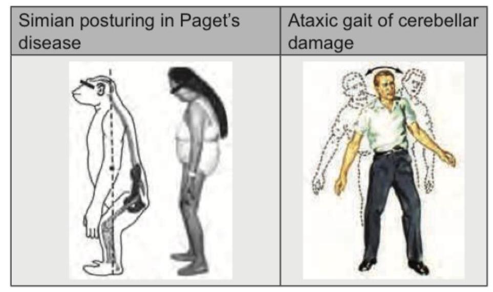
The appearance shown in the image is called as Simian Posturing. Notice the bent trunk, flexed legs and loss of spine normal axis. It occurs due to disordered bone turnover involving pelvis and vertebra. Which condition is most associated with this presentation?

A 60-year-old elderly female with a previous history of a Colles fracture is now complaining of backache. Which of the following statements regarding the treatment of this patient is incorrect?
Not seen in osteogenesis imperfecta
Albers Schonberg disease is also called as -
A 70-year-old female has been on alendronate for 7 years for osteoporosis and now complains of pain in her right thigh. What is the next investigation to be performed?
Wormian bones are seen in all except?
Brittle bone disease is -
A 45-year-old was given steroids after renal transplant. After 2 years he had difficulty in walking and pain in both hips. Which one of the following is most likely cause?
Avascular necrosis of head of femur occurs commonly at :
Avascular necrosis of bone is LEAST likely to be associated with?
Explanation: ***Paget's disease*** - **Paget's disease** is characterized by disordered bone turnover, leading to bone enlargement, deformity, and weakness. This can result in changes in posture, such as **simian posturing** due to involvement of the pelvis and vertebrae, as described. - The abnormal bone remodeling in Paget's disease can cause **bowing of long bones**, skull enlargement, and spinal changes, directly leading to the described posture. *Rickets* - **Rickets** is a childhood condition caused by a deficiency in vitamin D, calcium, or phosphate, leading to **softening and weakening of bones**. - While rickets can cause bone deformities like bowed legs and skeletal pain, it typically does not present with the specific **simian posturing** affecting the entire trunk and spine as described in an adult. *Achondroplasia* - **Achondroplasia** is a genetic disorder of bone growth resulting in **dwarfism**, characterized by short limbs and a normal-sized trunk. - While it causes skeletal abnormalities, it typically manifests differently from the described simian posturing, which involves advanced bone remodeling in the spine and pelvis, not primarily a growth disorder. *Osteoporosis* - **Osteoporosis** is a condition where bones become **brittle and fragile** due to loss of tissue, leading to an increased risk of fractures. - Although osteoporosis can lead to **kyphosis** (hunchback) due to vertebral compression fractures, it typically does not present with the global "simian posturing" involving the entire pelvis, spine, and flexed legs due to disordered bone turnover.
Explanation: ***Teriparatide should be started before supplementing bisphosphonates*** - This statement is incorrect because **bisphosphonates are typically the first-line treatment** for osteoporosis, especially in patients with a history of fragility fractures like a Colles fracture. - **Teriparatide**, an anabolic agent, is usually reserved for patients with very severe osteoporosis, those who have failed bisphosphonate therapy, or those with highly accelerated bone loss. *Oral vitamin D3 is given along with oral calcium* - This is a routine and **correct practice in osteoporosis management** as calcium and vitamin D are essential for bone health. - **Vitamin D** aids in calcium absorption from the gut, and both are crucial for bone mineralization and density. *Calcium requirement is 1200 mg per day* - The recommended daily **calcium intake for postmenopausal women** and elderly individuals with osteoporosis is typically around 1200 mg. - This amount helps to maintain skeletal health and reduce the risk of fractures. *Bisphosphonates can be given for 3-5 years depending on patient response and risk factors* - This statement is correct, as **bisphosphonates are commonly prescribed for 3-5 years** to reduce fracture risk in osteoporosis. - A **"drug holiday"** may be considered after this period, depending on the patient's fracture risk and bone mineral density.
Explanation: ***Thick cortical bone*** - Osteogenesis imperfecta (OI) is characterized by **fragile bones** due to defects in **Type I collagen** synthesis, leading to abnormally **thin cortical bone**. - **Thick cortical bone** would indicate increased bone density or strength, which is the opposite of the fundamental pathology in OI. *Wormian bones* - **Wormian bones** (intrasutural bones) are frequently seen in individuals with **osteogenesis imperfecta**, particularly in types I and III. - They are small, irregular bones that develop within the **cranial sutures**. *Coxa vara* - **Coxa vara**, a deformity where the angle between the femoral neck and shaft is decreased, is a common orthopedic complication of **osteogenesis imperfecta**. - This deformity is primarily due to the **bone fragility** and remodeling issues inherent to the condition. *Saber shin* - **Saber shin** refers to an anterior bowing of the tibia, which is a classic orthopedic manifestation in patients with **osteogenesis imperfecta**. - This bowing results from repeated microfractures and **abnormal bone remodeling** characteristic of the disease.
Explanation: ***Osteopetrosis*** - Albers-Schonberg disease is a common synonym for **osteopetrosis**, also known as **marble bone disease**. - This genetic disorder is characterized by abnormally **dense bones** due to a defect in **osteoclast function**, leading to unresorbed bone. *Osteoporosis* - **Osteoporosis** is a condition of **reduced bone density** and mass, leading to fragile bones and increased fracture risk, which is the opposite of Albers-Schonberg disease. - It results from an imbalance where **bone resorption** by osteoclasts outpaces bone formation by osteoblasts. *Osteitis punctata* - This term is not a recognized medical condition but might refer to a non-specific localized inflammation or lesion of bone characterized by "dots" or points. - It does not describe a systemic bone density disorder like Albers-Schonberg disease. *Osteodystrophy* - **Osteodystrophy** is a general term referring to a **bone disease** or malformation, often due to a metabolic disorder like **renal osteodystrophy**. - It is a broader category and not a specific synonym for Albers-Schonberg disease.
Explanation: **X-ray** - Alendronate, a **bisphosphonate**, is associated with **atypical femoral fractures** after prolonged use, and an X-ray is the most appropriate initial investigation to visualize such a fracture. - Complaints of thigh pain in a patient on long-term bisphosphonate therapy should prompt imaging to rule out this serious complication. *DEXA scan* - A DEXA scan assesses **bone mineral density** but does not provide information about acute fractures or structural integrity in response to specific pain. - While it's used for osteoporosis diagnosis and monitoring, it won't directly identify an atypical femoral fracture. *Serum vitamin D levels* - Maintaining adequate **vitamin D levels** is important for bone health, but its measurement won't explain acute thigh pain or identify a fracture. - Low vitamin D levels can contribute to osteoporosis but are not the primary cause of pain suggestive of an atypical femoral fracture. *Serum alkaline phosphate levels* - **Alkaline phosphatase** levels can be elevated in conditions involving increased bone turnover, such as healing fractures or certain bone diseases. - However, it is not a direct diagnostic tool for identifying atypical femoral fractures and would not be the first line investigation.
Explanation: ***Fibrous dysplasia*** - **Fibrous dysplasia** is a bone disorder where normal bone is replaced by fibrous tissue and immature woven bone, but it is not typically associated with the development of **Wormian bones**. - Its manifestations are usually localized to specific bones and include pain, deformity, and fractures, rather than abnormalities in cranial suture ossification. *Osteogenesis imperfecta* - **Osteogenesis imperfecta** (OI), or brittle bone disease, is a genetic disorder characterized by **fragile bones** and often includes **Wormian bones** due to defective collagen synthesis. - The presence of multiple Wormian bones is a common radiographic finding in individuals with OI. *Rickets* - **Rickets** is caused by a deficiency in vitamin D, calcium, or phosphate, leading to impaired bone mineralization and **softening of bones**. - While rickets primarily affects long bones, severe and prolonged cases, particularly in children, can lead to widespread defects in bone ossification, including the appearance of **Wormian bones** in the skull. *Cretinism* - **Cretinism** (congenital hypothyroidism) leads to delayed skeletal maturation and abnormal bone development. - One of the skeletal anomalies seen in cretinism is the presence of **Wormian bones**, reflecting impaired ossification of the skull.
Explanation: ***Osteogenesis imperfecta*** - **Osteogenesis imperfecta** is an inherited disorder characterized by **brittle bones** that fracture easily, due to a defect in **collagen type I** synthesis. - Patients often present with **blue sclera**, **dentinogenesis imperfecta**, and **hearing loss**, in addition to frequent fractures. *Osteoporosis* - **Osteoporosis** is a condition of **decreased bone density**, making bones fragile and prone to fracture, but it is not typically referred to as "brittle bone disease" in the same congenital sense. - It is more common in older adults and is often related to **hormonal changes** (e.g., post-menopause) or lifestyle factors. *Paget's disease* - **Paget's disease of bone** involves abnormal bone remodeling with excessive bone resorption followed by disorganized and expanded bone formation, leading to **enlarged, weakened bones**. - It typically affects older individuals and can lead to bone pain, deformities, and fractures, but it's not the primary condition associated with "brittle bone disease." *Osteopetrosis* - **Osteopetrosis** is characterized by **abnormally dense bones** due to impaired osteoclast function, leading to a buildup of bone. - While bones are dense, they are also **brittle** and prone to fracture, and the condition is also known as "marble bone disease" rather than "brittle bone disease."
Explanation: ***Avascular necrosis*** - Chronic **steroid use**, especially after organ transplantation, is a major risk factor for avascular necrosis (AVN) due to impaired blood supply to bone, particularly in the femoral head. - **Hip pain** and **difficulty walking** are classic symptoms of AVN, which can lead to collapse of the femoral head if untreated. *Tuberculosis* - While tuberculosis can affect bones and joints (**Pott's disease**), it typically presents with more systemic symptoms like fever, weight loss, and night sweats, which are not mentioned. - Skeletal TB often affects the spine more commonly and usually presents with granulomatous inflammation and bone destruction rather than isolated joint pain in the hips *Primary Osteoarthritis* - Primary osteoarthritis is typically an **age-related degenerative joint disease** occurring in older individuals, and while it causes hip pain, it is not directly linked to steroid use in a 45-year-old. - The onset of pain in this scenario, following long-term steroid use, strongly points away from primary osteoarthritis as the primary driving factor. *Aluminum toxicity* - Aluminum toxicity can occur in patients with **renal failure** and can cause **osteomalacia** or **dialysis encephalopathy**. - Its presentation typically involves bone pain, fractures, and neurological symptoms, but it does not specifically cause avascular necrosis of the femoral head as seen with steroid use.
Explanation: ***Subcapital region*** - The **subcapital region** of the femoral head is the most common site for avascular necrosis due to its precarious blood supply, especially after trauma or with certain medical conditions. - This area is particularly vulnerable because the main blood supply from the **medial femoral circumflex artery** passes through the femoral neck to reach the head. *Transcervical region* - While fractures in the **transcervical region** can compromise blood supply to the femoral head, avascular necrosis primarily affects the subcapital region itself, rather than the neck. - This region is more prone to fracture, which can subsequently lead to disruption of vascularity to the femoral head, but not the primary site of necrosis. *Subchondral region* - The **subchondral region** is the area directly beneath the articular cartilage, and while it is eventually impacted by necrosis as the bone collapses, it is not the initial or primary site of avascular changes. - Necrosis typically begins in the bone marrow and trabeculae before affecting the subchondral plate. *Trochanteric region* - The **trochanteric region** (both greater and lesser trochanters) is well-vascularized and is rarely affected by avascular necrosis. - This area is a site for muscle attachments and has a robust blood supply, making it resilient to ischemic events.
Explanation: ***Osgood-Schlatter disease*** - This condition is characterized by **inflammation of the patellar ligament** at its insertion into the tibial tuberosity, primarily due to repetitive stress in adolescents. - While it involves pain and swelling around the knee, it is a **traction apophysitis** and not a form of avascular necrosis. *Long-term use of corticosteroids* - **Corticosteroids** are a well-established risk factor for avascular necrosis, particularly in the femoral head, by affecting lipid metabolism and blood flow. - They can lead to **fat embolism** and increased intraosseous pressure, compromising blood supply to the bone. *Sickle-cell disease* - **Sickle cell disease** significantly increases the risk of avascular necrosis due to **vaso-occlusive crises**, where sickled red blood cells block small blood vessels. - This leads to **ischemia and infarction** in bone marrow, commonly affecting the femoral and humeral heads. *Legg-Perthes disease* - This is a specific type of **avascular necrosis of the femoral head** in children, causing a temporary interruption of blood supply to the epiphysis. - It results in the collapse of the femoral head and subsequent repair processes, consistent with the pathology of avascular necrosis.
Osteoporosis
Practice Questions
Osteomalacia and Rickets
Practice Questions
Paget's Disease of Bone
Practice Questions
Hyperparathyroidism
Practice Questions
Renal Osteodystrophy
Practice Questions
Fluorosis
Practice Questions
Osteogenesis Imperfecta
Practice Questions
Bone Mineral Density Assessment
Practice Questions
Pharmacological Management of Metabolic Bone Diseases
Practice Questions
Surgical Considerations in Metabolic Bone Diseases
Practice Questions
Fragility Fractures
Practice Questions
Prevention Strategies
Practice Questions
Get full access to all questions, explanations, and performance tracking.
Start For Free