A 60-year-old person suffering from myositis ossificans progressive, what is the usual cause of death?
Nodular growth of alveolus is seen in which of the following?
Which bone is most commonly involved in Paget's disease?
Which of the following are characteristic features of childhood osteopetrosis?
An X-ray of a patient shows bulbous ends of long bones, normal appositional bone growth, and failure of physiologic root resorption. Laboratory findings show myelophthisic anemia. What is the probable diagnosis?
Absence of lamina dura in the alveolus occurs in which of the following conditions?
Rickets in an infant present with which of the following signs, except?
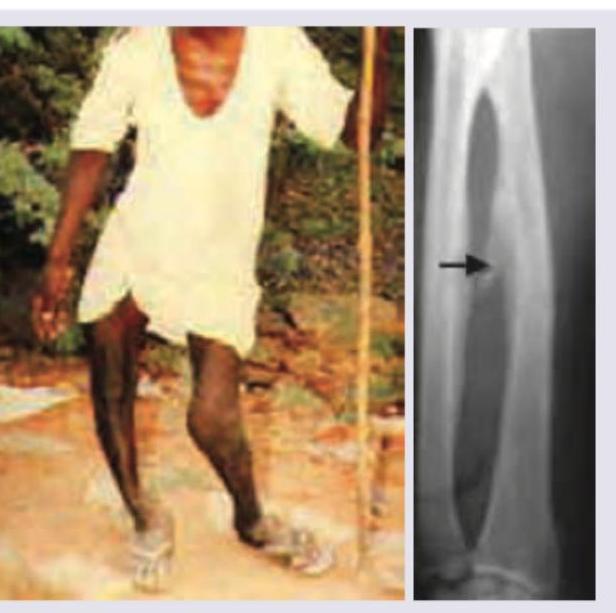
In a rural posting, a 35-year-old farmer comes to the OPD. Identify the condition shown:

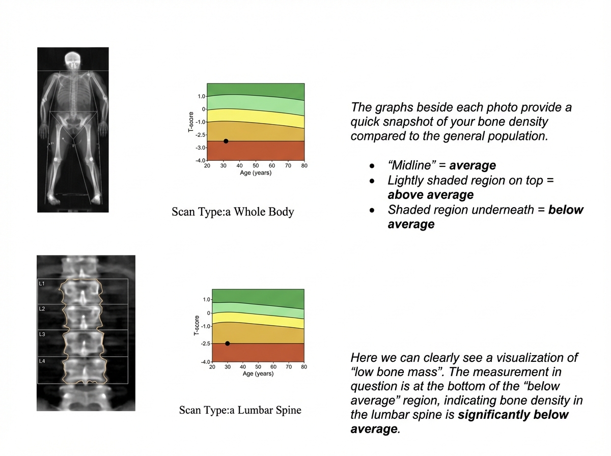
Which of the following is correct about the report of bone densitometry shown?

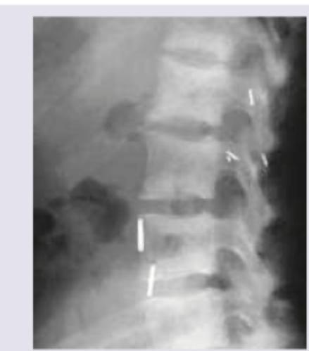
A 72-year-old woman presents with bone pain and tenderness in her lower extremities and left clavicle. Laboratory studies reveal elevated calcium, decreased phosphate, and elevated serum alkaline phosphatase. X-ray spine shows?

Explanation: **Explanation:** **Myositis Ossificans Progressiva**, also known as **Fibrodysplasia Ossificans Progressiva (FOP)**, is a rare genetic disorder characterized by the progressive transformation of soft tissues (muscles, tendons, and ligaments) into heterotopic bone. **Why Lung Disease is the Correct Answer:** The primary cause of mortality in FOP is **Thoracic Insufficiency Syndrome**. As the disease progresses, heterotopic ossification involves the intercostal muscles, paravertebral soft tissues, and the joints of the thoracic cage. This leads to: 1. **Chest Wall Restriction:** The rib cage becomes "frozen" or fused, severely limiting expansion during inspiration. 2. **Respiratory Failure:** Chronic restrictive lung disease develops, leading to pneumonia and eventually right-sided heart failure (Cor Pulmonale). Most patients succumb to respiratory complications by their 40s or 50s. **Analysis of Incorrect Options:** * **A. Nutritional deficiency:** While jaw involvement (ankylosis of the temporomandibular joint) can make eating difficult, it is rarely the primary cause of death due to modern nutritional support. * **B & D. Bed sore and Septicemia:** While immobility increases the risk of pressure sores and subsequent sepsis, these are secondary complications. The physiological hallmark that limits lifespan in FOP is the mechanical failure of the respiratory system. **Clinical Pearls for NEET-PG:** * **Genetic Mutation:** Caused by a mutation in the **ACVR1 gene** (encoding the ALK2 receptor). * **Classic Sign:** Congenital **malformation of the great toe** (short, hallux valgus) is a pathognomonic early diagnostic clue. * **Management Caution:** Avoid intramuscular injections, biopsies, or surgeries, as **trauma triggers "flare-ups"** and rapid new bone formation.
Explanation: **Explanation:** **Cementifying fibroma** (also known as ossifying fibroma) is a benign fibro-osseous neoplasm most commonly involving the mandible. It is characterized by the replacement of normal bone with fibrous tissue containing mineralized masses of cementum-like material or bone. Clinically, it presents as a slow-growing, painless, and well-demarcated **nodular expansion of the alveolus** (the tooth-bearing portion of the jaw). This localized, nodular growth is a hallmark feature that helps distinguish it from more diffuse bone pathologies. **Analysis of Incorrect Options:** * **Paget’s Disease:** While Paget’s can involve the maxilla (causing "leontiasis ossea"), it typically presents as a generalized, diffuse enlargement of the bone with a "cotton-wool" appearance on X-ray, rather than a localized nodular growth of the alveolus. * **Osteomas:** These are benign osteogenic tumors that appear as very dense, radiopaque masses. While they can occur in the jaw (often associated with Gardner’s syndrome), they are usually peripheral or endosteal masses rather than specific nodular growths of the alveolar ridge itself. * **All of the above:** Incorrect because the specific clinical descriptor "nodular growth of the alveolus" is the classic presentation specifically linked to cementifying fibromas in standard orthopaedic and oral pathology texts. **High-Yield Clinical Pearls for NEET-PG:** * **Radiology:** Cementifying fibroma appears as a well-circumscribed lesion with a radiolucent rim (fibrous capsule), which helps differentiate it from fibrous dysplasia (which has "ground-glass" appearance and ill-defined borders). * **Demographics:** Most common in the 3rd and 4th decades of life, with a predilection for females. * **Treatment:** Surgical enucleation or curettage is usually sufficient due to its well-demarcated nature.
Explanation: **Explanation:** Paget’s disease of bone (Osteitis Deformans) is a chronic disorder characterized by focal areas of increased and disorganized bone remodeling. The correct answer is **Pelvis**, as it is the most frequently involved site in this condition. **1. Why Pelvis is Correct:** Paget’s disease typically involves the axial skeleton. Epidemiological studies and clinical data consistently show that the **Pelvis** is involved in approximately **70–75%** of cases, making it the most common site. It is followed by the spine (vertebrae), femur, and skull. **2. Analysis of Incorrect Options:** * **Skull (Option A):** While iconic for clinical signs like "Cotton wool spots" on X-ray and increasing hat size, it is less frequently involved than the pelvis or femur. * **Femur (Option B):** The femur is the most common site in the **appendicular skeleton** (long bones), but it ranks below the pelvis in overall frequency. * **Vertebrae (Option C):** The lumbar spine is the second most common site overall, but it does not surpass the pelvis in incidence. **3. High-Yield Clinical Pearls for NEET-PG:** * **Pathophysiology:** Initial osteoclastic (lytic) phase → Mixed phase → Osteoblastic (sclerotic) phase. * **Biochemical Marker:** Characterized by **Isolated elevation of Serum Alkaline Phosphatase (ALP)** with normal Calcium and Phosphate levels. * **Radiological Signs:** "Picture frame" vertebrae, "Blade of grass" sign in long bones, and "Brim sign" in the pelvis. * **Complications:** The most dreaded complication is **Osteosarcoma** (seen in <1% of cases). High-output heart failure can occur due to increased vascularity in bone. * **Treatment:** Bisphosphonates (Zoledronic acid is the drug of choice).
Explanation: **Explanation:** **Osteopetrosis (Albers-Schönberg disease)**, also known as "Marble Bone Disease," is a genetic disorder characterized by **defective osteoclast function**. Because osteoclasts fail to resorb bone, the bone becomes excessively dense but structurally weak and brittle. 1. **Why Option A is Correct:** * **Multiple Fractures:** Despite the increased bone density (radiopacity), the lack of normal remodeling leads to poor bone quality, making them prone to "chalk-stick" fractures. * **Hepatosplenomegaly:** The excessive bone formation obliterates the medullary (bone marrow) cavity. To compensate for the loss of hematopoietic space, the body resorts to **extramedullary hematopoiesis** in the liver and spleen, leading to their enlargement. 2. **Why Other Options are Incorrect:** * **Option B:** While hepatosplenomegaly is present, it is an incomplete description. The hallmark clinical paradox of osteopetrosis is "dense but fragile" bones. * **Option C & D:** **Frontal bossing** is more characteristic of Rickets or Thalassemia. **Cataracts** are not a feature of osteopetrosis; however, patients may suffer from **optic atrophy** or blindness due to the narrowing of cranial nerve foramina (bone overgrowth compressing the nerves). **NEET-PG High-Yield Pearls:** * **Radiology:** Look for the "Bone within a bone" appearance (Endobone) and "Erlenmeyer flask deformity" of the distal femur. * **Rugger-Jersey Spine:** Characterized by dense bands at the superior and inferior endplates (also seen in Renal Osteodystrophy). * **Complications:** Pancytopenia (due to marrow space loss) and cranial nerve palsies (most commonly CN II, VII, and VIII). * **Treatment:** Bone marrow transplant is the definitive treatment for the infantile (malignant) form to provide functional osteoclasts.
Explanation: ### Explanation **Correct Answer: C. Osteopetrosis** **Mechanism and Clinical Presentation:** Osteopetrosis (Albers-Schönberg disease or Marble Bone Disease) is caused by **defective osteoclast function**, leading to a failure of normal bone resorption. * **Bulbous ends of long bones:** Failure of remodeling results in the characteristic **"Erlenmeyer flask deformity"** (metaphyseal widening). * **Normal appositional growth:** While longitudinal remodeling is impaired, subperiosteal bone formation continues, leading to increased bone density. * **Myelophthisic Anemia:** Because the medullary cavity is not resorbed, it becomes obliterated by calcified cartilage and bone. This "crowds out" the bone marrow, leading to pancytopenia and extramedullary hematopoiesis. * **Dental findings:** Failure of physiologic root resorption and delayed tooth eruption are classic features due to the dense alveolar bone. **Why other options are incorrect:** * **A. Fibrous Dysplasia:** Characterized by "Ground-glass appearance" on X-ray and replacement of bone with fibrous tissue; it does not cause generalized increased bone density or myelophthisic anemia. * **B. Osteomyelitis:** This is an infection showing bone destruction (sequestrum) and new bone formation (involucrum), usually localized rather than systemic. * **D. Paget’s Disease:** Involves disordered bone remodeling (high turnover). While it shows cortical thickening, it typically presents with a "mosaic pattern" on histology and elevated Alkaline Phosphatase, without obliterating the marrow cavity to cause anemia. **High-Yield NEET-PG Pearls:** * **Radiological Signs:** "Bone-within-a-bone" appearance (Endobone) and "Sandwich vertebrae" (Rugger-jersey spine). * **Complications:** Pathological fractures (despite high density, the bone is brittle) and cranial nerve palsies (due to narrowing of cranial foramina). * **Treatment:** Bone marrow transplant is the definitive treatment for the infantile (malignant) form to provide functional osteoclasts.
Explanation: **Explanation:** The **lamina dura** is a thin layer of compact bone that lines the tooth socket (alveolus). On a radiograph, it appears as a continuous, radio-opaque white line around the tooth root. Its integrity is a sensitive indicator of systemic bone metabolism. **1. Why Hyperparathyroidism is correct:** In **Hyperparathyroidism** (specifically primary or secondary), there is an excess of Parathyroid Hormone (PTH), which stimulates osteoclastic activity. This leads to generalized subperiosteal bone resorption. The lamina dura is one of the earliest sites to undergo resorption because it has a high turnover rate. The "loss of lamina dura" is a classic, high-yield radiographic sign of hyperparathyroidism, often occurring alongside "Salt and Pepper" appearance of the skull and Brown tumors. **2. Why the other options are incorrect:** * **Rickets & Osteomalacia:** While these involve defective mineralization of the bone matrix (osteoid), they typically present with features like Looser’s zones (pseudofractures) or bowing of long bones. While thinning of the lamina dura *can* rarely occur, it is not the classic or pathognomonic feature associated with these conditions in exams. * **Deficiency of Vitamin C (Scurvy):** Scurvy affects collagen synthesis. Radiographic features include the Wimberger ring sign, Frankel’s line, and Pelkan spurs. It typically causes gingival bleeding and tooth mobility, but not the specific resorption of the lamina dura. **3. NEET-PG High-Yield Pearls:** * **Most sensitive site for resorption in Hyperparathyroidism:** Radial aspect of the middle phalanx of the index and middle fingers. * **Differential Diagnosis for Loss of Lamina Dura:** Hyperparathyroidism (most common), Paget’s disease, and Systemic Sclerosis (Scleroderma). * **Rugger-Jersey Spine:** Seen in secondary hyperparathyroidism (Renal Osteodystrophy).
Explanation: **Explanation:** The correct answer is **D. Bow legs**. The clinical presentation of rickets depends significantly on the **age of the child** and the specific bones undergoing the most rapid growth at that time. 1. **Why "Bow legs" is the correct answer:** In an **infant** (non-ambulatory), the lower limbs are not yet weight-bearing. Bowing of the legs (Genu varum) typically develops only after the child starts walking and the softened osteoid is subjected to mechanical stress. Therefore, it is a sign seen in toddlers, not infants. 2. **Why the other options are incorrect:** * **Craniotabes (A):** This is the **earliest** clinical sign of rickets, seen in infants under 6 months. It is characterized by the softening of the skull bones (ping-pong ball sensation). * **Widened fontanel (B):** Delayed closure of the anterior fontanel and frontal bossing are classic cranial features of infantile rickets. * **Rachitic rosary (C):** This refers to the palpable/visible enlargement of the costochondral junctions. It is a hallmark sign of active rickets in infancy. **NEET-PG High-Yield Pearls:** * **Earliest Sign:** Craniotabes. * **Earliest Radiological Sign:** Rarefaction of the zone of provisional calcification (followed by cupping, splaying, and fraying of the metaphysis). * **Best Site for X-ray:** Wrist (specifically the distal ulna) is the most sensitive site for early diagnosis. * **Harrison’s Sulcus:** A horizontal groove along the lower border of the thorax corresponding to the insertion of the diaphragm, seen in severe rickets. * **Biochemical Profile:** Low/Normal Calcium, Low Phosphate, and **Elevated Alkaline Phosphatase (ALP)** (the most sensitive biochemical marker).
Explanation: ***Skeletal fluorosis*** - The image on the left shows a person with **bowing of legs** and **joint deformities**, consistent with the chronic bone and joint pain, and stiffness characteristic of skeletal fluorosis. - The accompanying X-ray on the right, showing increased bone density and periosteal new bone formation (indicated by the arrow), is a classic radiographic finding in **skeletal fluorosis** caused by *excess fluoride intake*, often from contaminated drinking water in rural areas. *Neurolathyrism* - This condition is caused by the neurotoxin **ODAP** found in *Lathyrus sativus* (grass pea) and primarily manifests as **spastic paraplegia** and weakness of the lower limbs, typically without significant bone deformities or characteristic X-ray changes seen here. - While it affects locomotion and is prevalent in rural farming communities due to dietary habits, the visual and radiological findings presented do not align with its typical presentation. *Tropical spastic paraparesis* - This is a chronic progressive **myelopathy** often associated with **HTLV-1 infection**, characterized by **spasticity** and weakness primarily in the lower limbs, bladder dysfunction, and sensory disturbances. - It does not present with the specific bone deformities or the radiographic findings of increased bone density and periosteal new bone formation observed in the image. *Guillian-Barre syndrome* - This is an **acute post-infectious demyelinating polyneuropathy** characterized by *rapidly progressive ascending paralysis* and areflexia, which is a medical emergency. - It has an acute onset and lacks the chronic bony changes and deformities depicted in the image.
Explanation: ***The patient was diagnosed with osteoporosis initially based on a T-score of -2.5*** - The image indicates "At Diagnosis -2.5" with an arrow pointing to the T-score of -2.5, which falls within the osteoporotic range (T-score ≤ -2.5) per WHO criteria. - Per WHO classification: **Normal** = T-score ≥ -1.0; **Osteopenia** = T-score between -1.0 and -2.5; **Osteoporosis** = T-score ≤ -2.5. - A T-score of exactly **-2.5 meets the threshold for osteoporosis**, placing it in the brown zone. *The initial T-score of -2.5 places the patient in the osteopenia (yellow) zone* - Incorrect. The osteopenia zone spans T-scores between -1.0 and -2.5 (exclusive). A T-score of **-2.5 is at the boundary of osteoporosis**, not osteopenia — the WHO cutoff is T-score ≤ -2.5 for osteoporosis. *The green zone on the report indicates osteopenia (T-score between -1 and -2.5)* - Incorrect. The **green zone represents normal bone density** (T-score above -1.0). The yellow zone represents osteopenia, and the brown/red zone represents osteoporosis. *A T-score above -1 indicates osteoporosis on this report* - Incorrect. A T-score **above -1.0 indicates normal bone density** (green zone). Osteoporosis is defined by a T-score **at or below -2.5** (brown zone).
Explanation: ***Osteitis fibrosa cystica*** - The combination of **elevated calcium**, **decreased phosphate**, and **elevated alkaline phosphatase** is classic for **primary hyperparathyroidism**, which leads to osteitis fibrosa cystica. - The X-ray image shows evidence of **bone resorption**, particularly **subperiosteal resorption** in the distal phalanges, which is highly characteristic of this condition. *Paget's disease of the bone* - Characterized by **disordered bone remodeling**, leading to localized areas of **bone enlargement** and **sclerosis**, often with a normal calcium and phosphate level. - The elevated alkaline phosphatase is consistent, but normal calcium and phosphate would be expected, and the X-ray findings for Paget's usually show pronounced **cortical thickening** or a **"cotton wool" appearance**. *Osteomalacia* - Results from **defective mineralization of bone due to vitamin D deficiency**, leading to low calcium or phosphate, and elevated alkaline phosphatase. - While laboratory findings can be similar, osteomalacia often presents with characteristic **pseudofractures (Looser zones)** on X-ray, which are not depicted here. *Osteoporosis* - Involves **decreased bone density** and increased fracture risk, typically with normal calcium, phosphate, and alkaline phosphatase levels. - The X-ray would show generalized **osteopenia**, but the specific findings of subperiosteal resorption and the given lab values (elevated calcium, decreased phosphate) point away from uncomplicated osteoporosis.
Osteoporosis
Practice Questions
Osteomalacia and Rickets
Practice Questions
Paget's Disease of Bone
Practice Questions
Hyperparathyroidism
Practice Questions
Renal Osteodystrophy
Practice Questions
Fluorosis
Practice Questions
Osteogenesis Imperfecta
Practice Questions
Bone Mineral Density Assessment
Practice Questions
Pharmacological Management of Metabolic Bone Diseases
Practice Questions
Surgical Considerations in Metabolic Bone Diseases
Practice Questions
Fragility Fractures
Practice Questions
Prevention Strategies
Practice Questions
Get full access to all questions, explanations, and performance tracking.
Start For Free