
Metabolic Bone Diseases — MCQs

On this page
Commonest deformity in Rickets is
Not seen in osteogenesis imperfecta
Albers Schonberg disease is also called as -
Which of the following is true about osteoporosis?
A 70-year-old female has been on alendronate for 7 years for osteoporosis and now complains of pain in her right thigh. What is the next investigation to be performed?
Brittle bone disease is -
Blue sclera is seen in:
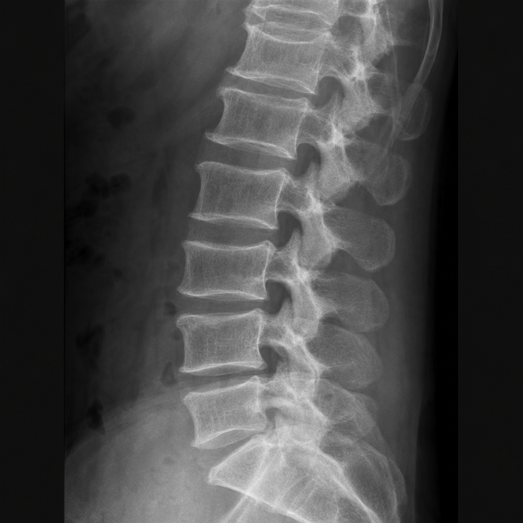
A 65-year-old female attended the Orthopaedics OPD with a chief complaint of chronic backache. The radiograph of the lumbar spine is obtained. What could be the most probable diagnosis?
An intrauterine scan at the 13th week of pregnancy showed a fetus with multiple long bone fractures. What is commonly associated with this finding?
Windswept deformity is seen in which condition?
Practice by Chapter
Osteoporosis
Practice Questions
Osteomalacia and Rickets
Practice Questions
Paget's Disease of Bone
Practice Questions
Hyperparathyroidism
Practice Questions
Renal Osteodystrophy
Practice Questions
Fluorosis
Practice Questions
Osteogenesis Imperfecta
Practice Questions
Bone Mineral Density Assessment
Practice Questions
Pharmacological Management of Metabolic Bone Diseases
Practice Questions
Surgical Considerations in Metabolic Bone Diseases
Practice Questions
Fragility Fractures
Practice Questions
Prevention Strategies
Practice Questions
Want unlimited practice?
Get full access to all questions, explanations, and performance tracking.
Scan to download app