Hyperextension of the Proximal Interphalangeal (PIP) joint and flexion of the Distal Interphalangeal (DIP) joint is seen in which deformity?
What does Tinel's sign indicate?
Stenosing tenosynovitis of the flexor tendon sheath is also known as:
Which of the following is not a recognized treatment for carpal tunnel syndrome?
Which of the following is a feature of Carpal tunnel syndrome?
Kanavel's sign is seen in which of the following conditions?
Which of the following structures is involved in Dupuytren's contracture?
The Card test or Book test is used to assess injury of which nerve?
What is the definition of a swan-neck deformity?
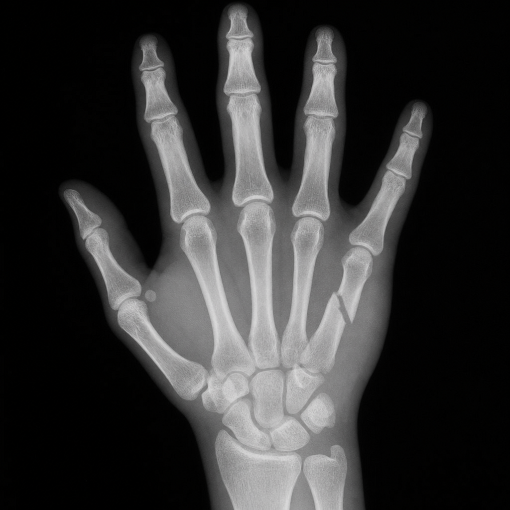
Finkelstein's test is used for which condition?
Explanation: ### Explanation **Swan Neck Deformity** is characterized by the classic combination of **hyperextension at the PIP joint** and **flexion at the DIP joint**. This occurs due to an imbalance in the extensor mechanism, often triggered by a volar plate rupture or dorsal displacement of the lateral bands. The resulting tension pulls the PIP joint into hyperextension, while the compensatory pull of the Flexor Digitorum Profundus (FDP) causes the DIP joint to flex. #### Analysis of Options: * **B. Mallet Finger:** This involves an injury to the extensor tendon at its insertion on the distal phalanx, resulting in **isolated flexion of the DIP joint** (inability to extend the tip of the finger). * **C. Lumbricals Paralysis:** Lumbricals normally flex the MCP joints and extend the IP joints. Their paralysis leads to the **"Claw Hand"** deformity (hyperextension of MCP and flexion of IP joints). * **D. Boutonniere’s Deformity:** This is the exact opposite of Swan Neck. It involves **flexion of the PIP joint** and **hyperextension of the DIP joint**, caused by a rupture of the central slip of the extensor tendon. #### NEET-PG High-Yield Pearls: * **Swan Neck Deformity** is most commonly associated with **Rheumatoid Arthritis** (due to synovitis) and Ehlers-Danlos syndrome. * **Boutonniere Deformity** is also seen in Rheumatoid Arthritis but is classically associated with direct trauma to the dorsum of the PIP joint. * **Mnemonic:** **"B"** comes before **"S"**; **B**outonniere starts with **flexion** (at PIP), **S**wan neck starts with **extension** (at PIP).
Explanation: **Explanation:** **Tinel’s sign** is a clinical physical examination finding used to identify irritated or regenerating nerves. It is elicited by **light percussion** (tapping) over the course of a nerve. A positive sign is defined as a **tingling sensation** or "pins and needles" (paresthesia) in the distal distribution of that nerve. **Why the correct answer is right:** The underlying physiological mechanism is **mechanical hyper-excitability**. When a nerve is compressed or regenerating (following axonal regrowth), the newly formed axons lack mature myelin sheaths, making them hypersensitive to mechanical stimulation. Percussing these fibers triggers ectopic nerve impulses, which the brain interprets as tingling in the nerve's cutaneous territory. **Analysis of incorrect options:** * **A & B:** Loss of sensation and loss of 2-point discrimination are signs of **advanced nerve dysfunction** or axonal death. Tinel’s sign, conversely, indicates that the nerve is either irritated or actively regenerating. * **D:** This is incorrect because Tinel’s sign is a classic clinical test for **Carpal Tunnel Syndrome (CTS)**, where percussion is performed over the median nerve at the flexor retinaculum. **NEET-PG High-Yield Pearls:** * **Hoffmann-Tinel Sign:** In peripheral nerve injuries, a "distally progressing" Tinel’s sign is a positive prognostic indicator, suggesting successful axonal regeneration (approx. 1mm/day). * **Phalen’s Test:** Often tested alongside Tinel’s; it involves maximal wrist flexion for 60 seconds and is generally more sensitive than Tinel’s for diagnosing CTS. * **Common Sites:** Median nerve (Wrist - CTS), Ulnar nerve (Cubital tunnel), and Common Peroneal nerve (Fibular head).
Explanation: **Explanation:** **1. Why Trigger Finger is Correct:** **Stenosing tenosynovitis** of the flexor tendons is the pathological basis of **Trigger Finger**. It occurs due to inflammation and thickening of the flexor tendon sheath (usually at the level of the **A1 pulley**). This creates a size mismatch where the tendon nodule struggles to glide through the narrowed pulley, causing the finger to "lock" or "catch" in flexion. When the patient forcefully extends the finger, it snaps open like a trigger being released. **2. Why the Other Options are Incorrect:** * **Tennis Elbow (Lateral Epicondylitis):** This is an overuse injury involving the common extensor origin (specifically the *Extensor Carpi Radialis Brevis*) at the lateral epicondyle of the humerus, not the flexor tendons. * **Carpal Tunnel Syndrome:** This is a compressive neuropathy of the **median nerve** as it passes through the carpal tunnel. While it involves the same anatomical region, it is not a primary stenosing tenosynovitis of the tendons. * **Dupuytren's Contracture:** This is a progressive fibrosis and thickening of the **palmar fascia**, leading to permanent flexion contractures (most commonly affecting the ring and little fingers). It does not involve the tendon sheath itself. **Clinical Pearls for NEET-PG:** * **Most common site:** The **A1 pulley** (located over the metacarpal head). * **Associated conditions:** Frequently seen in patients with **Diabetes Mellitus** and Rheumatoid Arthritis. * **Treatment:** Initial management includes NSAIDs and corticosteroid injections. Definitive surgical management involves the **surgical release of the A1 pulley**. * **Differential Diagnosis:** Do not confuse this with *De Quervain’s Tenosynovitis*, which is stenosing tenosynovitis of the **1st dorsal compartment** (APL and EPB tendons).
Explanation: **Explanation:** **Carpal Tunnel Syndrome (CTS)** is the most common entrapment neuropathy, caused by compression of the **median nerve** as it passes through the carpal tunnel. **Why Option A is correct:** **Nerve stimulation therapy** (such as TENS or electrical muscle stimulation) is **not** a recognized or evidence-based treatment for CTS. In fact, applying electrical stimulation to an already compressed and ischemic nerve can potentially exacerbate symptoms or delay definitive treatment. Management focuses on relieving pressure, not stimulating the nerve. **Why the other options are incorrect:** * **Steroid Injections (Option B):** These are a mainstay of conservative management. Local corticosteroid injections reduce inflammation and edema of the synovial sheaths, providing significant symptomatic relief in mild to moderate cases. * **Endoscopic Surgical Release (Options C & D):** When conservative management fails, surgical decompression by dividing the **transverse carpal ligament** is indicated. This can be performed via an open technique or endoscopically. * **One-portal (Agee technique):** Uses a single proximal incision at the wrist crease. * **Two-portal (Chow technique):** Uses both a proximal wrist incision and a distal palmar incision. Both are recognized surgical standards. **High-Yield Clinical Pearls for NEET-PG:** * **Contents of Carpal Tunnel:** Median nerve + 9 tendons (4 FDS, 4 FDP, 1 FPL). * **First-line treatment:** Night splinting in neutral position and NSAIDs. * **Gold Standard Diagnosis:** Nerve Conduction Study (NCS) showing increased distal latency and decreased conduction velocity. * **Phalen’s Test & Tinel’s Sign:** Classic clinical provocative tests. * **Ape Thumb Deformity:** Occurs in late stages due to thenar muscle atrophy (Opponens pollicis).
Explanation: **Explanation:** **Carpal Tunnel Syndrome (CTS)** is the most common entrapment neuropathy, caused by the **compression of the median nerve** as it passes through the carpal tunnel beneath the **flexor retinaculum** (transverse carpal ligament). 1. **Why the correct answer is right:** The carpal tunnel is a fibro-osseous space containing nine tendons and the median nerve. Any condition that increases pressure within this tunnel (e.g., hypothyroidism, pregnancy, RA, or repetitive trauma) leads to ischemia of the median nerve, resulting in paresthesia and pain in its distribution. 2. **Why the other options are wrong:** * **Option A:** Compression of the ulnar nerve occurs at the **Guyon’s canal** or the cubital tunnel, not the carpal tunnel. * **Option C:** Sensory loss occurs in the lateral 3.5 fingers. However, the **thenar eminence is spared** because its nerve supply (the palmar cutaneous branch of the median nerve) arises proximal to the flexor retinaculum and passes superficial to it. * **Option D:** The median nerve supplies the thenar muscles (O-A-F: Opponens pollicis, Abductor pollicis brevis, Flexor pollicis brevis). Therefore, chronic CTS leads to **thenar atrophy**, whereas hypothenar atrophy is a feature of ulnar nerve palsy. **High-Yield Clinical Pearls for NEET-PG:** * **Clinical Tests:** Phalen’s test (wrist flexion), Tinel’s sign (percussion over the retinaculum), and Durkan’s test (most sensitive). * **First muscle to show wasting:** Abductor Pollicis Brevis (APB). * **Management:** Conservative (Splints, NSAIDs, Steroids) or Surgical (Division of the flexor retinaculum). * **Associated Conditions:** Diabetes Mellitus, Acromegaly, and Amyloidosis.
Explanation: **Explanation:** **Kanavel’s Signs** are a clinical quartet used to diagnose **Acute Suppurative Flexor Tenosynovitis**, a surgical emergency involving an infection within the flexor tendon sheath of the finger. The correct answer is **Tenosynovitis (Option A)**. The four cardinal signs described by Kanavel are: 1. **Uniform swelling** of the entire finger (Sausage digit). 2. **Finger held in flexion** for comfort. 3. **Exquisite tenderness** along the course of the tendon sheath. 4. **Pain on passive extension** of the finger (the most important/earliest sign). **Why other options are incorrect:** * **Trigger Finger (B):** Caused by a mismatch between the flexor tendon and the A1 pulley. It presents with "snapping" or locking, not the diffuse inflammatory signs of Kanavel. * **Dupuytren’s Contracture (C):** A chronic fibroproliferative disorder of the palmar fascia leading to permanent flexion contractures (usually of the ring and little finger). It is painless and lacks acute inflammatory signs. * **Carpal Tunnel Syndrome (D):** A compression neuropathy of the median nerve. It presents with paresthesia and wasting of the thenar eminence, not signs of tendon sheath infection. **High-Yield Clinical Pearls for NEET-PG:** * **Most common organism:** *Staphylococcus aureus*. * **The "Horseshoe Abscess":** Infection can spread from the thumb (radial bursa) to the little finger (ulnar bursa) because these sheaths often communicate at the wrist. * **Treatment:** Early cases may respond to IV antibiotics; however, most require urgent surgical incision and drainage (washout) to prevent tendon necrosis due to increased pressure within the sheath.
Explanation: ### Explanation **Dupuytren’s Contracture** is a benign fibroproliferative disorder of the **palmar fascia**. The core pathology involves the transformation of normal fibroblasts into **myofibroblasts**, which produce excessive Type III collagen. This leads to the formation of nodules and cords that eventually shorten, causing fixed flexion deformities of the fingers (most commonly the ring and little fingers). #### Analysis of Options: * **A. Thickening of the palmar fascia (Correct):** The disease specifically targets the palmar aponeurosis and its digital extensions. The pathological shortening of these fascial bands pulls the MCP and PIP joints into flexion. * **B. Thickening of the dorsal fascia:** Dupuytren’s is localized to the volar (palmar) aspect of the hand. While Garrod’s pads (knuckle pads) can occur on the dorsum of the PIP joints in these patients, the primary contracture is palmar. * **C. Contracture of the flexor tendons:** This is a common distractor. In Dupuytren’s, the **tendons themselves are normal** and move freely within their sheaths; the restriction is entirely due to the overlying fascial cords. * **D. Post-burns contracture:** This is a secondary scarring process involving the skin and dermis, whereas Dupuytren’s is an idiopathic, often hereditary, primary fascial disease. #### High-Yield Clinical Pearls for NEET-PG: * **Risk Factors:** Strong association with **Diabetes Mellitus**, chronic alcoholism, smoking, epilepsy (anticonvulsant use), and Northern European ancestry (Viking disease). * **Clinical Sign:** **Hueston’s Table Top Test** — positive when the patient cannot flatten their palm against a flat surface. * **Anatomy:** The **Spiral Cord** (formed by the spiral band, lateral digital sheet, and Grayson’s ligament) is responsible for displacing the neurovascular bundle toward the midline, increasing the risk of iatrogenic injury during surgery. * **Treatment:** Indicated when the MCP joint contracture is >30° or any PIP joint contracture is present. Options include Fasciectomy or Collagenase (*Clostridium histolyticum*) injections.
Explanation: The **Card test** (and its variant, the **Book test**) is a classic clinical assessment for **Ulnar nerve** palsy. ### Why Ulnar Nerve is Correct The test specifically evaluates the strength of the **Palmar Interossei** muscles. These muscles are responsible for **adducting** the fingers (bringing them together). * **Mechanism:** The examiner places a card or book between the patient’s extended fingers and asks them to hold it tightly while the examiner tries to pull it away. * **Clinical Sign:** If the ulnar nerve is injured, the palmar interossei are paralyzed, and the patient cannot maintain a strong grip on the card, causing it to be easily pulled out. ### Why Other Options are Incorrect * **Radial Nerve:** Primarily supplies the extensors of the wrist and fingers. Injury leads to **Wrist Drop**. * **Median Nerve:** Supplies the muscles of the thenar eminence (LOAF muscles). Injury is tested via the **Pen test** (Abductor Pollicis Brevis) or by checking for "Ape Thumb" deformity. * **Tibial Nerve:** A nerve of the lower limb; injury results in loss of plantar flexion and intrinsic foot muscle function, unrelated to hand tests. ### High-Yield Clinical Pearls for NEET-PG * **Froment’s Sign:** Often confused with the Card test, this also tests the ulnar nerve (Adductor Pollicis). When asked to hold a piece of paper between the thumb and index finger, the patient flexes the thumb's IP joint (using the Median-innervated Flexor Pollicis Longus) to compensate for weak adduction. * **Wartenberg’s Sign:** Inability to adduct the little finger due to ulnar nerve palsy. * **Egawa’s Test:** Tests the **Dorsal Interossei** (abduction) by asking the patient to sidestep the middle finger. * **Mnemonic:** **PAD** (Palmar Adduct) and **DAB** (Dorsal Abduct).
Explanation: **Explanation:** The **Swan-neck deformity** is a characteristic finger deformity characterized by **hyperextension of the Proximal Interphalangeal (PIP) joint** and **flexion of the Distal Interphalangeal (DIP) joint**. It occurs due to an imbalance in the extensor mechanism, often triggered by a lax volar plate or overpull of the extrinsic extensor muscles, commonly seen in Rheumatoid Arthritis. **Analysis of Options:** * **Option B (Correct):** Accurately describes the PIP extension and DIP flexion. The "swan-neck" appearance is created by the arched PIP joint resembling the bird's neck. * **Option C (Incorrect):** This describes a **Boutonnière deformity** (PIP flexion and DIP hyperextension), which is caused by a rupture of the central slip of the extensor tendon. * **Options A & D (Incorrect):** These describe various positions of the Metacarpophalangeal (MCP) joints, which are not the primary diagnostic criteria for defining a swan-neck deformity. **High-Yield NEET-PG Pearls:** 1. **Etiology:** Most commonly associated with **Rheumatoid Arthritis** (synovitis leads to volar plate laxity) or untreated **Mallet finger** (leads to secondary PIP hyperextension). 2. **Boutonnière vs. Swan-neck:** These are "opposites." Remember: **B**outonnière = **B**ent (flexed) PIP. 3. **Bunnell-Littler Test:** Used to differentiate if PIP restriction is due to intrinsic muscle tightness or capsular contracture. 4. **Treatment:** Mild cases use Silver Ring splints; severe cases may require volar plate reconstruction or dermodesis.
Explanation: **Explanation:** **De Quervain’s Tenosynovitis** is a stenosing tenosynovitis of the **first dorsal compartment** of the wrist. This compartment contains the tendons of the **Abductor Pollicis Longus (APL)** and **Extensor Pollicis Brevis (EPB)**. Finkelstein’s test is the pathognomonic clinical maneuver used to diagnose this condition. * **Mechanism of the Test:** The patient places their thumb into the palm, closes their fingers over it (making a fist), and then the wrist is passively deviated towards the ulnar side. A positive result is indicated by sharp, excruciating pain over the radial styloid process, caused by the stretching of the inflamed APL and EPB tendons through the narrowed fibro-osseous tunnel. **Analysis of Incorrect Options:** * **Congenital Dislocation of Hip (CDH/DDH):** Diagnosed using **Barlow’s** (dislocating the hip) and **Ortolani’s** (reducing the hip) maneuvers. * **Trigger Finger:** Characterized by "locking" or "snapping" of the finger due to a nodule in the flexor tendon catching on the **A1 pulley**. It is diagnosed clinically by palpating a nodule that moves with the tendon. * **Lateral Epicondylitis (Tennis Elbow):** Diagnosed using **Cozen’s test** or **Mill’s maneuver**, which involve resisted wrist extension or passive wrist flexion. **High-Yield Clinical Pearls for NEET-PG:** * **Anatomy:** Remember the mnemonic "Apples (APL) and Peanut Butter (EPB)" for the first compartment contents. * **Demographics:** Most common in middle-aged women and "new mothers" (due to repetitive lifting of infants). * **Eichhoff’s Test:** Often confused with Finkelstein’s; Eichhoff’s is the version where the patient actively makes a fist, while the classic Finkelstein’s involves the examiner pulling the thumb distally. * **Treatment:** Initial management is conservative (NSAIDs, thumb spica splint); refractory cases require corticosteroid injection or surgical release of the first dorsal compartment.
Explanation: **Explanation:** The term **'No Man's Land'** (originally coined by Sterling Bunnell) refers to **Zone II** of the flexor tendon zones of the hand. It is anatomically defined as the region extending from the **distal palmar crease** to the **insertion of the Flexor Digitorum Superficialis (FDS)** on the middle phalanx (roughly corresponding to the area between the proximal and distal creases). **Why Option C is correct:** In Zone II, both the **Flexor Digitorum Profundus (FDP)** and **Flexor Digitorum Superficialis (FDS)** tendons are enclosed within a tight, fibro-osseous tunnel (the digital pulley system). Historically, primary repair in this zone was avoided because the restricted space leads to the formation of dense adhesions between the two tendons and the sheath, resulting in a "frozen finger" with poor functional outcomes. **Why other options are incorrect:** * **A & B (Proximal/Distal Phalanx):** While these bones form the floor of the zones, "No Man's Land" specifically refers to the complex relationship between the two tendons within the sheath in Zone II, not just a single bone. * **D (Wrist):** This corresponds to **Zone IV** (Carpal Tunnel). While space is tight here, the clinical prognosis for repairs is generally better than in Zone II. **High-Yield Clinical Pearls for NEET-PG:** * **Flexor Zones:** There are 5 zones in the hand. Zone I is distal to the FDS insertion (Jersey Finger occurs here). * **Current Management:** The "No Man's Land" rule has evolved; with modern microsurgical techniques and early active mobilization protocols, primary repair is now the standard of care. * **Blood Supply:** Tendons in Zone II receive nutrition via **Vincular vessels** and synovial diffusion. * **Prognosis:** Zone II injuries still carry the highest risk of post-operative adhesions.
Explanation: ### Explanation **Concept:** Trigger finger (Stenosing Tenosynovitis) is caused by a size mismatch between the flexor tendons (FDS and FDP) and the surrounding fibro-osseous tunnel. The primary site of constriction is the **A1 pulley**, which is located at the level of the **Metacarpophalangeal (MCP) joint**. Repetitive microtrauma or inflammation leads to thickening of the pulley and the formation of a nodule on the tendon. When the finger flexes, the nodule passes through the pulley; however, during extension, it gets stuck, causing a characteristic "snapping" or "triggering" sensation. **Analysis of Options:** * **Metacarpophalangeal joint (Correct):** The A1 pulley is anatomically situated over the volar aspect of the MCP joint. This is the narrowest point of the sheath and the most common site of entrapment. * **Proximal phalanx (Incorrect):** While the A2 pulley is located here, it is much wider and stronger; it is rarely the site of primary stenosis in trigger finger. * **Proximal interphalangeal joint (Incorrect):** Although the patient often feels the "click" or pain referred to the PIP joint, the actual pathology (the mechanical block) is proximal to it at the MCP level. * **Middle phalanx (Incorrect):** This level contains the A4 pulley; it is distal to the common site of pathology. **High-Yield Clinical Pearls for NEET-PG:** * **Most common digit involved:** Ring finger (followed by the thumb). * **Associated Conditions:** Diabetes Mellitus (most common), Rheumatoid Arthritis, and Hypothyroidism. * **Clinical Sign:** A palpable nodule can often be felt moving with the tendon at the level of the MCP joint. * **Treatment:** First-line is activity modification and NSAIDs. Refractory cases require **corticosteroid injection** or **surgical release** of the A1 pulley. * **Congenital Trigger Thumb:** Usually presents with a fixed flexion deformity at the IP joint (Notta’s node).
Explanation: **Explanation:** **Pollicization** is a surgical procedure where a functional finger (most commonly the **index finger**) is repositioned to replace an absent or severely damaged thumb. The goal is to restore the essential functions of the thumb, primarily **opposition and pinch grip**, which account for approximately 40–50% of total hand function. **Analysis of Options:** * **Option B (Correct):** Pollicization is the gold standard for **thumb reconstruction** in cases of congenital thumb aplasia/hypoplasia (e.g., Holt-Oram syndrome, VACTERL) or traumatic loss. It involves shortening the finger, rotating it on its neurovascular bundle, and reattaching muscles to mimic thumb kinetics. * **Option A:** While "Toe-to-thumb transfer" (Microsurgical transfer of the great toe or second toe) is a method of thumb reconstruction, it is not the definition of pollicization. Pollicization specifically refers to using an existing digit from the *same* hand. * **Option C:** "Finger shortening" (or recession) may be a step within the pollicization procedure to match thumb length, but it does not describe the procedure itself. * **Option D:** Amputation is the removal of the thumb, which is the pathology that pollicization aims to treat. **Clinical Pearls for NEET-PG:** * **Most common donor:** The **Index finger** is most frequently used due to its proximity and independent mobility. * **Indications:** Blauth Classification Grade IIIb, IV, and V of thumb hypoplasia. * **Key Anatomy:** The procedure relies on the preservation of the **digital arteries and nerves** to ensure the survival and sensation of the "new" thumb. * **Functional Outcome:** The index finger's MCP joint becomes the new thumb's CMC joint.
Explanation: **Explanation:** **Carpal Tunnel Syndrome (CTS)** is the most common entrapment neuropathy of the upper limb. It occurs due to the compression of the **Median nerve** as it passes through the carpal tunnel, a fibro-osseous space bounded by the carpal bones (floor) and the flexor retinaculum (roof). * **Why Median Nerve is Correct:** The carpal tunnel contains ten structures: the Median nerve, four tendons of Flexor Digitorum Superficialis (FDS), four tendons of Flexor Digitorum Profundus (FDP), and one tendon of Flexor Pollicis Longus (FPL). Compression leads to paresthesia in the lateral 3.5 fingers and atrophy of the thenar muscles. **Analysis of Incorrect Options:** * **Anterior Interosseous Nerve (AIN):** This is a motor branch of the median nerve that passes *deep* to the pronator teres, not through the carpal tunnel. AIN syndrome presents with an inability to make the "OK" sign (weakness of FPL and FDP to the index finger). * **Ulnar Nerve:** This nerve passes superficial to the flexor retinaculum through **Guyon’s canal**. Compression here leads to "Handlebar palsy" or ulnar claw hand. * **Radial Nerve:** This nerve primarily supplies the extensor compartment of the forearm. It does not enter the palm or the carpal tunnel. **High-Yield Clinical Pearls for NEET-PG:** * **Clinical Tests:** Phalen’s test (most sensitive), Tinel’s sign, and Durkan’s compression test (most specific). * **Sensory Sparing:** The **palmar cutaneous branch** of the median nerve arises proximal to the carpal tunnel; therefore, sensation over the thenar eminence is **spared** in CTS. * **Associated Conditions:** Pregnancy, Hypothyroidism, Diabetes Mellitus, Rheumatoid Arthritis, and Acromegaly. * **Treatment:** Splinting in neutral position is the first-line conservative management; surgical release of the flexor retinaculum is indicated for refractory cases.
Explanation: **Explanation:** **Phalen’s Test** is a clinical provocative maneuver used to diagnose **Carpal Tunnel Syndrome (CTS)**. The test involves the patient holding their wrists in complete and forced flexion (pushing the dorsal surfaces of the hands together) for 60 seconds. This action increases the pressure within the carpal tunnel, further compressing the **median nerve** against the transverse carpal ligament. A positive test is indicated by the reproduction of paresthesia (numbness or tingling) in the median nerve distribution (thumb, index, middle, and radial half of the ring finger). **Analysis of Incorrect Options:** * **De Quervain's Tenosynovitis:** Diagnosed using the **Finkelstein test**, where the patient makes a fist over their thumb and deviates the wrist ulnarly to stretch the abductor pollicis longus and extensor pollicis brevis tendons. * **Rotator Cuff Injury:** Assessed using tests like the **Empty Can test** (Supraspinatus), **Neer’s sign**, or **Hawkins-Kennedy test** for impingement. * **Tennis Elbow (Lateral Epicondylitis):** Diagnosed using **Cozen’s test** or **Mill’s test**, which involve resisted wrist extension or passive stretching of the extensor carpi radialis brevis. **High-Yield Clinical Pearls for NEET-PG:** * **Reverse Phalen’s Test:** Also known as the "Prayer Test" (wrists in forced extension); also used for CTS. * **Tinel’s Sign:** Percussion over the flexor retinaculum that produces a "pins and needles" sensation; less sensitive but more specific than Phalen’s. * **Durkan’s Test:** Manual compression of the carpal tunnel by the examiner; considered the **most sensitive** clinical test for CTS. * **Gold Standard Diagnosis:** Nerve Conduction Velocity (NCV) studies.
Explanation: ### Explanation In hand surgery, when a primary repair of a flexor tendon is not possible (due to extensive scarring or neglect), a **tendon graft** is required. **Why Plantaris is the Correct Answer:** The **Plantaris tendon** is considered the "gold standard" or the first choice for long tendon grafts in the hand. * **Length and Girth:** It provides a long, thin, and consistent cord-like structure (approx. 30-35 cm) that fits perfectly within the narrow fibro-osseous tunnels of the fingers. * **Vestigial Nature:** It is a vestigial muscle, meaning its removal causes no functional deficit in the lower limb. * **Anatomy:** It originates from the lateral supracondylar ridge of the femur and inserts into the calcaneus. However, it is absent in approximately 7–10% of the population. **Analysis of Incorrect Options:** * **Palmaris Longus (Option A):** While frequently used for shorter grafts (like pulley reconstruction), it is often too short for a full-length flexor reconstruction from the forearm to the fingertip. It is also absent in about 15% of people. * **Extensor Digitorum Communis (Option C):** These are primary functional tendons of the hand. Harvesting them would result in a significant loss of finger extension. * **Extensor Digiti Minimi (Option D):** This is a functional tendon for the little finger. While it can be used for tendon transfers, it is not a preferred source for free grafting due to its functional importance. **High-Yield Clinical Pearls for NEET-PG:** 1. **Order of Preference for Grafts:** Plantaris > Palmaris Longus > Extensor Digitorum Longus (toes) > Longus colli (rare). 2. **Donor Site Testing:** To check for the presence of Palmaris Longus, use **Schaeffer’s Test** (opposing thumb to little finger with wrist flexion). 3. **Bunnell’s Technique:** Often used for suturing these grafts. 4. **Zone II (No Man’s Land):** The most common site requiring complex reconstruction due to the risk of adhesions within the flexor sheath.
Explanation: **Explanation:** The term **'No Man's Land'** refers to **Zone II** of the flexor tendon zones of the hand. Anatomically, it extends from the **distal palmar crease** to the **insertion of the Flexor Digitorum Superficialis (FDS)** at the middle of the middle phalanx. **Why the correct answer is right:** In Zone II, both the Flexor Digitorum Profundus (FDP) and Flexor Digitorum Superficialis (FDS) pass through a tight, narrow fibro-osseous tunnel (the tendon sheath). Historically, Bunnell coined the term 'No Man's Land' because surgical repair in this region often resulted in severe scarring and adhesions between the two tendons and the sheath, leading to a stiff, non-functional finger. While modern microsurgical techniques have improved outcomes, it remains the most challenging area for tendon repair. **Analysis of Incorrect Options:** * **A & B (Proximal/Distal Phalanx):** These are bone-specific locations. While Zone II involves the proximal phalanx, the definition of 'No Man's Land' is specifically based on the relationship between the tendons and their pulleys/sheaths, not just the bone itself. * **D (Wrist):** This corresponds to **Zone V**, where the tendons are proximal to the carpal tunnel. Repairs here generally have a much better prognosis due to the lack of a restrictive fibro-osseous tunnel. **NEET-PG High-Yield Pearls:** * **Flexor Zones:** There are 5 zones for flexor tendons. Zone I is distal to the FDS insertion (FDP only). * **Blood Supply:** Tendons in Zone II receive nutrition via **Vincular vessels** and synovial diffusion. * **Prognosis:** Zone II injuries have the poorest prognosis for functional recovery. * **Verdan’s Classification:** This is the standard system used to classify these flexor tendon zones.
Explanation: **Explanation:** **Phalen’s test** is a provocative diagnostic maneuver used to identify **Carpal Tunnel Syndrome (CTS)**, which is the compression of the median nerve as it passes through the carpal tunnel at the wrist. 1. **Why the correct answer is right:** The test involves asking the patient to hold their wrists in complete, unforced flexion (pushing the dorsal surfaces of the hands together) for 60 seconds. This position increases the interstitial pressure within the carpal tunnel and further compresses the median nerve. A **positive test** is indicated by the reproduction of symptoms (numbness, tingling, or paresthesia) in the median nerve distribution (thumb, index, middle, and radial half of the ring finger). 2. **Why the incorrect options are wrong:** * **De Quervain Tenosynovitis:** Diagnosed using the **Finkelstein test** (ulnar deviation of the wrist with the thumb tucked into the palm). It involves the first dorsal compartment (APL and EPB tendons). * **Trigger Finger:** This is a clinical diagnosis characterized by "locking" or "snapping" of the finger during extension due to a nodule on the flexor tendon catching at the **A1 pulley**. No specific wrist flexion test is used. **High-Yield Clinical Pearls for NEET-PG:** * **Reverse Phalen’s Test (Prayer Test):** Extension of the wrists for 60 seconds; also used for CTS. * **Tinel’s Sign:** Percussion over the flexor retinaculum produces paresthesia in the median nerve distribution. * **Durkan’s Test:** Direct manual compression of the carpal tunnel by the examiner; it is considered the **most sensitive** clinical test for CTS. * **Gold Standard Investigation:** Nerve Conduction Studies (NCS) showing delayed conduction velocity across the wrist.
Explanation: **Explanation:** **Pollicization** is a surgical procedure where a functional finger (most commonly the **index finger**) is repositioned and reconstructed to function as a **thumb**. The goal is to restore the essential functions of the thumb, such as opposition, grasp, and pinch, in cases where the thumb is congenitally absent (e.g., radial club hand, hypoplasia) or lost due to trauma. Therefore, it is fundamentally a method of **thumb reconstruction**. **Analysis of Options:** * **Option A (Toe to thumb transfer):** While this is a method of thumb reconstruction, it is a microsurgical procedure involving a free tissue transfer from the foot. Pollicization specifically refers to using an existing digit from the same hand. * **Option C (Finger shortening):** This is a descriptive term for reducing the length of a digit, often part of a larger procedure, but it does not define pollicization. * **Option D (Amputation of thumb):** This is the removal of the thumb, which is the clinical problem that pollicization aims to solve, not the procedure itself. **High-Yield Clinical Pearls for NEET-PG:** * **Buck-Gramcko Technique:** The most widely used surgical technique for pollicization, particularly in congenital cases. * **Indications:** Blauth Grade IIIb, IV, and V thumb hypoplasia (where the CMC joint is unstable or absent). * **Key Step:** The index finger is transposed on its neurovascular bundle, and the metacarpophalangeal (MCP) joint of the index finger becomes the new carpometacarpal (CMC) joint of the reconstructed thumb. * **Nerve Supply:** The sensory and motor supply is preserved by meticulous dissection of the digital nerves and vessels.
Explanation: **Explanation:** **Mallet Finger** (also known as Baseball finger) is a common injury caused by sudden forced flexion of an extended finger, typically when a ball hits the fingertip. **1. Why the Correct Answer is Right:** The extensor mechanism of the finger divides into a **central slip** (inserting on the middle phalanx) and two **lateral/collateral slips** (lateral bands). These lateral slips converge to form a single terminal tendon that inserts into the base of the **distal phalanx**. Mallet finger occurs due to the disruption of this terminal extensor mechanism—either through a pure tendon rupture or an avulsion fracture at the dorsal base of the distal phalanx. This loss of extensor continuity allows the Flexor Digitorum Profundus (FDP) to act unopposed, resulting in a characteristic flexion deformity at the Distal Interphalangeal (DIP) joint. **2. Why the Other Options are Wrong:** * **Option A:** An avulsion fracture in Mallet finger involves the **distal phalanx**, not the middle phalanx. * **Option C:** Rupture of the **central slip** leads to a **Boutonnière deformity**, characterized by PIP joint flexion and DIP joint hyperextension. * **Option D:** While trauma can cause DIP dislocation, Mallet finger specifically refers to the extensor mechanism disruption, not a loss of joint congruity. **3. Clinical Pearls for NEET-PG:** * **Clinical Feature:** Patient cannot actively extend the DIP joint but passive extension is possible. * **Management:** Most cases are treated conservatively with a **Mallet splint** (holding the DIP in continuous extension/slight hyperextension) for 6–8 weeks. * **Boutonnière Deformity:** Remember the "Central Slip" rule for PIP joint pathology to distinguish it from Mallet finger.
Explanation: **Explanation:** Carpal Tunnel Syndrome (CTS) is a compressive neuropathy of the **median nerve** as it passes through the fibro-osseous carpal tunnel. It is caused by any condition that increases the volume of the tunnel contents or decreases the space within the tunnel. **Why Addison’s Disease is the Correct Answer:** Addison’s disease (primary adrenal insufficiency) is **not** associated with CTS. In contrast, **Cushing’s syndrome** (excess glucocorticoids) and **Acromegaly** (excess growth hormone) are known causes due to soft tissue hypertrophy and fluid retention. **Analysis of Incorrect Options:** * **Amyloidosis:** This is a classic cause where amyloid protein deposits (specifically $\beta_2$-microglobulin in dialysis patients) infiltrate the flexor retinaculum or synovium, narrowing the tunnel. * **Hypothyroidism:** Myxedematous tissue and the accumulation of glycosaminoglycans (hyaluronic acid) lead to increased interstitial fluid and pressure within the carpal tunnel. * **Diabetes Mellitus:** Diabetics have a lower threshold for nerve compression due to underlying metabolic polyneuropathy and increased glycosylation of collagen in the transverse carpal ligament. **NEET-PG High-Yield Pearls:** * **Most common cause:** Idiopathic. * **Most common associated systemic condition:** Obesity; followed by Pregnancy (due to edema). * **Clinical Signs:** Phalen’s test (most sensitive), Tinel’s sign, and Durkan’s compression test (most specific). * **Anatomy:** The carpal tunnel contains **10 structures**: 1 Median nerve, 4 Flexor Digitorum Superficialis tendons, 4 Flexor Digitorum Profundus tendons, and 1 Flexor Pollicis Longus tendon. * **Gold Standard Diagnosis:** Electrodiagnostic studies (Nerve Conduction Velocity).
Explanation: **Explanation:** **Boutonniere’s deformity** (also known as Buttonhole deformity) is a characteristic finger deformity resulting from the **rupture or avulsion of the central slip** of the extensor tendon from its insertion at the base of the middle phalanx. **1. Why Option C is Correct:** When the central slip is damaged, the lateral bands of the extensor mechanism slide volarly (towards the palm) past the axis of the Proximal Interphalangeal (PIP) joint. These lateral bands then act as flexors of the PIP joint rather than extensors. Simultaneously, the tension on the lateral bands increases at their distal insertion, causing compensatory **hyperextension of the Distal Interphalangeal (DIP) joint**. Thus, the classic presentation is **PIP flexion and DIP extension.** **2. Why Incorrect Options are Wrong:** * **Option A & B:** These describe uniform positions (both joints extended or both flexed), which do not account for the reciprocal mechanical imbalance caused by the displaced lateral bands. * **Option D:** This describes **Swan-neck deformity** (PIP hyperextension and DIP flexion), which is the clinical opposite of Boutonniere’s deformity and is often caused by volar plate laxity or rheumatoid arthritis. **3. Clinical Pearls for NEET-PG:** * **Etiology:** Most commonly seen in Rheumatoid Arthritis or trauma (e.g., a jammed finger). * **Elson’s Test:** The gold standard clinical test to diagnose early central slip injury before the deformity becomes fixed. * **Management:** Initial treatment is usually non-operative, involving splinting the PIP joint in full extension for 6–8 weeks while allowing active DIP motion. * **Mnemonic:** **B**outonniere = **B**ent (PIP) and **U**p (DIP).
Explanation: **Explanation:** **Phalen’s test** is a clinical provocative maneuver used to diagnose **Carpal Tunnel Syndrome (CTS)**. The test involves the patient holding their wrists in maximal forced flexion (pushing the dorsal surfaces of the hands together) for 60 seconds. This position increases the pressure within the carpal tunnel, further compressing the **median nerve** against the flexor retinaculum. A positive test is indicated by the reproduction of numbness or tingling (paresthesia) in the median nerve distribution (thumb, index, middle, and radial half of the ring finger). **Analysis of Incorrect Options:** * **Ulnar bursitis:** This involves inflammation of the synovial sheath of the flexor tendons on the medial side of the hand. It typically presents with pain and swelling along the ulnar aspect of the palm, not median nerve symptoms. * **Tennis elbow (Lateral Epicondylitis):** This is an overuse injury of the common extensor origin at the elbow. It is diagnosed via **Cozen’s test** or **Mill’s maneuver**, focusing on pain during resisted wrist extension. * **De Quervain’s disease:** This is a stenosing tenosynovitis of the first dorsal compartment (APL and EPB tendons). The pathognomonic clinical test is **Finkelstein’s test**. **NEET-PG High-Yield Pearls:** * **Reverse Phalen’s (Prayer Test):** Similar to Phalen’s but performed with maximal wrist extension; also used for CTS. * **Tinel’s Sign:** Percussion over the flexor retinaculum produces paresthesia; it is less sensitive but more specific than Phalen’s. * **Durkan’s Test (Carpet Compression Test):** The **most sensitive** clinical test for CTS, involving direct pressure over the carpal tunnel for 30 seconds. * **Gold Standard Diagnosis:** Nerve Conduction Velocity (NCV) studies.
Explanation: **Explanation:** The correct answer is **Osteochondrosis of the navicular** (also known as **Preiser’s disease**). 1. **Why it is correct:** Osteochondrosis refers to a group of disorders that affect the growing skeleton, involving idiopathic avascular necrosis (AVN) of a bone. When it affects the carpal navicular (scaphoid), it is termed Preiser’s disease. It typically presents in adults with progressive wrist pain, stiffness, and weakness, often exacerbated by repetitive stress or trauma. The **"humpback" deformity** is a classic radiological and clinical finding caused by the collapse of the scaphoid into flexion, leading to a prominent dorsal angulation at the fracture or necrosis site. 2. **Why the other options are incorrect:** * **A. Osteochondrosis of the lunate:** This is known as **Kienböck’s disease**. While it causes wrist pain and stiffness, it does not typically produce a "humpback" deformity; rather, it leads to lunate collapse and proximal migration of the capitate. * **C. Tuberculosis of the scaphoid:** While TB can affect small bones (carpal tunnel syndrome or dactylitis), it usually presents with systemic symptoms (fever, weight loss), "cold abscesses," and "rice bodies" in the synovium, rather than a specific humpback structural deformity. * **D. Syphilitic dactylitis:** This involves painless, spindle-shaped swelling of the phalanges (usually in infants) due to periostitis, not the carpal bones. **High-Yield Clinical Pearls for NEET-PG:** * **Preiser’s Disease:** AVN of the Scaphoid (Navicular). * **Kienböck’s Disease:** AVN of the Lunate. * **Humpback Deformity:** Specifically refers to the **dorsal angulation** of the scaphoid following a non-union or collapse. * **Terry Thomas Sign:** Widening of the scapholunate interval (>3mm), indicating ligamentous injury.
Explanation: **Explanation:** A **Felon** is a closed-space infection of the **terminal pulp space** of the fingertip. This space is anatomically unique because it is divided into multiple small, non-compliant compartments by tough **fibrous septa** that run from the periosteum of the distal phalanx to the skin. 1. **Why Terminal Pulp Space is correct:** When infection occurs here, the inflammatory edema causes a rapid rise in pressure within these tight compartments. This leads to intense, throbbing pain and can compress the local blood supply, potentially causing **ischemic necrosis** of the distal phalanx (osteomyelitis). 2. **Why other options are incorrect:** * **Epinychium:** Infection here is called **Paronychia**. It involves the soft tissue fold around the nail plate. * **Deep palmar space:** These include the thenar and mid-palmar spaces. Infections here present with generalized hand swelling and loss of the palmar concavity, rather than localized fingertip pain. * **Subcuticular space:** This refers to infections just beneath the epidermis (e.g., a blister or "collar-stud" abscess), which do not involve the deep fibrous compartments characteristic of a felon. **Clinical Pearls for NEET-PG:** * **Treatment:** Incision and drainage (I&D) are mandatory. The incision should be made where the tenderness is maximal, typically avoiding the "pinching" surface of the finger. * **Complication:** If untreated, a felon can lead to **sequestration** of the distal phalanx (diaphysis) because the pressure exceeds the capillary perfusion pressure of the bone. * **Anatomy:** Note that the **flexor tendon sheath** does not extend into the terminal pulp space; therefore, a felon rarely leads to tenosynovitis unless the injury is deep and proximal.
Explanation: In hand surgery, particularly in cases of complex trauma or replantation, the sequence of repair follows a specific surgical hierarchy to ensure stability and optimal outcomes. **Why Bone is Repaired First** The fundamental principle of hand reconstruction is **"Stability before Mobility."** The skeleton provides the structural framework for the hand. Rigid internal fixation of the bone (using K-wires, plates, or screws) is performed first because: 1. It provides a stable scaffold for the repair of soft tissue structures. 2. It prevents subsequent tension or disruption of delicate microvascular and nerve repairs that would occur if the bone were manipulated later. 3. It allows for accurate restoration of the limb's length. **Explanation of Incorrect Options** * **Tendon:** Tendons are repaired after bone fixation but usually before nerves and vessels. Repairing them too early without skeletal stability leads to gap formation or rupture during bone manipulation. * **Nerve:** Nerves are fragile and require a tension-free environment. They are repaired toward the end of the procedure to prevent accidental stretching during the orthopedic or vascular stages. * **Artery:** While restoring circulation is critical, vascular repair is typically performed after bone and tendon stabilization. If the artery is repaired before the bone, the subsequent manipulation required for fracture fixation can easily tear the delicate anastomosis. **NEET-PG High-Yield Pearls** * **Standard Sequence of Replantation:** Bone → Tendon → Artery → Nerve → Vein → Skin (Mnemonic: **B-T-A-N-V-S**). * **Exception:** If the warm ischemia time is nearing its limit, a temporary **vascular shunt** may be placed to restore perfusion before proceeding with bone fixation. * **Golden Rule:** Always stabilize the "chassis" (bone) before fixing the "cables" (tendons/nerves) and "pipes" (vessels).
Explanation: **Explanation:** The **Ulnar Paradox** refers to the clinical observation that a **High Ulnar Nerve Lesion** (at or above the elbow) results in a **less severe** claw hand deformity than a low lesion, despite the injury being more proximal. **1. Why Option A is Correct:** In a **Low Ulnar Nerve Lesion** (at the wrist), the Medial 2 Lumbricals are paralyzed, but the **Flexor Digitorum Profundus (FDP)** to the ring and little fingers remains intact (as it is supplied by the nerve in the forearm). The unopposed action of the intact FDP causes marked flexion at the Interphalangeal (IP) joints, making the "clawing" very prominent. In a **High Ulnar Nerve Lesion**, the FDP is also paralyzed. Since there is no long flexor force to pull the fingers into flexion, the clawing appears **milder**. The "paradox" is that a higher (worse) injury results in a better-looking hand. **2. Why Other Options are Incorrect:** * **Option B:** Low ulnar nerve lesions produce the most severe clawing (Main en Griffe). * **Option C & D:** Triple nerve or combined injuries result in complex deformities (like a "Total Claw Hand"), but they do not demonstrate the specific biomechanical trade-off between the FDP and lumbricals that defines the ulnar paradox. **Clinical Pearls for NEET-PG:** * **Claw Hand Mechanism:** Hyperextension at MCP joints (unopposed Extensor Digitorum) + Flexion at IP joints (paralyzed Lumbricals). * **Froment’s Sign:** Tests for Adductor Pollicis palsy (Ulnar nerve); patient compensates by flexing the thumb IP joint using the Median nerve (FPL). * **Mnemonic:** "The higher the lesion, the lesser the claw."
Explanation: ### Explanation The clinical presentation describes **De Quervain’s Tenosynovitis**, a common stenosing tenosynovitis affecting the **first dorsal compartment** of the wrist. The maneuver described is the **Finkelstein test**, which is pathognomonic for this condition. #### 1. Why Option C is Correct The first dorsal compartment of the extensor retinaculum contains two specific tendons: the **Abductor Pollicis Longus (APL)** and the **Extensor Pollicis Brevis (EPB)**. Chronic overuse (repetitive gripping or ulnar deviation) leads to thickening of the synovial sheath and narrowing of the fibro-osseous tunnel, causing pain during thumb and wrist movement. #### 2. Why Other Options are Wrong * **Option A & B:** The **Abductor Pollicis Brevis (APB)** is an intrinsic muscle of the hand (thenar eminence) and does not pass through the extensor compartments of the wrist. * **Option D:** The **Extensor Pollicis Longus (EPL)** passes through the **third dorsal compartment** and loops around Lister’s tubercle. It forms the ulnar border of the anatomical snuffbox, whereas De Quervain’s involves the radial border. #### 3. High-Yield Clinical Pearls for NEET-PG * **Demographics:** Most common in middle-aged females and "new mothers" (due to repetitive lifting of the infant). * **Anatomical Snuffbox:** The APL and EPB form the **radial (lateral) border**, while the EPL forms the **ulnar (medial) border**. * **Eichhoff’s Test:** Often confused with Finkelstein’s; it is the specific maneuver where the patient deviates the wrist while clutching the thumb. * **Management:** Conservative (NSAIDs, thumb spica splint, steroid injections). Surgical release of the first compartment is indicated if conservative measures fail. Watch out for a **septated first compartment** (a common cause of surgical failure).
Explanation: **Explanation:** **Dupuytren’s contracture** is a progressive fibroproliferative disorder of the palmar fascia, characterized by the formation of nodules and cords that lead to fixed flexion deformities of the fingers. **Why Option B is the Correct Answer (The False Statement):** In Dupuytren’s contracture, the **ring finger** is most commonly affected first, followed by the little finger. The index finger and thumb are rarely involved. Therefore, the statement that it typically affects the index finger first is incorrect. **Analysis of Other Options:** * **Option A:** It is frequently associated with other fibromatoses, including **Peyronie’s disease** (penile fibromatosis) and **Ledderhose disease** (plantar fibromatosis). This triad is often seen in patients with a strong genetic predisposition. * **Option C:** While primarily a palmar condition, the disease process involves myofibroblast proliferation leading to nodule formation. When it involves the feet (Ledderhose disease), it causes thickening of the **plantar fascia**. * **Option D:** While the primary treatments are fasciectomy or collagenase injection, **amputation** (specifically of the little finger) may be indicated in severe, recurrent cases where function cannot be restored or the finger is severely contracted and interfering with daily activities. **High-Yield Clinical Pearls for NEET-PG:** * **Risk Factors:** Male gender (M:F = 10:1), age >50, smoking, alcohol use, diabetes mellitus, and epilepsy (anticonvulsant use). * **Pathology:** Proliferation of **Type III collagen** (replacing Type I). * **Hueston’s Table Top Test:** Positive when the patient cannot flatten their palm against a table; indicates a need for surgical intervention. * **Treatment:** Indicated when the MCP joint contracture is >30° or any PIP joint contracture is present.
Explanation: **Diagnosis: Trigger Finger (Stenosing Tenosynovitis)** **Explanation:** The clinical presentation of a painful "clicking" sensation, a finger locked in flexion, and the requirement of passive assistance for extension is classic for **Trigger Finger**. This condition is caused by a size mismatch between the flexor tendon (often due to a nodule) and the **A1 pulley** at the metacarpophalangeal (MCP) joint. It is highly associated with **Diabetes Mellitus**. 1. **Why Corticosteroid Injection is Correct:** According to standard orthopedic guidelines (and high-yield for NEET-PG), **corticosteroid injection** into the tendon sheath is the **first-line treatment**. It reduces peritendinous inflammation and edema, resolving symptoms in up to 60-90% of non-diabetic patients. While diabetic patients may have higher recurrence rates, it remains the initial intervention before considering surgery. 2. **Why Other Options are Incorrect:** * **Splinting:** While an option for very mild/early cases, it has lower success rates than injections and is generally not the "first-line" choice in a patient already experiencing locking and painful clicks. * **Surgery (A1 Pulley Release):** This is the definitive treatment but is reserved for cases where conservative management (injections) has failed. * **Oral NSAIDs:** These may provide symptomatic pain relief but do not address the mechanical obstruction at the A1 pulley effectively. **High-Yield Clinical Pearls for NEET-PG:** * **Most common site:** Ring finger (followed by thumb and middle finger). * **Pathology:** Fibrocartilaginous metaplasia of the **A1 pulley**. * **Diabetic Association:** Patients with DM often have multiple digits involved and are more resistant to corticosteroid treatment. * **Pediatric Trigger Thumb:** Unlike adults, this is usually congenital and often requires surgical release rather than injections.
Explanation: **Explanation:** **Tinel’s sign** is a clinical test used to identify irritated or regenerating nerves. It is performed by lightly **percussing** over the course of a nerve. A positive result occurs when this percussion produces a **tingling sensation** (paresthesia) in the cutaneous distribution of that nerve. **Why the correct answer is right:** The underlying pathophysiology involves **mechanical hyper-excitability** of regenerating axonal sprouts or irritated nerve fibers. When the nerve is tapped, these sensitive fibers fire, sending signals to the brain that are interpreted as "pins and needles" or tingling in the nerve's territory. **Analysis of incorrect options:** * **A & B:** Loss of sensation and loss of two-point discrimination are signs of **nerve deficit** (anesthesia/hypesthesia). Tinel’s test is a provocative test for nerve *irritability* or *regeneration*, not a measure of sensory loss. * **D:** Tinel’s sign is classically associated with **Carpal Tunnel Syndrome (CTS)**, where percussion over the median nerve at the wrist crease reproduces symptoms. **High-Yield Clinical Pearls for NEET-PG:** * **Phalen’s Test:** The most sensitive provocative test for CTS (wrist flexion for 60 seconds). * **Regeneration Tracking:** In peripheral nerve injuries, a "distally progressing" Tinel’s sign is a positive prognostic indicator, suggesting that axons are regenerating (at a rate of approximately **1 mm/day**). * **Common Sites:** Median nerve (Carpal Tunnel), Ulnar nerve (Cubital Tunnel), and Common Peroneal nerve (at the fibular head).
Explanation: **Explanation:** **Dupuytren’s contracture** is a progressive fibroproliferative disorder of the **palmar fascia**, characterized by the formation of nodules and cords that lead to fixed flexion deformities of the fingers (most commonly the ring and little fingers). The exact etiology is idiopathic, but it is strongly associated with several systemic risk factors: 1. **Diabetes Mellitus (Option A):** There is a high prevalence of Dupuytren’s in diabetic patients. It is thought that microvascular changes and the accumulation of advanced glycation end-products (AGEs) in collagen fibers trigger myofibroblast proliferation. 2. **Alcohol Consumption (Option B):** Chronic alcohol intake and liver cirrhosis are well-documented risk factors. Alcohol may induce oxidative stress or alter local cytokine levels, promoting fibrogenesis. 3. **Smoking (Option C):** Smoking causes microvascular ischemia in the palmar fascia, creating a hypoxic environment that stimulates fibroblast activity and collagen deposition. Since all three factors—Diabetes, Alcohol, and Smoking—are established clinical associations that increase the risk of developing the condition, **Option D (All of the above)** is the correct answer. **High-Yield Clinical Pearls for NEET-PG:** * **Pathophysiology:** The hallmark is the transformation of fibroblasts into **myofibroblasts**, leading to an increase in **Type III collagen** (normally Type I predominates). * **Clinical Sign:** **Hueston’s Tabletop Test** is positive when the patient cannot place their palm flat on a table. * **Associations:** Often associated with other fibromatoses like **Ledderhose disease** (plantar fascia) and **Peyronie’s disease** (penile fascia). * **Treatment:** Surgical options include Fasciectomy (standard) or needle aponeurotomy. Non-surgical treatment includes Collagenase (*Clostridium histolyticum*) injections.
Explanation: **Explanation:** Carpal Tunnel Syndrome (CTS) is a compressive neuropathy of the **median nerve** as it passes through the carpal tunnel. It occurs when the space within the tunnel decreases or the volume of its contents increases. **Why Addison’s Disease is the Correct Answer:** Addison’s disease (primary adrenocortical insufficiency) is characterized by a deficiency of cortisol and aldosterone. It typically leads to weight loss and dehydration. In contrast, **Cushing’s syndrome** (excess glucocorticoids) or conditions causing fluid retention are associated with CTS. Addison’s does not cause the soft tissue edema or deposition required to compress the median nerve. **Why the other options are incorrect:** * **Colles’ Fracture:** This is a common bony cause. Malunion or acute displaced fractures of the distal radius can alter the anatomy of the carpal tunnel floor, leading to direct pressure on the nerve. * **Acromegaly:** Excess Growth Hormone causes **synovial hyperplasia** and overgrowth of the carpal bones and soft tissues, physically crowding the tunnel. * **Hypothyroidism:** This is a classic metabolic cause. It leads to the accumulation of **mucopolysaccharides (myxedema)** and fluid in the connective tissues, increasing interstitial pressure within the tunnel. **High-Yield Clinical Pearls for NEET-PG:** * **Most common cause:** Idiopathic. * **Most common canalicular compression syndrome:** Carpal Tunnel Syndrome. * **Associated Conditions:** Pregnancy (fluid retention), Diabetes Mellitus (decreased nerve threshold), Rheumatoid Arthritis (synovitis), and Chronic Renal Failure (Amyloidosis/AV fistula). * **Clinical Tests:** Phalen’s test, Durkan’s compression test (most sensitive), and Tinel’s sign. * **Gold Standard Diagnosis:** Nerve Conduction Velocity (NCV) studies showing delayed conduction across the wrist.
Explanation: ### Explanation: Boutonnière Deformity **The Core Concept:** Boutonnière deformity is a finger deformity characterized by **Flexion of the Proximal Interphalangeal (PIP) joint** and **Hyperextension of the Distal Interphalangeal (DIP) joint**. It results from the rupture or avulsion of the **central slip** of the extensor tendon from its insertion at the base of the middle phalanx. When the central slip is damaged, the lateral bands of the extensor mechanism slide volarly (towards the palm) past the axis of the PIP joint. These lateral bands then act as flexors of the PIP joint while exerting increased extensor force on the DIP joint, leading to the classic "buttonhole" appearance. **Analysis of Options:** * **Option B (Correct):** While the hallmark is PIP flexion and DIP hyperextension, the compensatory mechanism often involves extension at the MCP joint to maintain finger function. (Note: In many clinical contexts, the primary description is PIP flexion/DIP extension; however, based on the provided key, the focus is on the reciprocal extension at the proximal-most joint). * **Option A:** This is only half of the deformity. Flexion at the PIP must be accompanied by DIP hyperextension to be classified as Boutonnière. * **Option C & D:** These do not match the biomechanical failure of the extensor expansion seen in this condition. **High-Yield Clinical Pearls for NEET-PG:** * **Etiology:** Most commonly caused by rheumatoid arthritis or trauma (e.g., a jammed finger). * **Elson’s Test:** The gold standard clinical test for early diagnosis of central slip injury before the deformity becomes fixed. * **Swan Neck Deformity (The Opposite):** Characterized by **PIP hyperextension** and **DIP flexion** (due to volar plate laxity or intrinsic muscle contracture). * **Mallet Finger:** Deformity involving only the DIP joint (flexion) due to loss of the distal extensor tendon. * **Management:** Acute injuries are typically treated with **PIP splinting in full extension** for 6–8 weeks, leaving the DIP joint free to move.
Explanation: **Explanation:** **Gamekeeper’s Thumb** (also known as Skier’s Thumb in the acute setting) refers to an injury of the **Ulnar Collateral Ligament (UCL)** of the **Thumb Metacarpophalangeal (MCP) joint**. 1. **Why Option A is Correct:** The UCL is located on the medial (ulnar) aspect of the thumb MCP joint. It is the primary stabilizer against **valgus stress** (abduction of the thumb away from the hand). The injury typically occurs due to forceful abduction and hyperextension of the thumb. Historically, "Gamekeeper's" refers to chronic attrition of this ligament in Scottish gamekeepers, while "Skier's" refers to acute rupture from falling on an outstretched hand while holding a ski pole. 2. **Why Other Options are Incorrect:** * **Option B:** Radial collateral ligament (RCL) ruptures occur on the lateral side of the MCP joint due to varus stress; they are much less common than UCL injuries. * **Options C & D:** These involve the **Interphalangeal (IP) joint**. While IP joint dislocations occur, the classic "Gamekeeper’s" pathology is specifically localized to the MCP joint, which provides the critical stability required for a strong "pinch" grip. **High-Yield Clinical Pearls for NEET-PG:** * **Stener Lesion:** A crucial surgical concept where the torn UCL becomes displaced superficial to the **Adductor Pollicis aponeurosis**. This prevents the ligament ends from touching, making spontaneous healing impossible and requiring surgical intervention. * **Clinical Test:** Weakness of pinch grip and instability on valgus stress testing (>30 degrees of laxity or 15 degrees more than the uninjured side). * **X-ray:** May show an avulsion fracture at the base of the proximal phalanx.
Explanation: **Explanation:** The clinical presentation of an inability to extend the distal interphalangeal (DIP) joint following trauma is diagnostic of a **Mallet Finger** (also known as Baseball finger). This occurs due to the disruption of the terminal extensor tendon at its insertion on the base of the distal phalanx. **1. Why Splinting is Correct:** In this case, the X-ray is normal, indicating a **"Soft Tissue Mallet Finger"** (tendon rupture without a bony avulsion). The gold standard treatment for closed mallet finger injuries is **continuous immobilization** of the DIP joint in **neutral or slight hyperextension** using a Mallet splint (e.g., Stack splint) for 6–8 weeks. This allows the torn ends of the tendon to heal by primary intention. **2. Why Other Options are Incorrect:** * **Surgery:** Surgical intervention (e.g., K-wire fixation or ORIF) is generally reserved for "Bony Mallet" injuries involving >1/3rd of the articular surface or cases with volar subluxation of the distal phalanx. * **Wax Bath:** This is a form of thermotherapy used for chronic inflammatory conditions like Rheumatoid Arthritis to reduce stiffness; it has no role in treating acute tendon ruptures. * **Ignore:** Untreated mallet finger leads to a permanent extensor lag and may eventually progress to a **Swan-neck deformity** due to dorsal migration of the lateral bands. **Clinical Pearls for NEET-PG:** * **Mechanism:** Sudden forceful flexion of an actively extended finger. * **Deformity:** The DIP joint remains in a flexed position (extensor lag). * **Splinting Rule:** The splint must be worn **24/7**. If the patient removes the splint and the finger drops even once, the healing clock resets to zero. * **Associated Deformity:** Chronic mallet finger can lead to a **Swan-neck deformity** (DIP flexion with PIP hyperextension).
Explanation: **Explanation:** **Froment’s sign** is a classic clinical test used to diagnose **Ulnar nerve palsy**, specifically assessing the paralysis of the **Adductor Pollicis** muscle. 1. **Why Ulnar Nerve is Correct:** The Adductor Pollicis is supplied by the deep branch of the ulnar nerve. When a patient with ulnar nerve palsy attempts to hold a piece of paper between the thumb and index finger (key pinch), they cannot adduct the thumb. To compensate, the patient recruits the **Flexor Pollicis Longus (FPL)**, which is supplied by the **Median nerve (Anterior Interosseous branch)**. This results in compensatory **flexion of the thumb at the Interphalangeal (IP) joint**, which constitutes a positive Froment’s sign. 2. **Why Other Options are Incorrect:** * **Median Nerve:** Injury leads to "Ape thumb deformity" and loss of opposition. The FPL would be paralyzed, making the compensatory flexion seen in Froment's sign impossible. * **Radial Nerve:** Injury typically results in "Wrist drop" or "Finger drop" due to paralysis of the extensors. * **Axillary Nerve:** This supplies the Deltoid and Teres minor; injury results in loss of shoulder abduction and sensation over the "Regimental badge" area. **Clinical Pearls for NEET-PG:** * **Jeanne’s Sign:** If the thumb MP joint also shows hyperextension during Froment's test, it is called Jeanne’s sign (indicates ulnar nerve palsy). * **Wartenberg’s Sign:** Inability to adduct the little finger due to weakness of the 3rd Palmar Interosseous muscle (Ulnar nerve). * **Mnemonic:** **PAD** (Palmar Interossei Adduct) and **DAB** (Dorsal Interossei Abduct)—all supplied by the Ulnar nerve.
Explanation: **Explanation:** **Mallet Finger** (also known as Baseball finger) is a common sports injury characterized by the loss of active extension at the **Distal Interphalangeal (DIP) joint**. 1. **Why the Correct Answer is Right:** The extensor mechanism of the finger terminates as the **terminal extensor tendon**, which inserts into the dorsal base of the **distal phalanx**. An injury (either a formal tendon rupture or an avulsion fracture) at this specific insertion point prevents the patient from extending the DIP joint, resulting in a characteristic "droop" of the fingertip while the joint remains passively mobile. 2. **Why Incorrect Options are Wrong:** * **Middle Phalanx:** Injury to the extensor insertion here (the central slip) leads to a **Boutonnière deformity**, characterized by PIP joint flexion and DIP joint hyperextension. * **Proximal Phalanx:** This is the site of the insertion for the interossei and lumbricals (via the extensor expansion), but isolated injury here does not cause Mallet finger. * **Second Metacarpal:** This is the site of insertion for the Extensor Carpi Radialis Longus (ECRL). Injury here would affect wrist extension, not individual finger DIP joints. **High-Yield Clinical Pearls for NEET-PG:** * **Mechanism:** Sudden forceful flexion of an extended finger (e.g., a ball hitting the tip of the finger). * **Clinical Feature:** "Droop" at the DIP joint with inability to actively straighten the tip. * **Radiology:** May show a "Bony Mallet" (avulsion fracture of the dorsal base of the distal phalanx). * **Management:** Most cases are treated conservatively with **continuous splinting of the DIP joint in extension** (or slight hyperextension) for 6–8 weeks. If the splint is removed even once, the healing process restarts.
Explanation: **Explanation:** The term **"No Man's Land"** was coined by Sterling Bunnell to describe **Zone II** of the flexor tendon zones of the hand. This area extends from the **distal palmar crease** to the **insertion of the Flexor Digitorum Superficialis (FDS)** at the middle of the middle phalanx (clinically corresponding to the PIP joint crease). **Why Option C is correct:** In Zone II, both the Flexor Digitorum Profundus (FDP) and Flexor Digitorum Superficialis (FDS) tendons are enclosed within a tight, fibro-osseous tunnel (the digital pulley system). Historically, primary repair in this zone was avoided because surgical scarring and adhesions between the two tendons and the sheath often led to a "frozen" finger with poor functional outcomes. Modern microsurgical techniques have improved results, but it remains the most challenging area for tendon repair. **Why other options are incorrect:** * **A & B (Proximal/Distal Phalanx):** While Zone II involves the proximal phalanx, the definition specifically refers to the region where the two tendons coexist in the sheath. The distal phalanx (Zone I) contains only the FDP tendon, making repair significantly simpler. * **D (Wrist):** This corresponds to Zone IV (carpal tunnel) and Zone V (proximal to the carpal tunnel). These areas have more space and better vascularity, leading to fewer adhesion issues compared to Zone II. **High-Yield NEET-PG Pearls:** * **Flexor Zones:** There are 5 zones for flexor tendons. Zone II is "No Man's Land." * **Blood Supply:** Flexor tendons receive nutrition via **Vincular vessels** and **synovial diffusion**. * **Prognosis:** The most important factor for a successful repair in Zone II is **early controlled mobilization** (e.g., Kleinert or Duran protocol) to prevent adhesions.
Explanation: **Explanation:** **Dupuytren’s contracture** is a benign proliferative disorder of the palmar fascia, characterized by the formation of nodules and cords that lead to progressive flexion deformities of the fingers (most commonly the ring and little fingers). 1. **Why Autosomal Dominant is correct:** While many cases are sporadic, there is a strong genetic component often referred to as "Viking disease" due to its prevalence in Northern European populations. In familial cases, the inheritance pattern is **Autosomal Dominant** with variable penetrance. This means an affected parent has a 50% chance of passing the gene to their offspring, though the severity of the contracture can vary significantly among family members. 2. **Why the other options are incorrect:** * **X-linked recessive:** This would show a male-only predominance with transmission through carrier females (e.g., Hemophilia). While Dupuytren's is more common in males, it does not follow this specific genetic linkage. * **Autosomal recessive:** This would require two copies of the defective gene and often involves enzyme deficiencies. Dupuytren's is a structural/fibrotic disorder. * **Mitochondrial inheritance:** This involves transmission exclusively through the maternal line; Dupuytren's does not follow this pattern. **High-Yield Clinical Pearls for NEET-PG:** * **Pathophysiology:** The primary cell type involved is the **myofibroblast**. There is an increase in **Type III collagen** (normally Type I predominates in the fascia). * **Risk Factors:** Smoking, alcohol consumption, diabetes mellitus, and epilepsy (associated with phenytoin use). * **Clinical Sign:** **Hueston’s Tabletop Test** is positive when the patient cannot flatten their palm against a flat surface. * **Treatment:** Indications for surgery include a metacarpophalangeal (MCP) joint contracture of **>30°** or any proximal interphalangeal (PIP) joint contracture. Options include fasciectomy or collagenase (Clostridium histolyticum) injections.
Explanation: **Explanation:** **Bennett fracture** is an intra-articular fracture-dislocation at the base of the first metacarpal. The fracture involves a small volar-ulnar fragment that remains attached to the **anterior oblique ligament**, while the rest of the metacarpal shaft is displaced proximally, radially, and dorsally by the pull of the Abductor Pollicis Longus (APL). **Why Opponens Pollicis is the correct answer:** The **Opponens pollicis** is an intrinsic muscle of the thenar eminence that originates from the flexor retinaculum and trapezium and **inserts directly into the radial border of the first metacarpal shaft**. Because a Bennett fracture involves the displacement and instability of the first metacarpal shaft, the mechanical action and structural integrity of the Opponens pollicis are most directly compromised, leading to impaired opposition. **Analysis of Incorrect Options:** * **Abductor pollicis brevis (A) & Flexor pollicis brevis (B):** These muscles insert into the **proximal phalanx** of the thumb, not the metacarpal shaft. While their function may be indirectly affected by the instability of the base, they are not anatomically "injured" or disrupted by the metacarpal fracture itself. * **Adductor pollicis (D):** This muscle inserts into the ulnar side of the proximal phalanx. Interestingly, in a Bennett fracture, the adductor pollicis actually contributes to the deformity by pulling the distal fragment (the shaft) toward the palm (adduction). **High-Yield Clinical Pearls for NEET-PG:** * **Mechanism:** Axial loading on a partially flexed thumb (e.g., punching). * **Deforming Forces:** The **APL** (Abductor Pollicis Longus) pulls the shaft proximally/radially, while the **Adductor Pollicis** pulls it ulnarly. * **Rolando Fracture:** A comminuted (T or Y shaped) intra-articular fracture at the base of the first metacarpal (worse prognosis than Bennett). * **Treatment:** Most Bennett fractures require **ORIF** or percutaneous K-wire fixation because they are inherently unstable.
Explanation: **Explanation:** The goal of a tendon graft is to replace a damaged segment of a flexor tendon using a donor tendon that provides adequate length and strength without causing significant functional deficit at the donor site. **Why Extensor Indicis (EIP) is the Correct Answer:** The **Extensor Indicis Proprius (EIP)** is a preferred donor for flexor tendon repairs, particularly in the hand. Because the index finger has a dual nerve supply/musculature for extension (the Extensor Digitorum Communis and the EIP), harvesting the EIP allows for a long, high-quality graft while the index finger retains its ability to extend via the EDC. It is frequently used for **Extensor Pollicis Longus (EPL) reconstruction**. **Analysis of Incorrect Options:** * **Plantaris (A):** While the plantaris is a classic donor for long tendon grafts (e.g., in the leg or hand), it is absent in approximately 7–10% of the population. It is a vestigial muscle and not the primary choice when a local hand graft like EIP is specified in this context. * **Palmaris Longus (B):** This is the **most common** donor for tendon grafts because it is easily accessible and functionally redundant. However, it is absent in ~15% of people. While it is a "typical" source, in many board-style questions, if EIP is listed, it specifically tests the knowledge of redundant extensors in the hand. * **Extensor Digitorum (C):** Harvesting the EDC would result in a significant loss of extension for the respective finger, making it an unsuitable donor site. **NEET-PG High-Yield Pearls:** * **Most common donor for tendon graft:** Palmaris Longus. * **Test for Palmaris Longus:** Thompson’s test (opposition of thumb and little finger with wrist flexion). * **Donor for ACL reconstruction:** Semitendinosus and Gracilis. * **EIP Transfer:** Specifically used to treat **rupture of the Extensor Pollicis Longus (EPL)**, often seen secondary to distal radius fractures (Colles' fracture).
Explanation: **Explanation:** **Trigger Finger**, also known as **Stenosing Tenovaginitis**, is a common clinical condition characterized by the snapping or locking of a finger during flexion and extension. 1. **Why Option B is Correct:** The underlying pathology is a mismatch between the volume of the **flexor tendon** and its surrounding **fibro-osseous sheath**. Chronic inflammation or repetitive microtrauma leads to hypertrophy and thickening of the **A1 pulley** (the first annular pulley). This results in a narrowed (stenotic) canal. A nodule often forms on the flexor tendon (usually the Flexor Digitorum Superficialis); as the finger extends, the nodule gets momentarily trapped at the proximal edge of the A1 pulley before "snapping" through, mimicking the release of a trigger. 2. **Why Other Options are Incorrect:** * **Option A:** Despite the name, it is not an acute traumatic injury caused by operating a gun. The name refers to the mechanical "clicking" action of the finger. * **Option C:** While both are entrapment neuropathies/tendinopathies of the hand and can coexist in diabetic patients, trigger finger is not a feature or symptom of Carpal Tunnel Syndrome (which involves median nerve compression). **High-Yield Clinical Pearls for NEET-PG:** * **Most Common Site:** The **A1 Pulley** (located over the metacarpophalangeal joint). * **Most Commonly Involved Finger:** Ring finger, followed by the thumb (Trigger Thumb). * **Associations:** More common in females, diabetics, and patients with Rheumatoid Arthritis. * **Clinical Sign:** Palpable nodule at the base of the finger that moves with the tendon. * **Management:** Conservative (NSAIDs, splinting, steroid injections). Definitive treatment is **surgical release of the A1 pulley**.
Explanation: **Explanation:** **Froment’s sign** is a classic clinical test used to identify **Ulnar Nerve Palsy**, specifically assessing the paralysis of the **Adductor Pollicis** muscle [1]. 1. **Mechanism of the Correct Answer (Ulnar Nerve Palsy):** The Adductor Pollicis is the only muscle of the thumb innervated by the Ulnar nerve. When a patient with ulnar nerve palsy attempts to grip a piece of paper between the thumb and index finger (key pinch), they cannot adduct the thumb [1]. To compensate, the patient uses the **Flexor Pollicis Longus (FPL)**, which is innervated by the Median nerve. This results in **flexion of the Interphalangeal (IP) joint** of the thumb, signifying a positive Froment’s sign [1]. 2. **Analysis of Incorrect Options:** * **Median Nerve Palsy:** Characterized by "Ape Thumb" deformity and loss of opposition (Opponens pollicis) [2]. The FPL would be weak, making Froment's sign impossible to perform. * **Anterior Interosseous Nerve (AIN) Palsy:** A branch of the median nerve. Damage leads to the inability to flex the IP joint of the thumb and the DIP joint of the index finger, resulting in a positive **"Kiloh-Nevin" (OK sign) test**, not Froment's [3]. * **Radial Nerve Palsy:** Presents with **Wrist Drop** or finger drop due to paralysis of the extensors [4]. It does not affect the adduction or flexion of the thumb. **High-Yield Clinical Pearls for NEET-PG:** * **Jeanne’s Sign:** If the thumb MCP joint hyperextends along with IP flexion during the Froment's test, it is called Jeanne’s sign (also indicates Ulnar nerve palsy). * **Wartenberg’s Sign:** Inability to adduct the little finger due to weak third palmar interosseous muscle (Ulnar nerve). * **Mnemonic:** Ulnar nerve is the **"Musician’s Nerve"** (fine movements) and the **"Laborer’s Nerve"** (power grip).
Explanation: ### Explanation **Correct Answer: C. Recurrent branch of median nerve** The clinical presentation describes a classic iatrogenic injury following carpal tunnel release. The **recurrent branch of the median nerve** (also known as the "thenar branch") typically arises from the lateral side of the median nerve just distal to the flexor retinaculum. It provides motor innervation to the **"OAF"** muscles of the thenar eminence: **O**pponens pollicis, **A**bductor pollicis brevis, and superficial head of **F**lexor pollicis brevis. In this case, the loss of **thumb opposition** (mediated by the Opponens pollicis) without sensory loss confirms a pure motor injury distal to the sensory branching. During endoscopic or open release, this nerve is at risk due to anatomical variations, most notably the **pre-ligamentous** or **trans-ligamentous** variants where the nerve pierces or crosses the transverse carpal ligament. **Analysis of Incorrect Options:** * **A & B (Common digital branches):** These are mixed nerves but primarily provide **sensation** to the palmar aspect of the lateral 3.5 fingers. Injury would result in sensory deficits (numbness), which the patient does not have. * **D (Deep branch of the ulnar nerve):** This nerve innervates the adductor pollicis and interossei. Injury would cause weakness in finger abduction/adduction and thumb **adduction** (positive Froment’s sign), not a loss of opposition. **NEET-PG High-Yield Pearls:** * **Lanz Classification:** Used to describe anatomical variations of the recurrent branch; the "Trans-ligamentous" course is the most high-risk during surgery. * **Million’s Nerve:** Another name for the recurrent branch of the median nerve. * **Sensory Sparing:** In Carpal Tunnel Syndrome, sensation over the **thenar eminence** is spared because the **palmar cutaneous branch** of the median nerve arises proximal to the carpal tunnel. * **Ape Hand Deformity:** Long-term result of recurrent branch injury due to thenar atrophy and inability to oppose the thumb.
Explanation: **Explanation:** **De Quervain’s Tenosynovitis** is a stenosing tenosynovitis of the **first dorsal compartment** of the wrist. 1. **Why Option B is Correct:** The first dorsal compartment contains the tendons of the **Abductor Pollicis Longus (APL)** and the **Extensor Pollicis Brevis (EPB)**. Repetitive thumb movements or radial deviation cause thickening of the extensor retinaculum, leading to entrapment and inflammation of these specific tendons as they pass over the radial styloid. 2. **Why Other Options are Incorrect:** * **Option A:** Fingers are not held in extension. Patients typically present with pain and swelling over the radial styloid, exacerbated by thumb movement. * **Option C:** It involves the **thumb**, not the index finger. It is commonly seen in individuals performing repetitive gripping or wringing motions (e.g., new mothers lifting infants). * **Option D:** Surgery is not the first-line treatment. Management begins conservatively with **NSAIDs, thumb spica splinting, and corticosteroid injections**. Surgery (release of the first dorsal compartment) is reserved for refractory cases. **High-Yield Clinical Pearls for NEET-PG:** * **Finkelstein’s Test:** The pathognomonic clinical test where the patient makes a fist with the thumb tucked inside the fingers, followed by ulnar deviation of the wrist. Sharp pain over the radial styloid indicates a positive result. * **Differential Diagnosis:** Must be distinguished from **Intersection Syndrome** (inflammation where APL/EPB cross over the radial carpal extensors, located more proximally). * **Anatomy:** Remember the mnemonic **"APL is Longus, EPB is Brevis"**—both are in the 1st compartment.
Explanation: **Explanation:** **Carpal Tunnel Syndrome (CTS)** is the most common entrapment neuropathy, caused by the compression of the **median nerve** as it passes through the fibro-osseous carpal tunnel at the wrist. **Why Option A is the correct answer (The False Statement):** While pregnancy is a well-known physiological cause of CTS due to fluid retention and edema, it is **not the most common cause**. The most common cause of CTS is **idiopathic** (unknown origin). When an underlying pathology is present, it is often associated with repetitive stress, obesity, or systemic conditions like Diabetes Mellitus and Rheumatoid Arthritis. **Analysis of Incorrect Options:** * **Option B:** CTS is significantly **more common in women** (ratio approx. 3:1), likely due to smaller anatomical carpal tunnel dimensions. * **Option C:** The **median nerve** is the only nerve passing through the tunnel (along with nine tendons); its compression leads to the classic symptoms. * **Option D:** **Nocturnal symptoms** are a hallmark of CTS. Patients often wake up with numbness or pain, frequently relieved by shaking their hands (**"Flick sign"**). This occurs due to venous stasis and natural wrist flexion during sleep. **High-Yield Clinical Pearls for NEET-PG:** * **Sensory Loss:** Occurs over the lateral 3.5 fingers. Note: The **palmar cutaneous branch** of the median nerve arises proximal to the tunnel, so sensation over the **thenar eminence is preserved**. * **Motor Loss:** Weakness and atrophy of thenar muscles (LOAF: Lateral two lumbricals, Opponens pollicis, Abductor pollicis brevis, Flexor pollicis brevis). * **Provocative Tests:** Phalen’s test (most sensitive) and Tinel’s sign. * **Gold Standard Diagnosis:** Nerve Conduction Velocity (NCV) studies showing delayed conduction across the wrist.
Explanation: **Explanation:** **Bennett’s fracture** is a classic high-yield topic in Orthopaedics. It is defined as an **intra-articular fracture-dislocation** at the base of the **1st metacarpal** (the thumb). **Why Option C is Correct:** The injury occurs due to axial loading along the longitudinal axis of the thumb while the metacarpal is in slight flexion. The fracture pattern involves a small triangular fragment of the 1st metacarpal base remaining attached to the **trapezium** via the anterior oblique ligament. Crucially, the rest of the metacarpal shaft is displaced **proximally, dorsally, and radially** by the pull of the **Abductor Pollicis Longus (APL)** muscle. This instability makes it a fracture-dislocation rather than a simple fracture. **Why Other Options are Incorrect:** * **Options A, B, and D:** Fractures at the base of the 2nd, 3rd, or 4th metacarpals are rare and do not carry the eponym "Bennett’s." These metacarpals are more rigidly fixed at the carpo-metacarpal joints and lack the unique mobility and muscular pull (APL) that characterizes the 1st metacarpal injury. **Clinical Pearls for NEET-PG:** * **Rolando Fracture:** A comminuted (T or Y shaped) intra-articular fracture at the base of the 1st metacarpal. It has a worse prognosis than Bennett’s. * **Gamekeeper’s/Skier’s Thumb:** Injury to the Ulnar Collateral Ligament (UCL) of the 1st MCP joint. * **Management:** Bennett’s fracture is inherently unstable; therefore, it usually requires **Closed Reduction and Internal Fixation (CRIF)** with K-wires or Open Reduction (ORIF).
Explanation: **Explanation:** In flexor tendon reconstruction, when a direct primary repair is not possible (often due to neglect or extensive scarring), a **tendon graft** is required. **Why Palmaris Longus is the Correct Answer:** The **Palmaris Longus (PL)** is the most common donor site for hand surgery. It is considered the "ideal" graft because: 1. **Anatomical Proximity:** It is located in the same surgical field (forearm). 2. **Length and Diameter:** It provides sufficient length and a caliber that closely matches the digital flexor tendons. 3. **Vestigial Nature:** It is absent in approximately 15% of the population; however, its removal results in no functional deficit to the hand or wrist, making it an expendable structure. **Analysis of Incorrect Options:** * **A. Plantaris tendon:** While a common donor for long grafts (e.g., multi-stage reconstructions), it is located in the leg. It is the second choice if the PL is absent but requires a separate surgical site. * **C. Extensor digitorum tendon:** These are essential for finger extension and are not typically sacrificed as grafts due to the significant functional morbidity that would result. * **D. Extensor indicis tendon:** Though it can be used for tendon *transfers* (e.g., for Pollicis Longus rupture), it is rarely used as a free graft for flexor repairs. **High-Yield Clinical Pearls for NEET-PG:** * **Mannerfelt’s Sign:** Used to clinically identify the presence of the Palmaris Longus (opposition of thumb and little finger with slight wrist flexion). * **Donor Hierarchy:** Palmaris Longus (1st choice) > Plantaris (2nd choice) > Long toe extensors (3rd choice). * **Bunnell’s Technique:** A classic suture technique used for tendon repairs. * **Nutrition:** Flexor tendons receive nutrition via **vincula brevia and longa** (blood supply) and **synovial fluid diffusion** (primary source).
Explanation: **Explanation:** Carpal Tunnel Syndrome (CTS) is a compressive neuropathy of the median nerve as it passes through the carpal tunnel. The underlying pathophysiology involves any condition that increases the volume of the tunnel contents or decreases the tunnel's capacity. **Why "Post-menopausal females" is the correct answer:** While CTS is more common in females due to smaller anatomical dimensions of the carpal tunnel, it is specifically associated with **pregnancy** and **oral contraceptive use** (due to fluid retention). There is no direct hormonal or physiological link between the post-menopausal state itself and the development of CTS. In fact, if a female develops CTS during menopause, it is usually attributed to age-related degenerative changes or other comorbidities rather than the menopausal state itself. **Analysis of Incorrect Options:** * **Acromegaly:** Excess growth hormone causes soft tissue hypertrophy and bony overgrowth, leading to significant narrowing of the carpal tunnel. * **Myxedema (Hypothyroidism):** This leads to the deposition of mucopolysaccharides (glycosaminoglycans) within the synovial sheath, increasing pressure on the median nerve. * **Pregnancy:** Hormonal changes lead to generalized edema and fluid retention, which increases the interstitial pressure within the carpal tunnel. It often resolves postpartum. **NEET-PG High-Yield Pearls:** * **Most common cause:** Idiopathic. * **Most common systemic cause:** Diabetes Mellitus. * **Clinical Tests:** Phalen’s test (most sensitive) and Tinel’s sign. * **Gold Standard Diagnosis:** Nerve Conduction Velocity (NCV) studies (shows increased latency). * **First muscle to atrophy:** Abductor Pollicis Brevis (APB). * **Other associations:** Rheumatoid Arthritis (synovitis), Amyloidosis (especially in chronic dialysis patients), and Colles’ fracture (malunion).
Explanation: ### Explanation **Correct Answer: B. Terminal pulp space infection** A **Felon** (also known as a Whitlow) is an acute, painful abscess involving the **terminal pulp space** of the finger. The pulp space is a closed compartment located on the palmar aspect of the distal phalanx. It is anatomically unique because it contains numerous **vertical fibrous septa** that run from the skin to the periosteum. When infection occurs (usually due to *Staphylococcus aureus* following a minor prick), the resulting inflammatory edema causes a rapid rise in pressure within these non-compliant compartments. This high pressure can lead to: 1. **Ischemic Necrosis:** Compression of the digital artery branches. 2. **Osteomyelitis:** If untreated, the infection can spread to the distal phalanx (sequestrum of the diaphysis). --- ### Analysis of Incorrect Options: * **A. Midpalmar space infection:** This involves the deep fascial space of the palm (bounded by the midpalmar septum and hypothenar muscles). It typically presents with loss of the normal palmar concavity and "ballooning" of the hand. * **C & D. Infection of the Ulnar/Radial Bursa:** These are types of **Suppurative Tenosynovitis**. The radial bursa surrounds the Flexor Pollicis Longus, while the ulnar bursa surrounds the flexor tendons of the little finger. Infection here presents with **Kanavel’s Signs**. --- ### High-Yield Clinical Pearls for NEET-PG: * **Treatment:** Early **Incision and Drainage** (I&D) is mandatory. The incision should be longitudinal (unilateral) to avoid damaging the digital nerves and to break the fibrous septa. * **Complication:** The most common complication of a neglected Felon is **Osteomyelitis of the distal phalanx**. * **Anatomy Note:** The epiphysis of the distal phalanx is usually spared in children because its blood supply arises proximal to the pulp space. * **Differential:** Do not confuse a bacterial Felon with **Herpetic Whitlow** (caused by HSV). Herpetic Whitlow is characterized by vesicles and is treated conservatively; incision is contraindicated.
Explanation: **Explanation:** Carpal Tunnel Syndrome (CTS) is a compressive neuropathy of the **median nerve** as it passes through the carpal tunnel. Any condition that decreases the volume of the tunnel or increases the volume of its contents can lead to CTS. **1. Why Addison’s Disease is the Correct Answer:** Addison’s disease (primary adrenocortical insufficiency) is characterized by a deficiency of cortisol and aldosterone, leading to weight loss and dehydration. It does **not** cause fluid retention or soft tissue hypertrophy. In contrast, **Cushing’s syndrome** (excess glucocorticoids) can lead to CTS due to increased fat deposition and fluid retention. **2. Why the other options are incorrect:** * **Colles’ Fracture:** This is a common bony cause. Malunion or acute edema/hematoma following a distal radius fracture alters the anatomy of the carpal tunnel, leading to direct pressure on the median nerve. * **Acromegaly:** Excess growth hormone causes **soft tissue hypertrophy** and bony overgrowth of the carpal bones, significantly narrowing the tunnel space. * **Hypothyroidism:** This is a classic metabolic cause. It leads to the deposition of **mucopolysaccharides** (myxedema) and fluid within the synovial sheaths, increasing pressure on the nerve. **Clinical Pearls for NEET-PG:** * **Most common cause:** Idiopathic. * **Most common associated systemic disease:** Diabetes Mellitus. * **Pregnancy:** A high-yield cause due to generalized edema (usually resolves post-partum). * **Clinical Tests:** Phalen’s test (most sensitive) and Tinel’s sign. * **Gold Standard Diagnosis:** Nerve Conduction Velocity (NCV) studies showing delayed conduction across the wrist.
Explanation: **Explanation:** **Phalen’s test** is a diagnostic maneuver for **Carpal Tunnel Syndrome (CTS)**, which is the most common entrapment neuropathy caused by compression of the **median nerve** within the carpal tunnel. 1. **Mechanism (Why B is correct):** The test involves asking the patient to hold their wrists in complete, unforced flexion (pushing the dorsal surfaces of the hands together) for 60 seconds. This position increases the pressure within the carpal tunnel and further compresses the median nerve. A positive test is the reproduction of paresthesia (tingling/numbness) in the median nerve distribution (thumb, index, middle, and radial half of the ring finger). 2. **Why other options are incorrect:** * **De Quervain Tenosynovitis:** Diagnosed using the **Finkelstein test** (ulnar deviation of the wrist with the thumb tucked into the palm). * **Trigger Finger:** Diagnosed clinically by palpating a nodule at the A1 pulley and observing "locking" or "catching" of the finger during flexion/extension. * **Ulnar Nerve Injury:** Assessed using the **Froment’s sign** (testing adductor pollicis) or checking for sensory loss in the little finger and ulnar half of the ring finger. **High-Yield Clinical Pearls for NEET-PG:** * **Reverse Phalen’s Test:** Also used for CTS; performed by holding the wrists in forced extension (the "prayer" position). * **Tinel’s Sign:** Percussion over the flexor retinaculum that elicits a "pins and needles" sensation; it is less sensitive but more specific than Phalen’s for CTS. * **Durkan’s Test:** (Manual Compression Test) Applying firm pressure directly over the carpal tunnel; it is considered the **most sensitive** clinical test for CTS. * **Gold Standard Diagnosis:** Nerve Conduction Studies (NCS).
Explanation: **Explanation:** Carpal Tunnel Syndrome (CTS) is a compressive neuropathy of the **median nerve** as it passes through the carpal tunnel. The pathophysiology involves any condition that increases the volume of the tunnel contents or decreases the capacity of the canal. **Why Dupuytren's Contracture is the correct answer:** Dupuytren’s contracture is a fibroproliferative disorder of the **palmar fascia**, leading to thickening and shortening of the palmar aponeurosis and permanent flexion of the fingers. While it occurs in the hand, it is a superficial pathology and does not involve the carpal tunnel or increase pressure on the median nerve. Therefore, there is no direct causal link between Dupuytren’s and CTS. **Analysis of other options:** * **Myxedema (Hypothyroidism):** This is a classic systemic cause. It leads to the accumulation of glycosaminoglycans (mucopolysaccharides) in the connective tissues, causing fluid retention and swelling within the carpal tunnel. * **Idiopathic:** This is actually the **most common cause** of CTS. In many patients, no specific underlying systemic or local pathology is identified. * **Rheumatoid Arthritis:** This is the most common inflammatory cause. Synovitis of the flexor tendons (tenosynovitis) increases the volume of the tunnel contents, leading to secondary median nerve compression. **NEET-PG High-Yield Pearls:** * **Most common nerve compressed** in the upper limb: Median nerve. * **Clinical Tests:** Phalen’s test (most sensitive) and Tinel’s sign. * **Gold Standard Diagnosis:** Nerve Conduction Velocity (NCV) studies. * **Other common associations:** Pregnancy (fluid retention), Diabetes Mellitus, Acromegaly, and Amyloidosis (especially in dialysis patients). * **First muscle to atrophy:** Abductor Pollicis Brevis (APB).
Explanation: **Explanation:** Dupuytren’s contracture is a progressive fibroproliferative disorder of the **palmar fascia**, leading to permanent flexion contractures of the fingers. **Why Option B is the Correct Answer (The Exception):** While Dupuytren’s contracture can be bilateral, it is **typically unilateral** in its early stages or asymmetric in presentation. In the context of standard textbook descriptions and NEET-PG patterns, the disease is more frequently noted as starting in one hand. (Note: Some clinical sources suggest high rates of eventual bilaterality, but in classic MCQ patterns, "unilateral" is the traditional teaching compared to the definitive truths of the other options). **Analysis of Other Options:** * **Option A:** The **4th (ring) finger** is the most commonly involved digit, followed by the 5th (little) finger. This is a classic diagnostic hallmark. * **Option C:** Surgical release (Fasciectomy) or procedures like needle aponeurotomy and collagenase injection are the mainstays of treatment when the **metacarpophalangeal (MCP) joint contracture exceeds 30°** or any PIP joint involvement is present. * **Option D:** It is part of a systemic fibromatosis diathesis. It is frequently associated with **Peyronie’s disease** (penile fibromatosis), **Lederhose disease** (plantar fibromatosis), and Garrod’s pads (knuckle pads). **High-Yield Clinical Pearls for NEET-PG:** * **Risk Factors:** Strong genetic predisposition (Autosomal Dominant), male gender (older age), diabetes mellitus, chronic alcoholism, and smoking. * **Pathology:** Proliferation of **myofibroblasts** and deposition of Type III collagen. * **Hueston’s Table Top Test:** Positive when the patient cannot place their palm flat on a table; this indicates a need for surgical intervention. * **Key Anatomy:** The disease involves the **pretendinous cords** of the palmar fascia; it does *not* involve the tendons themselves.
Explanation: **Explanation:** The **Palmaris longus** is the most commonly used tendon graft for flexor tendon reconstruction in the hand. This is due to several anatomical and clinical factors: it is easily accessible, has an appropriate length and diameter for hand surgery, and its absence does not result in any significant functional deficit. It is a vestigial muscle, absent in approximately 15% of the population. **Analysis of Options:** * **A. Plantaris:** While the plantaris is a common donor site for long tendon grafts (providing up to 30-35 cm of length), it is considered the second choice after the Palmaris longus. It is often used when multiple grafts are required or when the Palmaris longus is absent. * **C. Extensor digitorum:** These are primary extensors of the fingers. Using them as grafts would cause significant functional loss (inability to extend fingers), making them unsuitable for harvest. * **D. Extensor indicis:** This is sometimes used as a **tendon transfer** (e.g., to restore thumb extension in EHL rupture), but it is not a standard choice for a free tendon graft in flexor repairs. **High-Yield Clinical Pearls for NEET-PG:** * **Testing for Palmaris Longus:** The **Schaeffer’s Test** is used to identify the presence of the tendon (opposing the thumb to the little finger while flexing the wrist). * **Other Donor Sites:** If Palmaris longus and Plantaris are unavailable, the **Extensor Digitorum Longus (to the 2nd, 3rd, and 4th toes)** or the **Extensor Indicis Proprius** can be used. * **Flexor Tendon Zones:** Remember that **Zone II** is known as "Bunnell’s No Man’s Land" because of the poor prognosis following primary repair due to adhesions within the fibro-osseous tunnel.
Explanation: ### Explanation **Concept:** Trigger finger (Stenosing Tenosynovitis) occurs due to a size mismatch between the flexor tendons (FDS and FDP) and the surrounding pulley system. The primary pathology is the thickening and narrowing of the **A1 pulley**, which is located at the level of the **Metacarpophalangeal (MCP) joint**. This constriction prevents the smooth gliding of the tendon; the tendon often develops a nodule distal to the pulley, which becomes "trapped" during extension, causing the characteristic snapping or "triggering" sensation. **Analysis of Options:** * **D. Metacarpophalangeal joint (Correct):** The A1 pulley originates from the volar plate and the base of the proximal phalanx at the MCP joint level. This is the most common site of entrapment. * **A & C. Middle and Proximal phalanx:** While the tendon sheath extends along these bones (containing A2, A3, and A4 pulleys), these are not the primary sites of constriction in trigger finger. The A2 and A4 pulleys are essential for preventing "bowstringing" but are rarely involved in stenosing tenosynovitis. * **B. Proximal interphalangeal (PIP) joint:** Although the patient often feels the "click" or pain over the PIP joint (referred pain), the actual mechanical obstruction is proximal to it, at the MCP joint. **High-Yield Clinical Pearls for NEET-PG:** * **Most common digit involved:** Ring finger (followed by the thumb, known as "Trigger Thumb"). * **Clinical Sign:** A palpable nodule can often be felt at the level of the MCP joint in the palm. * **Associated Conditions:** Diabetes Mellitus (most common), Rheumatoid Arthritis, and Hypothyroidism. * **Treatment:** First-line is often conservative (NSAIDs, splinting, or corticosteroid injection). Definitive treatment is **surgical release of the A1 pulley**.
Explanation: **Explanation:** **Dupuytren’s contracture** is a progressive fibroproliferative disorder of the **palmar fascia**, characterized by the formation of nodules and cords that lead to fixed flexion deformities of the fingers (most commonly the ring and little fingers). The exact etiology is idiopathic, but it is strongly associated with several systemic risk factors: * **Diabetes Mellitus:** Hyperglycemia leads to the formation of Advanced Glycation End-products (AGEs), which increase collagen cross-linking and fibroblast proliferation. * **Alcohol Consumption:** Chronic intake is linked to microvascular changes and altered metabolism that predispose the palmar fascia to fibrosis. * **Smoking:** Nicotine causes localized ischemia and microvascular damage, stimulating myofibroblast activity. **Why "All of the above" is correct:** While the condition has a strong genetic component (Autosomal Dominant with variable penetrance, linked to Northern European/Viking descent), Diabetes, Alcohol, and Smoking are all well-documented environmental triggers and exacerbating factors. Therefore, all three options contribute to the pathogenesis. **Clinical Pearls for NEET-PG:** * **Pathology:** It involves the transformation of fibroblasts into **myofibroblasts**, with a shift from Type I to **Type III collagen**. * **Clinical Test:** **Hueston’s Tabletop Test** is positive when the patient cannot place their palm flat on a table. * **Management:** Surgical intervention (Fasciectomy) is indicated if the MCP joint contracture is **>30°** or any PIP joint contracture is present. * **Associations:** Often associated with other fibromatoses like **Ledderhose disease** (plantar fascia) and **Peyronie’s disease** (penile fascia).
Explanation: **Explanation:** **Carpal Tunnel Syndrome (CTS)** is the most common entrapment neuropathy of the upper limb. It occurs due to the compression of the **Median nerve** as it passes through the carpal tunnel, a narrow osteofibrous canal bounded by the carpal bones (floor) and the flexor retinaculum (roof). **Why the Correct Answer is Right:** The carpal tunnel contains ten structures: the Median nerve and nine tendons (4 Flexor Digitorum Superficialis, 4 Flexor Digitorum Profundus, and 1 Flexor Pollicis Longus). Any condition that increases pressure within this space (e.g., synovitis, pregnancy, hypothyroidism, or trauma) compresses the Median nerve, leading to paresthesia in its sensory distribution (lateral 3.5 fingers) and weakness of the thenar muscles. **Why Other Options are Wrong:** * **Anterior Interosseous Nerve (AIN):** This is a pure motor branch of the median nerve. Compression (AIN syndrome) causes weakness of the "OK" sign (FPL and FDP to index finger) but involves no sensory loss and does not occur in the carpal tunnel. * **Radial Nerve:** This nerve passes posteriorly to the humerus and through the radial tunnel in the forearm. Compression typically leads to wrist drop or sensory loss on the dorsum of the hand. * **Ulnar Nerve:** This nerve passes through **Guyon’s canal**, not the carpal tunnel. Compression here causes sensory loss in the medial 1.5 fingers and clawing. **High-Yield Clinical Pearls for NEET-PG:** * **Clinical Tests:** Phalen’s test (most sensitive), Durkan’s test (most specific), and Tinel’s sign. * **Muscle Sparing:** The **Palmar Cutaneous Branch** of the median nerve passes *superficial* to the flexor retinaculum; therefore, sensation over the thenar eminence is **spared** in CTS. * **First-line Treatment:** Night splinting in neutral position; definitive treatment is surgical release of the flexor retinaculum.
Explanation: **Explanation:** **Phalen’s test** is a provocative clinical maneuver used to diagnose **Carpal Tunnel Syndrome (CTS)**, which is the compression of the median nerve as it passes through the carpal tunnel at the wrist. **Why the correct answer is right:** In Phalen’s test, the patient is asked to hold their wrists in complete, forced flexion (pushing the dorsal surfaces of the hands together) for 60 seconds. This position increases the pressure within the carpal tunnel and further compresses the median nerve. A positive test is indicated by the reproduction of symptoms—numbness, tingling, or paresthesia—in the median nerve distribution (thumb, index, middle, and radial half of the ring finger). **Why the incorrect options are wrong:** * **De Quervain Tenosynovitis:** Diagnosed using **Finkelstein’s test** (ulnar deviation of the wrist with the thumb tucked into the palm). It involves the 1st dorsal compartment (APL and EPB tendons). * **Trigger Finger:** A clinical diagnosis based on "locking" or "snapping" of the finger during extension due to a nodule in the flexor tendon at the A1 pulley. * **Ulnar Nerve Injury:** Associated with tests like **Froment’s sign** (adductor pollicis weakness) or Wartenberg’s sign, not wrist flexion maneuvers. **High-Yield Clinical Pearls for NEET-PG:** * **Reverse Phalen’s Test:** Performed by holding the wrists in forced extension ("prayer position"); also used for CTS. * **Tinel’s Sign:** Percussion over the flexor retinaculum that elicits electric-like shocks in the median nerve distribution. * **Durkan’s Test:** (Manual Compression Test) Pressing the thumb over the carpal tunnel for 30 seconds; it is considered the **most sensitive** clinical test for CTS. * **Gold Standard Investigation:** Nerve Conduction Velocity (NCV) studies.
Explanation: ### Explanation **Correct Answer: B. Carpal tunnel syndrome (CTS)** The clinical presentation is classic for **Carpal tunnel syndrome**, the most common entrapment neuropathy. It results from compression of the **median nerve** as it passes through the fibro-osseous carpal tunnel. * **Sensory Distribution:** The median nerve provides sensation to the palmar aspect of the thumb, index, middle, and radial half of the ring finger. * **Nocturnal Symptoms:** Patients typically report "night cries"—waking up with numbness—and the **"Flick sign"** (shaking the hand for relief). * **Provocative Tests:** The diagnosis is supported by **Phalen’s test** (wrist hyperflexion) and **Tinel’s sign** (percussion over the flexor retinaculum), both of which reproduce paresthesia in the median nerve distribution. --- ### Why the other options are incorrect: * **A. C5 cervical nerve root compression:** This typically presents with neck pain radiating to the shoulder and lateral arm, with weakness in shoulder abduction (deltoid) and elbow flexion (biceps). It does not cause isolated hand numbness. * **C. Cubital tunnel syndrome:** This involves compression of the **ulnar nerve** at the elbow. It causes numbness in the small finger and the ulnar half of the ring finger, not the thumb or index finger. * **D. Radial tunnel syndrome:** This is primarily a motor/pain syndrome involving the posterior interosseous nerve. It presents with lateral elbow pain (mimicking tennis elbow) and does not cause sensory loss in the palmar fingers. --- ### NEET-PG High-Yield Pearls: 1. **Most common cause:** Idiopathic; however, associated with Pregnancy, Hypothyroidism, Diabetes, and Rheumatoid Arthritis. 2. **Early sign:** Sensory loss in the median distribution. **Late sign:** Thenar atrophy (Ape-thumb deformity). 3. **Sensation over the thenar eminence:** This is usually **spared** in CTS because the **palmar cutaneous branch** of the median nerve arises proximal to the carpal tunnel. 4. **Gold Standard Investigation:** Nerve Conduction Velocity (NCV) studies (showing increased latency). 5. **First-line treatment:** Wrist splinting in neutral position (especially at night) and NSAIDs.
Explanation: **Explanation:** Carpal Tunnel Syndrome (CTS) is a compressive neuropathy of the median nerve within the carpal tunnel. The pathophysiology involves any condition that reduces the volume of the tunnel or increases the volume of its contents (tenosynovium, tendons, or nerve). **Why Hyperparathyroidism is the correct answer:** While several endocrine disorders are linked to CTS, **Hyperparathyroidism** is not classically associated with the condition. In contrast, **Hypothyroidism** is a well-known cause due to the deposition of mucopolysaccharides (myxedema) within the carpal canal, leading to increased pressure. **Analysis of other options:** * **Rheumatoid Arthritis:** This is a common cause of CTS. Chronic inflammation leads to **proliferative tenosynovitis** of the flexor tendons, which increases pressure within the rigid fibro-osseous tunnel. * **Wrist Osteoarthritis:** Degenerative changes, including the formation of **osteophytes** or joint space narrowing, can alter the anatomy of the carpal floor, effectively reducing the space available for the median nerve. * **Acromegaly:** Excess Growth Hormone causes **soft tissue hypertrophy** and bony overgrowth, which significantly narrows the carpal tunnel. **High-Yield Clinical Pearls for NEET-PG:** * **Most common cause:** Idiopathic. * **Most common associated endocrine cause:** Diabetes Mellitus and Hypothyroidism. * **Pregnancy:** CTS is common in the third trimester due to fluid retention (edema). * **Clinical Tests:** Phalen’s test (most sensitive) and Durkan’s compression test (most specific). * **Gold Standard Diagnosis:** Nerve Conduction Velocity (NCV) studies showing increased latency.
Explanation: **Explanation:** **Trigger Finger (Stenosing Tenosynovitis)** occurs due to a size mismatch between the flexor tendon and its surrounding pulley system, most commonly at the **A1 pulley**. **1. Why Trauma is correct:** The primary etiology of trigger finger is **repetitive micro-trauma** or overuse. Chronic irritation leads to inflammation and hypertrophy of the A1 pulley and the formation of a nodule on the flexor tendon (usually the Flexor Digitorum Superficialis). This creates a mechanical obstruction where the tendon "catches" or "locks" during extension. While systemic conditions like Diabetes Mellitus and Rheumatoid Arthritis are significant risk factors, among the provided options, mechanical/repetitive trauma is the fundamental causative mechanism. **2. Why other options are incorrect:** * **Alcohol, Smoking, and Drug Abuse:** These are general lifestyle factors that do not have a direct, evidence-based causal link to the mechanical thickening of the A1 pulley or the development of stenosing tenosynovitis. While smoking can impair tendon healing, it is not a primary cause of trigger finger. **High-Yield Clinical Pearls for NEET-PG:** * **Most common site:** The **A1 pulley** (located at the level of the MCP joint). * **Most common finger involved:** Ring finger, followed by the thumb (Trigger Thumb). * **Clinical Presentation:** Painful "snapping" or "locking" of the finger in flexion; a palpable nodule may be felt at the base of the finger. * **Associated Conditions:** Highly associated with **Diabetes Mellitus** (often multiple digits involved) and Rheumatoid Arthritis. * **Management:** First-line is activity modification and NSAIDs; second-line is **Corticosteroid injection** (most effective non-operative treatment). Surgical release of the A1 pulley is indicated for refractory cases.
Explanation: **Explanation:** Carpal Tunnel Syndrome (CTS) is a compressive neuropathy of the **median nerve** as it passes through the fibro-osseous carpal tunnel. It occurs when the space within the tunnel decreases or the volume of its contents increases. **Why Addison’s Disease is the Correct Answer:** Addison’s disease (primary adrenocortical insufficiency) is characterized by a deficiency of cortisol and aldosterone. It typically leads to weight loss and dehydration, which does not cause fluid retention or soft tissue hypertrophy. In contrast, **Cushing’s syndrome** (excess cortisol) and **Acromegaly** are associated with CTS due to increased fat deposition and soft tissue overgrowth, respectively. **Analysis of Incorrect Options:** * **Amyloidosis:** Deposition of amyloid proteins (especially $\beta_2$-microglobulin in dialysis patients) within the flexor retinaculum or synovium directly compresses the median nerve. * **Hypothyroidism:** Causes the accumulation of **glycosaminoglycans** (myxedematous tissue) and fluid in the carpal tunnel, leading to increased pressure. * **Diabetes Mellitus:** Hyperglycemia leads to the glycation of collagen, making the transverse carpal ligament stiffer and thicker. Diabetics also have a lower threshold for nerve compression (double crush syndrome). **High-Yield Clinical Pearls for NEET-PG:** * **Most common cause:** Idiopathic. * **Most common associated systemic condition:** Obesity; followed by Pregnancy (due to edema). * **Clinical Tests:** Phalen’s test (most sensitive), Tinel’s sign, and Durkan’s compression test (most specific). * **Gold Standard Diagnosis:** Nerve Conduction Velocity (NCV) studies showing increased latency. * **Anatomy:** The median nerve lies superficial to the flexor tendons; the **Flexor Pollicis Longus** is the most radial structure in the tunnel.
Explanation: ### Explanation **Correct Option: A. Mallet Finger** Mallet finger (also known as "Baseball finger") is a deformity caused by the disruption of the **extensor digitorum tendon** at its insertion into the base of the distal phalanx. This can occur due to a sudden forceful flexion of the extended finger (e.g., being struck by a ball) or an avulsion fracture. * **Mechanism:** Because the terminal extensor mechanism is lost, the patient cannot actively extend the Distal Interphalangeal (DIP) joint. * **Clinical Feature:** The DIP joint remains in a flexed position at rest. However, since the joint itself is not fused or locked, it can be extended **passively** by the examiner. **Analysis of Incorrect Options:** * **B. Trigger Finger (Stenosing Tenosynovitis):** This involves inflammation of the flexor tendon sheath at the A1 pulley. It presents as "locking" or "catching" during finger extension/flexion, not a loss of terminal extension. * **C. Butter Finger:** This is a colloquial term for clumsiness and is not a recognized medical or orthopedic diagnosis. * **D. Ring Finger:** This is an anatomical name for the fourth digit and does not describe a pathological condition. **NEET-PG High-Yield Pearls:** * **Treatment:** Most cases are managed conservatively with a **Mallet splint** (holding the DIP joint in continuous slight hyperextension) for 6–8 weeks. * **Boutonnière Deformity:** Often confused with Mallet finger; it involves disruption of the **central slip** of the extensor tendon, leading to PIP flexion and DIP hyperextension. * **Jersey Finger:** The opposite of Mallet finger; it is an avulsion of the **Flexor Digitorum Profundus (FDP)**, where the patient cannot actively *flex* the DIP joint.
Explanation: **Explanation:** **Zone II of the hand**, famously termed **"No Man's Land"** by Sterling Bunnell, refers to the region extending from the **distal palmar crease** (insertion of the lumbricals) to the **insertion of the Flexor Digitorum Superficialis (FDS)** at the middle of the middle phalanx. **Why Option C is Correct:** In this specific zone, both the **Flexor Digitorum Profundus (FDP)** and **Flexor Digitorum Superficialis (FDS)** tendons are enclosed within a tight, fibro-osseous synovial sheath. Historically, primary repair in this area was avoided because surgical trauma led to dense adhesions between the two tendons and the sheath, resulting in a "frozen finger." While modern microsurgical techniques have improved outcomes, it remains the most challenging area for tendon repair. **Why Other Options are Incorrect:** * **Option A & B:** These refer to specific bony segments. While Zone II involves the proximal phalanx, the "No Man's Land" definition is based on the relationship between the flexor tendons and their pulleys, not just the bone itself. Zone I is distal to the FDS insertion (distal phalanx), and Zones III-V are proximal to the distal palmar crease. * **Option D:** The wrist corresponds to **Zone IV**, where the tendons pass through the carpal tunnel. Adhesions here are less restrictive compared to the narrow fibro-osseous tunnel of Zone II. **High-Yield Clinical Pearls for NEET-PG:** * **Classification:** Hand flexor tendons are divided into **5 Zones** (Verdan’s classification). * **Prognosis:** Zone II has the worst prognosis for tendon healing due to poor vascularity (vincula) and space constraints. * **Management:** Modern protocol for Zone II injuries involves meticulous primary repair followed by **early protected mobilization** (e.g., Kleinert or Duran protocol) to prevent adhesions. * **Campbells' Fact:** The most important pulleys to preserve during repair are **A2 and A4** to prevent "bowstringing."
Explanation: **Explanation:** De Quervain’s tenosynovitis is a stenosing tenosynovitis of the **first dorsal compartment** of the wrist. This compartment contains two specific tendons: the **Abductor Pollicis Longus (APL)** and the **Extensor Pollicis Brevis (EPB)**. The condition is caused by repetitive friction or overuse, leading to thickening of the extensor retinaculum and narrowing of the fibro-osseous tunnel, which results in pain over the radial styloid. * **Why Option C is correct:** Both APL and EPB pass through the same sheath in the first compartment. Inflammation typically affects both, making "Both of the above" the most accurate clinical description. * **Why Options A & B are incorrect:** While both tendons are involved, selecting only one would be incomplete. In NEET-PG, when both constituents of a compartment are listed, the combined option is the preferred answer. * **Why Option D is incorrect:** It contradicts the established anatomy of the condition. **High-Yield Clinical Pearls for NEET-PG:** * **Finkelstein’s Test:** The pathognomonic clinical test where the patient makes a fist with the thumb tucked inside the fingers and deviates the wrist toward the ulnar side. Sharp pain over the radial styloid indicates a positive result. * **Anatomy Tip:** Remember the mnemonic **"Apple (APL) and Peanut Butter (EPB)"** for the first compartment. * **Differential Diagnosis:** Must be distinguished from **Intersection Syndrome** (involving the 1st and 2nd compartments) and **Wartenberg’s Syndrome** (compression of the superficial radial nerve). * **Management:** Initial treatment is conservative (NSAIDs, thumb spica splint, steroid injections). Surgical release of the first dorsal compartment is reserved for refractory cases.
Explanation: ### Explanation The clinical presentation of a diabetic patient with palmar nodules and finger flexion deformity is classic for **Dupuytren’s Contracture**. This condition involves pathological thickening and shortening of the palmar fascia, leading to fixed flexion deformities. **Why Option B is the Correct Answer (The "Incorrect" Management):** Surgical intervention (like subtotal fasciectomy) is indicated based on specific severity thresholds. For the **Proximal Interphalangeal (PIP) joint**, surgery is indicated if there is **any** degree of contracture (often cited as >0°) because PIP joints develop irreversible changes and stiffness very quickly. Waiting for 15 degrees of deformity is inappropriate; early intervention is preferred for PIP involvement to prevent permanent loss of function. **Analysis of Other Options:** * **Option A (Wait and watch):** This is appropriate for early, non-progressive disease where there is no functional impairment and only nodules are present. * **Option C (Subtotal fasciectomy for >30° MCP joint):** This is a standard surgical indication. Unlike the PIP joint, the MCP joint can tolerate more deformity before surgery is mandatory; 30 degrees is the widely accepted threshold. * **Option D (Collagenase injection):** *Clostridium histolyticum* collagenase is a modern, FDA-approved enzymatic treatment used to "dissolve" the cords (enzymatic fasciotomy) as a non-surgical alternative. **High-Yield Clinical Pearls for NEET-PG:** * **Associations:** Strongly linked with **Diabetes Mellitus**, smoking, alcohol use, and epilepsy (phenytoin use). * **Pathology:** Proliferation of **myofibroblasts** and a shift from Type I to **Type III collagen**. * **Hueston’s Table Top Test:** Positive when the patient cannot flatten their palm against a table; this indicates a need for surgical consultation. * **Most Common Finger:** Ring finger (4th), followed by the little finger (5th).
Explanation: The **Pen Test** is a clinical examination used to assess the motor function of the **Median nerve**, specifically the integrity of the **Abductor Pollicis Brevis (APB)** muscle. ### Why Median Nerve is Correct: The APB is one of the thenar muscles exclusively supplied by the recurrent branch of the Median nerve. Its primary action is **palmar abduction** (moving the thumb perpendicular to the plane of the palm). * **Procedure:** The patient’s hand is placed flat on a table (supinated). A pen is held horizontally above the thumb, and the patient is asked to lift the thumb to touch the pen. * **Positive Result:** In Median nerve palsy (especially in Carpal Tunnel Syndrome), the patient cannot touch the pen due to paralysis of the APB. ### Why Other Options are Wrong: * **Ulnar Nerve:** Tested via **Froment’s Sign** (Adductor Pollicis) or the **Card Test** (Palmar Interossei). Ulnar injury leads to "Claw Hand." * **Radial Nerve:** Tested by checking for **Wrist Drop** or **Finger Extension** at the MCP joints. The characteristic test is the ability to extend the wrist and thumb against resistance. * **Axillary Nerve:** Supplies the Deltoid; tested by assessing shoulder abduction and sensation over the "Regimental Badge" area. ### High-Yield Clinical Pearls: * **Ape Thumb Deformity:** Seen in chronic Median nerve injury due to thenar atrophy and loss of thumb opposition. * **Pointing Index (Benediction Gesture):** Occurs in high Median nerve palsy when the patient attempts to make a fist. * **Ochsner’s Clasping Test:** Another test for Median nerve (specifically FDP of index finger); when clasping hands, the index finger remains extended.
Explanation: ### **Explanation** **Diagnosis: Carpal Tunnel Syndrome (CTS)** The patient presents with the classic triad of **Carpal Tunnel Syndrome**: nocturnal paresthesia, relief by shaking the hand (**"Flick sign"**), and thenar atrophy (indicating chronic compression of the **Median Nerve** within the carpal tunnel). **1. Why Option C is Correct:** Conservative management is the first-line treatment for mild-to-moderate CTS. **Nocturnal wrist splinting in a neutral position** is the most effective initial intervention. It prevents wrist flexion during sleep, which minimizes intracarpal pressure and prevents further ischemia of the median nerve. While thenar atrophy suggests advanced disease where surgery (Carpal Tunnel Release) is often eventually required, clinical guidelines dictate a trial of splinting and activity modification before invasive procedures. **2. Why the Other Options are Incorrect:** * **Option A:** A firm grip increases the pressure within the carpal tunnel and exacerbates nerve compression. Patients are advised to avoid repetitive gripping and vibrating tools. * **Option B:** While ice may provide temporary symptomatic relief for inflammation, it does not address the mechanical compression or the nocturnal nature of the symptoms. * **Option D:** Physical therapy (nerve gliding exercises) may be an adjunct, but it is not the primary "best advice" compared to the proven efficacy of neutral splinting. **3. NEET-PG High-Yield Pearls:** * **Most common nerve entrapped:** Median Nerve. * **Boundaries:** Carpal bones (floor) and Flexor Retinaculum/Transverse Carpal Ligament (roof). * **Clinical Tests:** **Phalen’s test** (most sensitive), **Tinel’s sign** (percussion over the nerve), and **Durkan’s compression test** (most specific). * **Sensory Sparing:** The **palmar cutaneous branch** of the median nerve passes superficial to the flexor retinaculum; therefore, sensation over the thenar eminence itself is usually preserved. * **Gold Standard Diagnosis:** Nerve Conduction Studies (NCS) showing increased latency and decreased conduction velocity.
Explanation: **Explanation:** **Trigger Finger (Stenosing Tenosynovitis)** occurs due to a size mismatch between the flexor tendon and the **A1 pulley** (most common site). This leads to a mechanical "snapping" or "locking" of the finger during flexion and extension. **Why Trauma is the Correct Answer:** Repetitive **micro-trauma** or chronic irritation to the palm (often from occupational overuse or tools) is the primary etiology. This trauma causes inflammation and hypertrophy of the A1 pulley and the formation of a nodule on the flexor tendon. While systemic conditions like Diabetes Mellitus are strongly associated, localized mechanical trauma is a classic precipitating factor in clinical practice and standard textbook descriptions for this condition. **Analysis of Incorrect Options:** * **A. Rheumatoid Arthritis:** While RA can cause tenosynovitis, it more typically leads to "triggering" due to rheumatoid nodules within the tendon or diffuse synovial thickening, rather than the classic isolated A1 pulley stenosis seen in primary trigger finger. * **C. Osteosarcoma:** This is a malignant bone-forming tumor. It does not involve the flexor tendon sheath or cause mechanical triggering of the digits. * **D. Osteoarthritis:** OA primarily affects the articular cartilage of joints (like the DIP or CMC joints). It does not involve the stenosing tenosynovitis of the flexor pulleys. **High-Yield Clinical Pearls for NEET-PG:** * **Most common site:** A1 Pulley at the level of the MCP joint. * **Most commonly involved digit:** Ring finger (followed by the thumb). * **Associated Conditions:** Diabetes Mellitus (most common systemic association), Hypothyroidism, and Amyloidosis. * **Clinical Sign:** A palpable nodule that moves with the tendon; the finger "locks" in flexion. * **Management:** Conservative (NSAIDs/Splinting), Steroid injection (first-line medical), or **Surgical release of the A1 pulley** (definitive).
Explanation: **Explanation:** Dupuytren’s contracture is a progressive fibroproliferative disorder of the **palmar fascia**, characterized by the formation of nodules and cords that lead to permanent flexion deformities of the fingers. 1. **Why the Ring Finger is Correct:** Statistically and clinically, the **ring finger** is the most common and earliest finger to be involved in Dupuytren’s contracture. The pathological process typically begins in the palm as a nodule along the longitudinal pretendinous bands of the palmar aponeurosis, most frequently at the base of the ring finger. 2. **Why Other Options are Incorrect:** * **Little Finger:** This is the second most common finger involved. While it is frequently affected, it usually follows the involvement of the ring finger. * **Middle Finger:** This is less commonly involved than the ring and little fingers. * **Index Finger/Thumb:** These are rarely involved in the typical presentation of the disease. **High-Yield Clinical Pearls for NEET-PG:** * **Pathophysiology:** It involves the transformation of fibroblasts into **myofibroblasts**, leading to increased **Type III collagen** (normally Type I dominates). * **Risk Factors:** Strong genetic predisposition (Autosomal Dominant with variable penetrance), male gender (M:F = 7:1), smoking, alcohol use, and diabetes mellitus. * **Clinical Sign:** **Hueston’s Tabletop Test** is positive when the patient cannot place their palm flat on a table. * **Management:** Surgery (Fasciectomy) is indicated if the Metacarpophalangeal (MCP) joint contracture is **>30°** or if there is any **Proximal Interphalangeal (PIP) joint** involvement. Non-surgical options include Collagenase (*Clostridium histolyticum*) injections.
Explanation: **Explanation:** **Kanavel’s Signs** are a clinical quartet used to diagnose **Acute Flexor Tenosynovitis**, a surgical emergency involving infection of the synovial sheath surrounding the flexor tendons. The correct answer is **Tenosynovitis** because these signs specifically indicate increased pressure and inflammation within the confined space of the tendon sheath. The four cardinal Kanavel’s signs are: 1. **F**lexed posture of the finger at rest. 2. **U**niform (fusiform) swelling of the entire digit (Sausage digit). 3. **T**enderness along the course of the tendon sheath (Percussion tenderness). 4. **P**ain on passive extension of the finger (the earliest and most sensitive sign). **Why other options are incorrect:** * **Trigger Finger (Stenosing Tenosynovitis):** Characterized by "locking" or "snapping" of the finger due to a nodule at the A1 pulley; it lacks the diffuse swelling and exquisite pain on passive extension seen in Kanavel’s signs. * **Dupuytren’s Contracture:** A chronic fibroproliferative disorder of the palmar fascia leading to permanent flexion contractures (usually the ring and little finger). It is painless and does not involve the tendon sheath. * **Carpal Tunnel Syndrome:** A compressive neuropathy of the median nerve. It presents with paresthesia and wasting of the thenar eminence, not signs of acute infection. **Clinical Pearls for NEET-PG:** * **Most sensitive sign:** Pain on passive extension is usually the first sign to appear. * **Common Organism:** *Staphylococcus aureus* is the most common causative agent. * **Horseshoe Abscess:** Infection in the thumb (radial bursa) and little finger (ulnar bursa) can communicate in the palm, forming a "horseshoe" shaped infection. * **Treatment:** Early cases may respond to IV antibiotics, but advanced cases require urgent surgical incision and drainage (washout).
Explanation: **Explanation:** The clinical presentation is a classic case of **Carpal Tunnel Syndrome (CTS)**, the most common entrapment neuropathy. It occurs due to compression of the **median nerve** as it passes through the carpal tunnel under the flexor retinaculum. 1. **Why Option A is Correct:** * **Distribution:** The median nerve provides sensation to the thumb, index, middle, and radial half of the ring finger. * **Nocturnal Paresthesia:** This is a hallmark symptom, often due to wrist flexion during sleep increasing canal pressure. * **Thenar Atrophy:** The median nerve supplies the **LOAF** muscles (Lateral two lumbricals, Opponens pollicis, Abductor pollicis brevis, Flexor pollicis brevis). Chronic compression leads to wasting of the thenar eminence. * **Tinel’s Sign:** Percussion over the flexor retinaculum produces paresthesia in the median nerve distribution. 2. **Why Other Options are Incorrect:** * **B. De Quervain’s Tenosynovitis:** Involves the 1st dorsal compartment (APL and EPB tendons). It presents with radial-sided wrist pain, not paresthesia, and is diagnosed via **Finkelstein’s test**. * **C. Amyotrophic Lateral Sclerosis (ALS):** A motor neuron disease causing progressive weakness and atrophy. While it causes hand wasting, it **never** presents with sensory symptoms (paresthesia). * **D. Rheumatoid Arthritis:** While RA can *cause* CTS due to synovitis, the specific neurological findings and positive Tinel’s sign point directly to the entrapment syndrome itself as the primary diagnosis. **High-Yield Clinical Pearls for NEET-PG:** * **Phalen’s Test:** Most sensitive provocative test (forced wrist flexion for 60 seconds). * **Gold Standard Diagnosis:** Nerve Conduction Velocity (NCV) studies (shows increased latency). * **First-line Treatment:** Wrist splinting in neutral position (especially at night). * **Surgical Landmark:** The incision for carpal tunnel release is made on the ulnar side of the thenar crease to avoid injuring the **recurrent branch of the median nerve**.
Explanation: **Explanation:** **Dupuytren’s contracture** is a benign proliferative disorder of the palmar fascia characterized by the formation of nodules and cords, leading to progressive flexion deformities of the fingers. **1. Why Option C is Correct:** The pathophysiology involves the transformation of fibroblasts into **myofibroblasts**, which produce excessive Type III collagen. This leads to the characteristic **nodule formation and thickening of the palmar fascia**. These nodules eventually mature into longitudinal cords that contract, pulling the fingers into permanent flexion. **2. Analysis of Other Options:** * **Option A (Associated with Peyronie’s disease):** While this statement is clinically **true** (Dupuytren’s is part of a "fibromatosis" spectrum including Peyronie’s and Ledderhose disease), it is not the *defining* pathological feature described in the primary answer choice. In many MCQ formats, the most direct pathological description is preferred. * **Option B (First affects the index finger):** This is **incorrect**. It most commonly affects the **ring finger**, followed by the little finger. The index and thumb are rarely involved. * **Option D (Amputation may be required):** While salvage amputation is a theoretical last resort for severe, recurrent cases in elderly patients, it is not a standard or diagnostic feature of the disease. **Clinical Pearls for NEET-PG:** * **Risk Factors:** Strong genetic component (Autosomal Dominant with variable penetrance), smoking, alcohol, diabetes, and epilepsy (anticonvulsant use). * **Hueston’s Table Top Test:** Positive when the patient cannot flatten their palm against a table; indicates a need for surgical intervention. * **Management:** * Non-surgical: Collagenase Clostridium Histolyticum (Xiaflex) injections. * Surgical: Fasciectomy (Partial/Total) is the gold standard. * **Key Anatomy:** The disease involves the **Pretendinous cords** (MCP joint contracture) and **Spiral cords** (PIP joint contracture). Note that the spiral cord can displace the neurovascular bundle medially.
Explanation: This patient presents with classic features of **Carpal Tunnel Syndrome (CTS)**, the most common entrapment neuropathy. ### **Explanation of the Correct Answer** The **median nerve** passes through the carpal tunnel along with nine tendons. Chronic repetitive stress (common in carpenters) leads to inflammation and increased pressure within the tunnel. * **Sensory Loss:** The median nerve provides sensation to the palmar aspect of the **lateral 3.5 digits** (thumb, index, middle, and radial half of the ring finger). * **Motor Loss:** It supplies the **thenar muscles** (LOAF: Lateral two lumbricals, Opponens pollicis, Abductor pollicis brevis, Flexor pollicis brevis). Long-standing compression leads to **thenar atrophy**, causing difficulty with grip and opposition. ### **Why Other Options are Incorrect** * **Option B:** Ulnar nerve compression (Cubital Tunnel Syndrome) causes numbness in the **medial 1.5 digits** (little and ulnar half of ring finger) and atrophy of the **hypothenar eminence** and interossei (Claw hand). * **Option C:** The brachial plexus is typically compressed by the scalene muscles or a cervical rib (Thoracic Outlet Syndrome), not the triceps. It would present with more diffuse symptoms involving the whole arm or C8-T1 distribution. * **Option D:** Cervical spondylosis (C6-C7 radiculopathy) can mimic CTS, but it usually presents with neck pain, radiation (radiculopathy), and weakness in proximal muscles (like triceps or wrist extensors) rather than isolated thenar atrophy. ### **High-Yield Clinical Pearls for NEET-PG** * **Phalen’s Test & Tinel’s Sign:** Key provocative tests for CTS diagnosis. * **Sensory Sparing:** The **palmar cutaneous branch** of the median nerve branches *before* the carpal tunnel; thus, sensation over the central palm is usually **spared** in CTS. * **Gold Standard Investigation:** Nerve Conduction Velocity (NCV) studies showing increased latency. * **First-line Treatment:** Wrist splinting in neutral position (especially at night) and NSAIDs. Surgical release of the **flexor retinaculum** is indicated for thenar atrophy.
Explanation: **Explanation:** **Froment’s sign** is a classic clinical test used to identify **Ulnar nerve palsy**, specifically assessing the paralysis of the **Adductor Pollicis** muscle. **Why Ulnar Nerve is correct:** The Adductor Pollicis is the only muscle of the thumb innervated by the Ulnar nerve. Its primary function is to adduct the thumb against the index finger (e.g., when holding a piece of paper). When the ulnar nerve is injured, this muscle becomes paralyzed. To compensate and maintain a grip on the paper, the patient uses the **Flexor Pollicis Longus (FPL)**, which is innervated by the **Median nerve** (Anterior Interosseous branch). This results in compensatory **flexion of the thumb at the Interphalangeal (IP) joint**, which is the positive Froment’s sign. **Why other options are incorrect:** * **Intercostobrachial nerve:** A sensory nerve supplying the skin of the axilla and upper medial arm; it has no motor function in the hand. * **Radial nerve:** Supplies the extensors of the wrist and fingers. Injury leads to "Wrist Drop," not thumb adduction deficits. * **Median nerve:** Supplies the FPL and the thenar muscles. Injury would cause "Ape Thumb" deformity and an inability to flex the thumb IP joint, making a positive Froment's sign impossible. **Clinical Pearls for NEET-PG:** * **Jeanne’s Sign:** If the thumb MCP joint also shows hyperextension during the Froment’s test, it is called Jeanne’s sign (due to loss of stability from the adductor pollicis). * **Mannerfelt-Camitz Syndrome:** Another name for the compensatory FPL recruitment. * **Wartenberg’s Sign:** Inability to adduct the little finger (due to palmar interossei weakness), also seen in Ulnar nerve palsy. * **Ulnar Paradox:** A higher lesion (at the elbow) results in less clawing than a lower lesion (at the wrist).
Explanation: **Explanation:** **Dupuytren’s contracture** is a benign fibroproliferative disorder of the palmar fascia. It results in the formation of nodules and cords, leading to progressive, permanent flexion contractures of the fingers (most commonly the ring and little fingers). **1. Why Peyronie’s Disease is Correct:** Dupuytren’s contracture is part of a systemic fibromatosis diathesis. It is strongly associated with other ectopic fibromatoses, most notably **Peyronie’s disease** (fibromatosis of the tunica albuginea of the penis). Both conditions involve abnormal collagen deposition (Type III collagen replacing Type I) and myofibroblast proliferation. Other associated conditions include **Ledderhose disease** (plantar fibromatosis) and **Garrod’s pads** (knuckle pads over the PIP joints). **2. Why the Other Options are Incorrect:** * **Hypospadias & Epispadias:** These are congenital anatomical malformations of the male urethra (abnormal location of the urethral meatus). They are developmental defects, not fibrotic disorders, and have no association with palmar fascia pathology. * **Exotropia:** This is a form of strabismus (eye misalignment) where one or both eyes turn outward. It is a neuromuscular or refractive issue of the extraocular muscles, unrelated to systemic fibromatosis. **Clinical Pearls for NEET-PG:** * **Risk Factors:** Strong genetic predisposition (Autosomal Dominant with variable penetrance), male gender (older age), diabetes mellitus, chronic alcoholism, smoking, and epilepsy (associated with phenytoin use). * **Pathology:** Proliferation of **myofibroblasts** is the hallmark. * **Clinical Sign:** **Hueston’s Tabletop Test** is positive when the patient cannot flatten their palm against a flat surface. * **Management:** Surgical options include fasciectomy (gold standard) or needle aponeurotomy. Non-surgical treatment includes collagenase *Clostridium histolyticum* injections.
Explanation: **Explanation:** **Dupuytren’s contracture** is a progressive fibroproliferative disorder of the **palmar fascia**. It involves the pathological thickening and shortening of the palmar aponeurosis, leading to the formation of nodules and cords that result in fixed flexion deformities of the MCP (metacarpophalangeal) and PIP (proximal interphalangeal) joints. **Why the Ring Finger is Correct:** Epidemiological studies and clinical data consistently show that the **ring finger (4th digit)** is the most frequently involved digit, followed closely by the little finger. The disease typically begins with a painless nodule in the palm along the distal palmar crease, eventually progressing to a longitudinal cord. **Analysis of Other Options:** * **A. Little Finger:** This is the second most common digit affected. While frequently involved (often simultaneously with the ring finger), it is statistically less common as the primary or initial site compared to the ring finger. * **C & D. Middle and Index Fingers:** These are rarely involved in the early stages of the disease. The radial side of the hand is generally spared in typical Dupuytren’s contracture. **High-Yield Clinical Pearls for NEET-PG:** * **Risk Factors:** Strong association with **Diabetes Mellitus**, chronic alcoholism, smoking, epilepsy (anticonvulsant use), and Northern European (Viking) ancestry. * **Hueston’s Table Top Test:** A clinical test where the patient is unable to place their palm flat on a table; a positive test indicates a need for surgical intervention. * **Ectopic Manifestations:** * **Garrod’s pads:** Knuckle pads (PIP joints). * **Ledderhose disease:** Plantar fascia involvement. * **Peyronie’s disease:** Penile fascia involvement. * **Treatment:** Surgery (Fasciectomy) is indicated if the MCP joint contracture is >30° or any degree of PIP joint contracture exists. Non-surgical options include Collagenase (*Clostridium histolyticum*) injections.
Explanation: **Explanation:** Carpal Tunnel Syndrome (CTS) is the most common entrapment neuropathy, caused by compression of the **median nerve** as it passes through the carpal tunnel beneath the flexor retinaculum. **Why Tuberculosis is the Correct Answer:** While chronic infections can theoretically cause swelling, **Tuberculosis (Option B)** is not a standard or recognized systemic cause of Carpal Tunnel Syndrome. In the context of NEET-PG, CTS is associated with systemic conditions that cause fluid retention, metabolic changes, or space-occupying lesions within the tunnel. TB typically presents as a "cold abscess" or dactylitis in the hand, but it is not a classic etiology for CTS. **Analysis of Incorrect Options:** * **Hypothyroidism (Option A):** Causes the accumulation of glycosaminoglycans (myxedematous tissue) in the carpal tunnel, increasing pressure on the nerve. * **Pregnancy (Option C):** A very common cause due to generalized **fluid retention** and hormonal changes, typically resolving postpartum. * **Acromegaly (Option D):** Excess growth hormone leads to the overgrowth of soft tissues and bone (synovial edema), narrowing the carpal tunnel space. **High-Yield Clinical Pearls for NEET-PG:** * **Most common cause:** Idiopathic. * **Clinical Tests:** **Phalen’s test** (most sensitive) and **Tinel’s sign** (percussion over the nerve). * **Gold Standard Diagnosis:** Nerve Conduction Velocity (NCV) studies showing delayed conduction. * **Muscle Wasting:** Occurs in the **Thenar eminence** (L-O-A-F muscles: Lateral two Lumbricals, Opponens pollicis, Abductor pollicis brevis, Flexor pollicis brevis). * **Sensory Sparing:** The palm's skin is often spared because the **palmar cutaneous branch** of the median nerve passes *superficial* to the flexor retinaculum.
Explanation: **Explanation:** The **Scaphoid** is the most commonly fractured carpal bone. Due to its unique anatomy and its 45-degree angulation relative to the long axis of the forearm, it is often obscured by other carpal bones on standard Anteroposterior (AP) and Lateral views. To visualize the scaphoid clearly and profile its waist (the most common site of fracture), a **Scaphoid series** is required. This includes a **PA view with ulnar deviation** and an **Oblique view** (typically 45 degrees). The oblique view projects the scaphoid away from the overlying trapezoid and trapezium, making it essential for diagnosing subtle cortical disruptions. **Analysis of Options:** * **Capitate (A):** The largest carpal bone, situated centrally. It is best visualized on standard AP and Lateral views; an oblique view is not the primary diagnostic requirement. * **Navicular (C):** This is an outdated anatomical term for the scaphoid. While technically the same bone, in modern medical examinations, "Scaphoid" is the preferred clinical term. * **Hamate (D):** While the body is seen on AP views, the **Hook of Hamate** requires specific views like the **Carpal Tunnel view** or a supinated oblique view, but it is not the classic answer for a standard oblique hand X-ray requirement. **High-Yield Clinical Pearls for NEET-PG:** * **Blood Supply:** The scaphoid receives its blood supply distally via the radial artery. Therefore, proximal pole fractures have a high risk of **Avascular Necrosis (AVN)** and non-union. * **Clinical Sign:** Tenderness in the **Anatomical Snuffbox** is the most sensitive clinical sign for a scaphoid fracture. * **Management:** If a fracture is clinically suspected but X-rays are negative, the wrist should be immobilized in a **thumb spica cast** and re-X-rayed after 10–14 days, or an MRI should be performed.
Explanation: **Explanation:** Klumpke’s paralysis is a lower brachial plexus injury involving the **C8 and T1 nerve roots** (lower trunk). It typically occurs due to hyperabduction of the arm (e.g., a person falling from a height and clutching a tree branch or birth trauma). **1. Why Option A is the correct answer (The "Except"):** The statement "Claw hand is never seen" is false. In fact, a **total claw hand** is the hallmark clinical feature of Klumpke’s paralysis. This occurs because the C8 and T1 fibers supply all the intrinsic muscles of the hand. The loss of these muscles leads to an imbalance where the long extensors (unopposed) cause hyperextension at the metacarpophalangeal (MCP) joints, and the long flexors cause flexion at the interphalangeal (IP) joints. **2. Analysis of other options:** * **Option B:** True. The T1 root primarily supplies the **intrinsic muscles** (interossei, lumbricals, thenar, and hypothenar muscles). Their paralysis leads to the characteristic clawing and loss of fine motor functions. * **Option C:** True. **Horner’s syndrome** (ptosis, miosis, anhidrosis) can be associated if the T1 preganglionic sympathetic fibers are avulsed near the spinal cord. * **Option D:** True. Klumpke’s specifically involves the **lower trunk** (C8-T1), distinguishing it from Erb’s palsy, which involves the upper trunk (C5-C6). **Clinical Pearls for NEET-PG:** * **Erb’s Palsy:** "Waiter’s tip" or "Policeman’s tip" deformity (C5-C6). * **Klumpke’s Palsy:** "Total Claw Hand" (C8-T1). * **Sensory Loss:** Occurs along the ulnar aspect of the forearm and hand. * **Differential:** If only the ulnar nerve is involved, it causes a "partial" claw hand (medial two fingers); Klumpke’s involves both ulnar and median-derived intrinsics, causing a "total" claw hand.
Explanation: ***Bennett’s fracture***- This is a characteristic **intra-articular fracture** at the base of the **first metacarpal**, which extends into the carpometacarpal (CMC) joint.- The main metacarpal shaft is typically pulled proximally and radially by the **Abductor Pollicis Longus** tendon, leading to the obligatory **subluxation** described.*Scaphoid fracture*- This injury involves one of the **carpal bones** (the scaphoid) in the wrist, not the metacarpal bases or MCP/CMC joints.- Although commonly caused by a fall onto an outstretched hand (FOOSH), its primary presentation is tenderness in the **anatomical snuffbox**.*Reverse Colle’s fracture*- This fracture, also known as **Smith’s fracture**, affects the **distal radius** in the forearm.- It is defined by the **volar** (palmar) displacement of the distal radial fragment, opposite to the displacement seen in a typical Colle's fracture.*Colle’s fracture*- This common FOOSH injury affects the **distal radius** bones in the forearm, not the hand joints.- It is defined by the characteristic **dorsal** (posterior) displacement of the distal radial fragment, often creating a 'dinner fork' deformity.
Explanation: ***Ulnar*** - The image displays a classic **Ulnar Claw Hand**, a deformity that occurs at rest due to ulnar nerve palsy. It specifically affects the 4th and 5th digits. - This is caused by paralysis of the medial two **lumbricals** (3rd and 4th) and the **interossei** muscles, leading to unopposed extension at the metacarpophalangeal (MCP) joints and flexion at the interphalangeal (IP) joints of the ring and little fingers. *Median* - A **median nerve** injury typically results in an **“Ape Hand”** deformity (thenar atrophy) or a **“Hand of Benediction”** when the patient tries to make a fist, affecting the 1st, 2nd, and 3rd digits. - It does not cause the clawing of the 4th and 5th digits seen in the image. *Musculocutaneous* - The **musculocutaneous nerve** innervates the muscles of the anterior compartment of the arm, such as the **biceps brachii** and **brachialis**. - Injury to this nerve would lead to weakness in elbow flexion and supination, not a deformity of the hand. *None* - The deformity shown is a well-known clinical sign directly linked to a specific peripheral nerve injury. - As the presentation is characteristic of an ulnar nerve palsy, this option is incorrect.
Explanation: ***Thumb spica cast and follow-up after 10-14 days*** - The combination of a **fall on an outstretched hand (FOOSH)** and pain in the **anatomical snuff box** is highly suggestive of a **scaphoid fracture**, even if initial X-rays are negative. - The most appropriate initial management is immobilization with a **thumb spica cast** (to prevent non-union) and re-evaluation with a repeat X-ray or advanced imaging in **10–14 days**, as the fracture line often becomes visible after bone resorption. ***NSAIDs and discharge*** - Discharging the patient with only NSAIDs is inappropriate as it risks missing a potentially serious injury like an occult scaphoid fracture, which can lead to complications such as **non-union** and **avascular necrosis (AVN)**. - Scaphoid fractures are the most common carpal fracture and require prompt immobilization due to their precarious blood supply. ***MRI of the wrist*** - While MRI is the **most sensitive and specific** imaging modality for diagnosing an occult scaphoid fracture, it is often not the first line of management in resource-limited or non-critical settings due to cost and availability. - Immobilization and delayed X-ray is the standard, cost-effective initial approach, reserving MRI for cases where early definitive diagnosis is surgically important or when repeat X-rays are inconclusive. ***CT scan of the wrist*** - CT scans are excellent for evaluating **complex fractures**, **comminution**, and defining fragment displacement, but they are less sensitive than MRI or bone scan for detecting acute, undisplaced, occult fractures. - CT is typically used pre-operatively to better plan fixation or distinguish between acute and chronic non-union, rather than as the immediate next step for presumed occult scaphoid injury.
Explanation: ***Tests flexor digitorum superficialis*** - This image demonstrates the **Ochsner Clasp test**, which is used to assess **ulnar nerve function** by differentiating the activity of the FDP from the FDS. - The test involves asking the patient to clasp their hands together, and in ulnar nerve injury, one or more fingers cannot flex completely at the **distal interphalangeal (DIP) joint**, indicating FDP palsy. *Ochsner clasp test* - This is the **correct name** for the test being performed, where the patient is asked to clasp their hands together. - It is used to assess for **Froment's sign** or **Ochsner's sign**, which are characteristic of ulnar nerve palsy. *Performed in ulnar nerve injury* - The Ochsner clasp test is indeed performed to evaluate for **ulnar nerve injury**, specifically looking for signs like the inability to fully flex the ring or little finger. - In ulnar nerve lesions, the **flexor digitorum profundus (FDP)** muscles to the ring and little fingers are weakened, leading to incomplete flexion at the DIP joints. *Tests flexor digitorum profundus* - The Ochsner clasp test primarily evaluates the function of the **flexor digitorum profundus (FDP)**, especially for the ring and little fingers, which are innervated by the ulnar nerve. - A positive Ochsner's sign indicates paralysis or weakness of the **FDP of the ring and little fingers**.
Explanation: ***Wrist is held in forced flexion for 60 sec eliciting pain*** - The image depicts **Phalen's test**, used to diagnose **carpal tunnel syndrome**. In this test, the patient's wrists are held in maximal sustained **flexion** for 30-60 seconds. - The reproduction of **tingling or pain** in the median nerve distribution (thumb, index, middle, and radial half of the ring finger) within this time frame indicates a positive test. *Wrist is held in forced extension for 60 sec* - Holding the wrist in **forced extension** for 60 seconds describes **reverse Phalen's test**, not the standard Phalen's test shown. - While reverse Phalen's test also assesses for **carpal tunnel syndrome**, it typically involves holding the wrists in **extension**. *Wrist is held in forced flexion for 45 sec eliciting pain* - While **flexion** is correct for Phalen's test, the standard duration is up to **60 seconds**, not specifically 45 seconds to determine a positive result. - Pain should be elicited within this timeframe, but the 45-second duration is not the most accurate statement regarding the full range of the test's timing. *Wrist is held in forced extension for 45 sec* - This option incorrectly states **forced extension** rather than flexion for Phalen's test, and the specific duration of 45 seconds is not universally cited as the definitive endpoint for a positive result. - **Forced extension** is part of the reverse Phalen's maneuver, not the test shown.
Explanation: ***Tapping over the median nerve to elicit symptoms*** - The image depicts the **Tinel's sign** test, where light tapping is performed over the median nerve at the wrist. - This maneuver is used to elicit neurological symptoms like **tingling, numbness, or pain** in the median nerve distribution, indicative of conditions such as **carpal tunnel syndrome**. *Circular friction of the flexors* - **Circular friction** is a massage technique typically applied to muscles or tendons, not directly over a nerve in this manner for diagnostic purposes. - It would be used for muscle relaxation or to address **adhesions**, not to diagnose nerve compression. *Effleurage of the extensors* - **Effleurage** is a light, gliding massage stroke used for relaxation and increasing circulation, typically applied to larger muscle groups. - It is not a diagnostic test for nerve entrapment and is generally performed on the **skin/superficial tissues**, not deep structures like nerves. *Petrissage of the extensor* - **Petrissage** involves kneading, lifting, and wringing of muscles, aiming to affect deeper tissues. - This technique is therapeutic in nature, used to increase **circulation** and **tissue flexibility**, not a diagnostic maneuver for nerve compression.
Explanation: ***Bennett fracture*** - The X-ray image shows an **intra-articular fracture** at the base of the **first metacarpal bone**, extending into the carpometacarpal (CMC) joint of the thumb. - This fracture is often **unstable** due to the pull of the abductor pollicis longus muscle, leading to dorsal and radial displacement of the metacarpal shaft. *Boxer fracture* - A Boxer fracture involves a break in the **neck of the fifth metacarpal bone**, typically occurring after punching a hard object. - While it is a common hand injury, the image clearly shows the fracture at the base of the thumb's metacarpal, not the fifth metacarpal. *Gamekeeper thumb* - Also known as skier's thumb, this injury is a tear or rupture of the **ulnar collateral ligament** of the metacarpophalangeal (MCP) joint of the thumb. - This condition is a soft tissue injury and would not typically present as a bone fracture on an X-ray, although avulsion fractures can sometimes be associated. *Colle's fracture* - A Colles' fracture is a fracture of the **distal radius** with dorsal displacement of the distal fragment, occurring about 1 inch proximal to the radiocarpal joint. - The fracture shown in the image is in the hand, specifically at the base of the thumb's metacarpal, and not in the distal forearm.
Explanation: ***Median nerve*** - The image depicts the **Phalen's test**, where prolonged forced wrist flexion compresses the **median nerve** within the **carpal tunnel**. - The lightning bolt symbol indicates the characteristic **paresthesia** (tingling, numbness) experienced in the distribution of the median nerve, affecting the **thumb, index finger, middle finger, and radial half of the ring finger**. *Radial nerve* - The **radial nerve** primarily innervates the **extensor muscles** of the forearm and hand and provides sensation to the posterior aspect of the forearm and hand, as well as the dorsal side of the lateral 3.5 digits; it is not compressed by Phalen's maneuver. - Injury to the radial nerve typically causes **wrist drop** and sensory loss in a different distribution. *Axillary nerve* - The **axillary nerve** innervates the **deltoid** and **teres minor** muscles and provides sensation over the lateral shoulder. - It is not involved in conditions affecting the wrist or hand tested by maneuvers like Phalen's. *Ulnar nerve* - The **ulnar nerve** provides sensation to the **little finger** and **ulnar half of the ring finger**, and innervates most of the intrinsic hand muscles. - Compression of the ulnar nerve is typically tested by **Tinel's sign** at the **cubital tunnel** or Guyon's canal, not Phalen's test.
Explanation: ***Ulnar nerve*** - The image on the right depicts **Froment's sign**, which is a clinical test for **ulnar nerve palsy**. The patient attempts to hold a piece of paper between the thumb and index finger. - When the adductor pollicis (innervated by the ulnar nerve) is weak, the patient compensates by flexing the **interphalangeal joint of the thumb** using the flexor pollicis longus (innervated by the median nerve). *Median nerve* - Damage to the median nerve would typically affect **thumb opposition**, abduction, and sensation over the first three and a half digits, but would not cause the compensatory action seen in Froment's sign. - While the median nerve compensates for ulnar nerve weakness in Froment's sign, the primary deficit points to the ulnar nerve. *Radial nerve* - Radial nerve injury primarily affects **wrist extension** and **finger extension**, leading to **wrist drop**. It does not typically present with the inability to hold paper between the thumb and index finger. - Sensory deficits for the radial nerve affect the dorsum of the hand, which is unrelated to the motor function tested here. *Axillary nerve* - The axillary nerve innervates the **deltoid muscle** and **teres minor**, responsible for shoulder abduction and external rotation. - Injury to the axillary nerve would cause weakness in shoulder movements and sensory loss over the lateral shoulder, not hand intrinsic muscle weakness.
Explanation: ***Ulnar nerve*** - The image shows a patient with inability to adequately flex the **ring and little fingers**, a classic sign of **ulnar nerve palsy**. This is known as the **"ulnar claw"** when the patient attempts to make a fist or extend the fingers. - The ulnar nerve innervates most of the **intrinsic hand muscles**, including the **interossei** and the **medial two lumbricals (ring and little fingers)**, which are responsible for flexion at the metacarpophalangeal joints and extension at the interphalangeal joints of these digits. *Median nerve* - **Median nerve injury** typically results in inability to **oppose the thumb** (ape hand deformity) and sensory loss over the radial three and a half digits. - It affects the **flexor muscles of the forearm** and the **thenar muscles**, not primarily the ring and little finger flexion shown. *Radial nerve* - **Radial nerve injury** causes **wrist drop** and **finger drop** due to paralysis of the extensors of the wrist and fingers. - It primarily affects **extension** of the wrist and fingers, rather than the intrinsic hand function of flexion shown in the image. *Axillary nerve* - The **axillary nerve** primarily innervates the **deltoid muscle** and **teres minor**, responsible for shoulder abduction and external rotation. - Injury to the axillary nerve would result in **shoulder weakness** and sensory loss over the lateral shoulder, with no direct impact on hand or finger function.
Explanation: ***Table top test is negative*** - The image shows a hand affected by **Dupuytren's contracture**, characterized by fixed flexion deformities of the finger joints. - In Dupuytren's contracture, the **tabletop test** is typically **positive** (meaning the hand cannot be laid flat on a table) when the metacarpophalangeal (MCP) or proximal interphalangeal (PIP) joint contracture exceeds 30 degrees, indicating significant fascial thickening and shortening. *Seen in cirrhosis* - **Dupuytren's contracture** is indeed associated with conditions like **alcoholism** and **liver cirrhosis**, though the exact pathophysiological link is not fully understood. - Other associated conditions include diabetes mellitus, manual labor, and certain anti-epileptic medications. *Involves the ring and little finger* - Dupuytren's contracture most commonly affects the **ulnar side of the hand**, particularly the **ring finger** and **little finger**, due to fibrosis and shortening of the palmar fascia. - It can also involve the middle finger and rarely the index finger and thumb. *Clostridial collagenase for resolution* - **Collagenase clostridium histolyticum (CCH)**, derived from *Clostridium histolyticum*, is an approved non-surgical treatment for Dupuytren's contracture. - It works by injecting the enzyme directly into the palmar cord to **dissolve the collagen fibers** responsible for the contracture, allowing for manipulation to release the contracture.
Explanation: ***Mallet finger*** - The image shows a **rupture of the extensor tendon** at its insertion into the distal phalanx, or an avulsion fracture of the distal phalanx, causing an inability to extend the distal interphalangeal (DIP) joint. - This injury results in the characteristic **flexion deformity** of the DIP joint, known as mallet finger. *Jersey finger* - This injury involves the **rupture of the flexor digitorum profundus tendon** from its insertion at the base of the distal phalanx. - Patients cannot actively **flex their DIP joint**, unlike the extensor tendon injury shown. *Jammed finger* - This is a general term for an **axial load injury** to the finger, usually causing a sprain or mild fracture without a specific tendon rupture as depicted. - It typically results from impact on the fingertip, leading to **ligamentous injury** or joint capsule damage. *Boxer knuckle* - **Boxer's knuckle** refers to a sagital band rupture at the metacarpophalangeal (MCP) joint, affecting the central slip of the extensor tendon. - This injury primarily involves the **MCP joint** and not the DIP joint, as shown in the image.
Explanation: ***A= Bennett fracture, B= Rolando fracture*** * **Bennett fracture** (A) is an **intra-articular fracture** of the base of the first metacarpal bone with a characteristic **two-part fracture** pattern. It involves a small fragment of the metacarpal base remaining attached to the carpometacarpal joint, while the rest of the metacarpal is dislocated. * **Rolando fracture** (B) is a **comminuted intra-articular fracture** at the base of the first metacarpal. It is characterized by a **Y or T-shaped fracture pattern**, indicating a more complex, three-part or more fragment involvement compared to a Bennett fracture. *A= Rolando fracture, B= Bennett fracture* * This option incorrectly identifies A as a Rolando fracture; Image A clearly shows a **two-part fracture** typical of a Bennett fracture, not the comminuted pattern of a Rolando. * Similarly, B is incorrectly identified as a Bennett fracture as it depicts a **multi-fragmented, Y-shaped fracture**, which defines a Rolando fracture. *A= Boxer's fracture, B= Bennett fracture* * **Boxer's fracture** is a fracture of the **neck of the fifth metacarpal**, which is not depicted in either image A or B. Both images show fractures at the base of the first metacarpal. * Image A is a **Bennett fracture**, not a Boxer's fracture, and B is a **Rolando fracture**, not a Bennett fracture. *A= Bennett fracture, B= Boxer's fracture* * While A is correctly identified as a **Bennett fracture**, B is incorrectly identified as a Boxer's fracture. * As previously stated, a **Boxer's fracture affects the fifth metacarpal**, whereas image B clearly shows a fracture of the first metacarpal base with multiple fragments.
Explanation: **C** - The symptoms described (inability to move the 4th and 5th digits and difficulty with adduction/abduction of fingers, e.g., holding a pen) are characteristic of **ulnar nerve injury**. - The ulnar nerve passes behind the **medial epicondyle** of the humerus, which corresponds to location **C** in the image, making it vulnerable to injury here. *A* - Location **A** represents the surgical neck of the humerus. Injury here primarily affects the **axillary nerve**, leading to weakness in shoulder abduction and loss of sensation over the deltoid. - This does not explain the specific loss of function in the 4th and 5th digits. *B* - Location **B** represents the mid-shaft of the humerus. Fractures here commonly injure the **radial nerve**, leading to "wrist drop" and sensory loss over the posterior forearm and hand. - This injury pattern does not match the patient's symptoms affecting the ulnar side of the hand. *D* - Location **D** represents the lateral epicondyle of the humerus. This area is associated with conditions like **tennis elbow** (lateral epicondylitis), which involves inflammation of the common extensor origin. - Nerve injuries related to this area typically involve the deep branch of the **radial nerve** (posterior interosseous nerve), leading to weakness in finger and thumb extension, not ulnar nerve symptoms.
Explanation: ***Mallet finger*** - This image clearly shows a **flexion deformity of the distal interphalangeal (DIP) joint**, where the fingertip is bent downwards, characteristic of a mallet finger. - This deformity results from an injury to the **extensor tendon** at its insertion on the distal phalanx, preventing full extension of the fingertip. *Swan neck* - A swan neck deformity involves **hyperextension of the proximal interphalangeal (PIP) joint** and flexion of the DIP joint, which is the opposite of what is seen in the image for the PIP joint. - It often results from conditions like **rheumatoid arthritis** or a ruptured flexor digitalis superficialis tendon. *Boutonniere deformity* - This deformity is characterized by **flexion of the PIP joint** and **hyperextension of the DIP joint**, resembling a buttonhole. - It occurs due to a rupture of the central slip of the **extensor tendon** over the PIP joint. *Jersey finger* - Jersey finger is an injury to the **flexor digitorum profundus tendon**, preventing the patient from flexing the DIP joint against resistance. - The digit would typically appear extended or unable to actively flex, not in a *flexed* position as shown in the image.
Explanation: ***Sclerodactyly*** - The image shows **thickening and tightening of the skin** of the fingers, particularly noticeable over the joints, characteristic of sclerodactyly. - This condition is often associated with **systemic sclerosis (scleroderma)**, where excessive collagen deposition leads to skin hardening. *Prayer sign* - The **prayer sign** refers to the inability to approximate the palmar surfaces of the hands, fingers, and thumbs due to **limited joint mobility**, typically seen in patients with diabetes mellitus. - The image does not depict the hands in a prayer posture or show evidence of generalized joint immobility that would define this sign. *Boutonniere deformity* - A **Boutonniere deformity** is characterized by **flexion of the proximal interphalangeal (PIP) joint** and **hyperextension of the distal interphalangeal (DIP) joint** of a finger. - The image exhibits diffuse skin tightening and swelling rather than the specific joint angulation seen in a Boutonniere deformity. *Arachnodactyly* - **Arachnodactyly** is a condition where the fingers and toes are unusually **long and slender**, often described as "spider-like." - The fingers in the image appear swollen and thickened, which is the opposite of the slender appearance in arachnodactyly.
Explanation: ***Ganglion cyst*** - A **ganglion cyst** is the most common mass of the hand and wrist. It presents as a **smooth, firm, mobile cystic mass** that transilluminates. - It classically appears on the **dorsum of the wrist** and often becomes more prominent with wrist flexion (or plantar flexion of the wrist, as stated in the question, though dorsiflexion/flexion are more common terms for the wrist). *Dermoid cyst* - **Dermoid cysts** are congenital and result from entrapment of ectodermal and mesodermal elements. They are typically found in areas of **embryonic fusion lines** (e.g., face, scalp). - While they can be cystic, they are usually **immobile** and do not typically fluctuate in prominence with wrist movement. *Sebaceous cyst* - Also known as an **epidermoid cyst** (when arising from epidermis) or a **pilar cyst** (when arising from hair follicle), these are typically filled with keratin and sebum. - They tend to occur in areas with **hair follicles** (e.g., scalp, trunk, face) and often have a visible central punctum; they are less common on the dorsum of the wrist. *Epidermoid cyst* - **Epidermoid cysts** are subepidermal nodules formed by the cystic enclosure of epidermal cells. They are typically firm and mobile. - While they can occur anywhere, they are less characteristic of the dorsum of the wrist, and their prominence is generally **not affected by wrist movement**.
Explanation: ***Traumatised or sutured nerve*** - **Tinel's sign** is a clinical test used to assess for nerve regeneration or irritation following injury or repair. - Percussion over the course of a damaged nerve elicits a **tingling sensation** or "pins and needles" in its distribution if regeneration is occurring or if the nerve is compressed. *Ruptured artery* - A ruptured artery is assessed through signs like **pulsatile bleeding**, **hematoma formation**, or **loss of distal pulses**, not by eliciting tingling sensations. - Management typically involves **surgical repair** or **ligation** to control hemorrhage and restore blood flow. *Vein rupture* - **Vein ruptures** typically present with non-pulsatile bleeding, hematoma, or visible bruising. - They are usually assessed with clinical signs and sometimes imaging, not through neurological percussion tests. *All of the options* - **Tinel's sign** is specifically associated with nerve pathology and regeneration, not vascular injuries. - The distinct clinical presentations and diagnostic approaches for arterial and venous injuries differ significantly from those for nerve damage.
Explanation: ***Finkelstein's test*** - **Finkelstein's test** is used to diagnose **De Quervain's tenosynovitis**, which involves inflammation of the tendons on the thumb side of the wrist. - The patient's symptoms (nocturnal pain in the thumb, index, and middle fingers) are classic for **carpal tunnel syndrome (CTS)**, not De Quervain's. *Phalen's test* - **Phalen's test** is a provocative test for **carpal tunnel syndrome (CTS)**, where the patient forcefully flexes their wrists for 60 seconds. - A positive result, indicated by numbness or tingling in the median nerve distribution, supports a diagnosis of CTS. *Tourniquet test* - While not as commonly used as Phalen's or Tinel's, the **tourniquet test** (or **pressure test**) can provoke symptoms of **carpal tunnel syndrome (CTS)** by applying pressure to the median nerve. - It involves applying a blood pressure cuff to compress the wrist above the carpal tunnel, which can elicit median nerve symptoms. *Tinel sign* - The **Tinel sign** is a classic test for **carpal tunnel syndrome (CTS)**, performed by tapping directly over the median nerve at the wrist. - A positive sign is indicated by tingling or shock-like sensations radiating into the thumb, index, and middle fingers.
Explanation: ***Boutonniere deformity*** - This deformity is characterized by **flexion of the proximal interphalangeal (PIP) joint** and **hyperextension of the distal interphalangeal (DIP) joint**, a classic presentation resulting from damage to the central slip of the extensor tendon. - The injury to the finger from the door slam likely caused trauma to the **extensor mechanism**, leading to this specific joint posture. *Mallet finger* - This condition involves an inability to **extend the distal interphalangeal (DIP) joint**, resulting in a constant flexed posture of the DIP joint. - It usually occurs due to a rupture or avulsion of the **extensor tendon at its insertion on the distal phalanx**, which is different from the described PIP flexion and DIP hyperextension. *Dupuytren's contracture* - This involves a progressive **fibrosis of the palmar fascia**, leading to fixed flexion deformities primarily in the metacarpophalangeal (MCP) and PIP joints. - It most commonly affects the **ring and little fingers** and is generally an idiopathic or inherited condition, not typically caused by acute trauma like a door slam. *Swan-neck deformity* - This deformity presents with **hyperextension of the PIP joint** and **flexion of the DIP joint**, which is the inverse of the described clinical picture. - It is often seen in conditions like **rheumatoid arthritis** or following trauma that disrupts the flexor tendons.
Explanation: ***Proximal*** - The **scaphoid's blood supply** enters primarily through its distal pole and then flows proximally; a fracture at the **waist** disrupts blood flow to the **proximal segment**. - Without adequate blood supply, the **proximal fragment** is highly susceptible to **avascular necrosis**. *Middle* - A fracture at the **waist** *is* a fracture in the middle third of the scaphoid. The question asks which *segment* of the scaphoid is most susceptible following such a fracture. - While the fracture is in the middle, the **proximal segment** is the piece of bone that loses its blood supply due to the **retrograde blood flow**. *Distal* - The **distal segment** of the scaphoid receives its blood supply directly from branches that enter the distal pole, meaning blood flow to this part is usually preserved even with a waist fracture. - Therefore, the **distal segment** is less likely to suffer from **avascular necrosis** compared to the proximal segment. *Scaphoid tubercle* - The **scaphoid tubercle** is part of the distal pole and receives its own robust blood supply. - A fracture at the **waist** generally does not compromise the blood supply to the **scaphoid tubercle**.
Explanation: ***Observation*** - A 15-degree flexion deformity of the **metacarpophalangeal (MCP) joint** is considered mild and does not significantly impair hand function, making observation the appropriate initial management. - The goal of intervention in **Dupuytren's contracture** is to improve hand function, and surgical or medical treatment is typically reserved for deformities of **30 degrees or more** at the MCP joint or any **proximal interphalangeal (PIP) joint contracture**. *Complete fasciectomy* - This procedure involves the **excision of all diseased palmar fascia**, including tissue not directly involved in the contracture. - Due to its **aggressiveness** and higher rates of complications and recurrence, complete fasciectomy is generally **not recommended** for Dupuytren's contracture. *Partial (selective) fasciectomy* - This involves the **excision of only the diseased and contracted fascia**, aiming to release the finger contracture. - While an effective treatment for advanced Dupuytren's contracture, it is **overtreatment** for a mild, 15-degree MCP joint deformity, which typically does not require surgical intervention. *Percutaneous fasciotomy* - This minimally invasive procedure involves **dividing the contracted cords** using a needle, typically performed in an outpatient setting. - It's mainly suitable for **isolated cord contractures** at the MCP joint without extensive fascial involvement and for patients who prefer less invasive options, but similar to fasciectomy, it is reserved for more significant contractures to improve function.
Explanation: ***Boutonniere deformity*** - This deformity is characterized by **flexion of the proximal interphalangeal (PIP) joint** and **hyperextension of the distal interphalangeal (DIP) joint**, often due to damage to the central slip of the extensor tendon. - It is commonly seen in conditions like **rheumatoid arthritis** or following trauma to the finger. *Z deformity* - This deformity typically affects the **thumb**, presenting as **hyperflexion of the metacarpophalangeal (MCP) joint** and **hyperextension of the interphalangeal (IP) joint**, giving a "Z" shape. - It primarily involves the thumb and not the PIP and DIP joints in the described manner. *Wind swept deformity* - This refers to a deformity in which the **knees are angled in opposite directions**, resembling how trees might bend in a strong wind, where one knee is in valgus and the other in varus. - This is a lower limb deformity and is not related to finger joint deformities. *Swan neck deformity* - This deformity is characterized by **hyperextension of the proximal interphalangeal (PIP) joint** and **flexion of the distal interphalangeal (DIP) joint**, which is the opposite of the given description. - It also commonly occurs in conditions like **rheumatoid arthritis** but presents with a distinct pattern of joint involvement.
Explanation: ***Weakness of flexor digitorum profundus to index finger*** - The **flexor digitorum profundus** muscle has dual innervation: the ulnar half (to the ring and little fingers) is supplied by the ulnar nerve, but the radial half (to the index and middle fingers) is supplied by the **median nerve**. - Therefore, an ulnar nerve injury would not affect the function of the flexor digitorum profundus to the index finger. *Claw hand* - A low ulnar nerve injury (at the wrist) typically results in **ulnar claw hand**, where the 4th and 5th digits are hyperextended at the MCP joints and flexed at the IP joints. - This is due to paralysis of the **lumbricals** to the 4th and 5th digits, leading to unopposed action of the extensor digitorum and flexor digitorum profundus. *Weakness of flexor carpi ulnaris* - The **flexor carpi ulnaris** is innervated exclusively by the ulnar nerve. - An ulnar nerve injury in the arm would therefore lead to weakness or paralysis of this muscle, affecting wrist flexion and ulnar deviation. *Sensory loss over medial aspect of hand* - The ulnar nerve provides sensory innervation to the **medial 1 and 1/2 fingers** (little finger and medial half of the ring finger) as well as the corresponding palmar and dorsal aspects of the hand. - An ulnar nerve injury would result in sensory deficits in this distribution.
Explanation: ***Autoimmune etiology*** - De Quervain's tenosynovitis is primarily an **overuse injury** or **repetitive strain injury** involving the tendons on the thumb side of the wrist, not an autoimmune disease. - It is often seen in individuals performing repetitive tasks involving wrist and thumb movements, or in new mothers due to lifting infants. *Pain at radial styloid process* - This is a very common and hallmark symptom of De Quervain's tenosynovitis, as the affected tendons (abductor pollicis longus and extensor pollicis brevis) pass over the **radial styloid process**. - Tenderness and pain in this specific area, especially with movement of the thumb, are key diagnostic findings. *Positive Finkelstein's test* - The **Finkelstein's test** is a classic provocative maneuver used to diagnose De Quervain's tenosynovitis, by causing sharp pain at the radial styloid. - This test involves making a fist with the thumb tucked inside, and then deviating the wrist towards the ulnar side, stretching the affected tendons. *Thickening of first dorsal compartment* - **Thickening** and inflammation of the synovial sheath surrounding the **abductor pollicis longus** and **extensor pollicis brevis** tendons within the first dorsal compartment are characteristic pathological features. - This thickening contributes to the symptoms of pain and restricted movement as the tendons rub against the sheath.
Explanation: ***All of the options*** - **Dupuytren's contracture** has been associated with several risk factors, including **alcoholism**, **diabetes mellitus**, and certain medications like **phenytoin (Eptoin)**. - The exact mechanism is not fully understood, but these conditions are thought to contribute to the **fibroproliferative process** in the palmar fascia. *Alcoholism* - **Chronic alcohol abuse** is a known risk factor and can exacerbate the fibrotic changes in the palmar fascia. - Alcoholism is linked to increased oxidative stress and altered collagen metabolism, which may contribute to the development of **Dupuytren's contracture**. *Diabetes* - Patients with **diabetes mellitus**, particularly those with poorly controlled glucose levels, have a higher incidence of Dupuytren's contracture. - Diabetic microvascular complications and **advanced glycation end products (AGEs)** are believed to play a role in the fibrotic process. *Eptoin* - **Phenytoin**, often marketed as Eptoin, is an anticonvulsant medication that has been implicated in the development of Dupuytren's contracture. - The mechanism is thought to involve alterations in **collagen metabolism** and fibroblast activity.
Explanation: ***Claw hand*** - A **claw hand** (specifically an **ulnar claw**) is a classic sign of low ulnar nerve palsy, resulting from paralysis of the **interossei** and **medial two lumbricals**. - This leads to hyperextension at the **metacarpophalangeal (MCP) joints** and flexion at the **interphalangeal (IP) joints** of the 4th and 5th digits. *Sensory loss of lateral three digits* - Sensory loss in the **lateral three digits** (thumb, index, middle fingers) is characteristic of **median nerve palsy**, not ulnar nerve palsy. - The ulnar nerve supplies sensation to the **medial 1.5 digits** (half of the ring finger and the little finger). *Weakness of wrist flexion* - While the ulnar nerve contributes to wrist flexion via the **flexor carpi ulnaris**, significant weakness in overall wrist flexion alone is not its most characteristic distinguishing feature. - The median nerve and radial nerve also play crucial roles in wrist flexion and extension, respectively. *Inability to oppose the thumb* - The inability to **oppose the thumb** (touch the thumb to the tips of the other fingers) is a hallmark of **median nerve palsy**, specifically affecting the **opponens pollicis** muscle. - The ulnar nerve primarily affects adduction of the thumb via the **adductor pollicis**. *Inability to extend at M.C.P. joint* - The inability to extend at the **metacarpophalangeal (MCP) joint** is more characteristic of **radial nerve palsy**, which affects the **extensor muscles** of the fingers. - Ulnar nerve palsy causes increased extension at the MCP joints due to paralysis of the lumbricals and interossei.
Explanation: ***De Quervain's disease*** - This condition involves **tenosynovitis** of the **extensor pollicis brevis** and **abductor pollicis longus** tendons, not a rupture of the extensor pollicis longus. - The pathology is an inflammation and thickening of the tendon sheaths, distinct from a tendon tear. *Rheumatoid arthritis* - **Chronic inflammation** in rheumatoid arthritis can lead to weakening and eventual rupture of tendons, including the **extensor pollicis longus**, often due to synovitis eroding the tendon. - The condition creates an environment where tendons are vulnerable to **attrition** and damage, making rupture a recognized complication. *Drummers* - Repetitive, high-force movements involved in drumming can cause significant **stress** and microscopic damage to tendons, including the **extensor pollicis longus**. - Over time, this cumulative trauma can lead to inflammation, degeneration, and eventual **rupture** due to overuse. *Colles' fracture* - A **Colles' fracture** of the distal radius can cause a delayed rupture of the **extensor pollicis longus (EPL)** tendon. - This occurs due to attrition of the tendon as it rubs over the **roughened fracture site** or due to *avascular necrosis* of the tendon as it passes through a narrow osteofibrous tunnel.
Explanation: ***Cubitus valgus*** - **Cubitus valgus** is an increased carrying angle at the elbow, often a result of a childhood elbow fracture (e.g., **supracondylar humerus fracture**). - This deformity causes chronic stretching and friction on the **ulnar nerve** as it passes behind the medial epicondyle, leading to delayed onset (tardy) neuropathy. *Cubitus varus* - **Cubitus varus** is a decreased carrying angle (gunstock deformity), which does not typically predispose to ulnar nerve compression or palsy. - While it is also often a sequela of elbow fractures, it alters the nerve's path differently. *Excision of elbow joint* - Excision of the elbow joint is a severe surgical procedure, usually performed for conditions like severe **arthritis** or **infection**. - While it could potentially damage the ulnar nerve intraoperatively or due to scarring, it is not a classic cause of "tardy" (delayed onset) ulnar nerve palsy in the same chronic mechanical way as cubitus valgus. *Fracture of olecranon process* - An **olecranon fracture** could cause acute injury to the ulnar nerve due to direct trauma or swelling. - However, it is not a common cause of *tardy* (delayed) ulnar nerve palsy unless it leads to significant deformity impacting the ulnar groove or chronic instability, which is less common than with cubitus valgus.
Explanation: ***Finkelstein test*** - The **Finkelstein test** is performed to diagnose **De Quervain's tenosynovitis**, which involves inflammation of the **abductor pollicis longus** and **extensor pollicis brevis** tendons. - The test involves making a fist with the thumb tucked inside the fingers, followed by **ulnar deviation** of the wrist. Pain along the **radial styloid** is a positive sign. *Phalen test* - The **Phalen test** is used to diagnose **carpal tunnel syndrome**, which is compression of the **median nerve**. - This test involves holding the wrists in maximal **flexion** for 30-60 seconds, which exacerbates median nerve symptoms like **numbness** and **tingling**. *Cozen test* - The **Cozen test** is used to diagnose **lateral epicondylitis**, also known as "tennis elbow." - It involves resisted **wrist extension** and **radial deviation** with the elbow extended, causing pain at the **lateral epicondyle**. *Kanavel's sign* - **Kanavel's signs** (pain on passive extension, uniform swelling, flexed posture of digit, tenderness along the tendon sheath) are clinical indicators for **flexor tenosynovitis** in the hand. - These signs suggest a severe infection of the **flexor tendon sheath**, requiring urgent surgical intervention.
Explanation: ***Give a knuckle bender splint*** - This patient presents with features of **Saturday Night Palsy** (radial nerve compression from prolonged pressure), which is typically a **neurapraxia**. - Management for neurapraxia usually involves **conservative measures** like splinting to support the wrist and fingers, protecting the nerve, and allowing for spontaneous recovery, which typically occurs within weeks to months. *Neurolysis* - **Neurolysis** (surgical freeing of a nerve from scar tissue) is an invasive procedure generally reserved for cases of **nerve entrapment** or persistent compression that have failed conservative therapy or show signs of ongoing nerve damage. - Given the acute presentation and typical course of Saturday Night Palsy, it is too premature and often unnecessary for this type of injury, where spontaneous recovery is common. *Instant exploration* - **Instant surgical exploration** of the nerve is usually only indicated in cases of **acute, severe trauma** where nerve transection or severe crush injury is suspected, or when there are clear signs of progressive nerve dysfunction. - In Saturday Night Palsy, the injury is typically a **mild compression (neurapraxia)**, making immediate surgery unwarranted and potentially more harmful than beneficial. *Electromyography after 2 days and decide after results* - **Electromyography (EMG)** and **nerve conduction studies (NCS)** are valuable diagnostic tools but have limitations in the very acute phase of a nerve injury. - **EMG changes (denervation potentials)** typically take 2-3 weeks to develop after an injury, so performing it after only two days would likely yield normal results and not provide useful information for immediate management.
Explanation: ***Cubitus valgus and lateral condylar fracture*** - **Cubitus valgus**, often a sequela of a **lateral condylar fracture** in childhood, is the most common cause of **tardy ulnar nerve palsy**. - The increased valgus angle stretches the ulnar nerve behind the medial epicondyle over time, leading to demyelination and eventual palsy. *Cubitus varus and medial epicondylitis* - **Cubitus varus** is not typically associated with ulnar nerve compression; it can cause elbow instability but less commonly affects the ulnar nerve directly. - **Medial epicondylitis** (golfer's elbow) is an inflammation of the common flexor tendon origin and does not primarily cause ulnar nerve compression or palsy through anatomical deformity. *Cubitus varus* - **Cubitus varus**, also known as **gunstock deformity**, is a decrease in the carrying angle of the elbow. - It usually does not directly cause ulnar nerve compression, but rather can lead to other issues like elbow instability. *Medial epicondylitis and lateral epicondylitis* - **Medial epicondylitis** (golfer's elbow) involves inflammation at the medial epicondyle, and **lateral epicondylitis** (tennis elbow) involves inflammation at the lateral epicondyle; neither is a direct cause of tardy ulnar nerve palsy. - While prolonged inflammation or swelling around the medial epicondyle in some severe cases *might* indirectly affect the ulnar nerve, these conditions are not the primary cause of **tardy palsy** linked to a long-standing anatomical deformity. *Lateral condylar fracture* - A **lateral condylar fracture** itself, particularly if it heals with a **cubitus valgus** deformity, is an indirect cause. - The immediate fracture does not cause tardy palsy; rather, the *consequence* of the fracture (the deformity) causes the delayed onset of symptoms.
Explanation: ***Palmar fascia*** - **Dupuytren's contracture** is a fibromatosis affecting the palmar fascia, causing gradual flexion contractures of the fingers. - The condition leads to thickening and shortening of the **fibrous tissue** in the palm, particularly affecting the fourth and fifth digits. *Plantar fascia* - The plantar fascia is located on the sole of the foot; its inflammation or degeneration leads to **plantar fasciitis**, characterized by heel pain. - While it is a type of fibromatosis, it is distinct from Dupuytren's contracture, which specifically affects the hand. *Shoulder fascia* - The shoulder fascia is not typically associated with contractures in the same way Dupuytren's affects the hand. - Conditions affecting the shoulder, like **adhesive capsulitis** (frozen shoulder), involve the joint capsule, not primarily the fascia in this context. *Hip joint fascia* - The fascia around the hip joint can be involved in various conditions, but it does not develop **Dupuytren's-like contractures**. - Hip problems often involve the joint itself, muscles, or tendons, not a localized fascial contracture similar to that seen in the palm.
Explanation: ***Repetitive use*** - **Repetitive gripping** activities are the most common cause of trigger finger (stenosing tenosynovitis), leading to inflammation and thickening of the flexor tendon sheath. - This inflammation restricts the smooth gliding of the tendon, causing it to catch, particularly at the **A1 pulley**. *Diabetes* - While **diabetes** is a common risk factor for trigger finger, it is not the direct cause but rather predisposes individuals to the condition due to microvascular changes and increased tendon thickness. - Diabetic patients often experience more severe or multiple digit involvement, but the immediate precipitating factor is often overuse. *Trauma* - **Acute trauma** can sometimes lead to trigger finger if it directly injures the tendon sheath, but it is a less common cause than chronic repetitive strain. - Direct impact or lacerations are typically required for trauma to be the sole cause. *Rheumatoid arthritis* - **Rheumatoid arthritis** can cause tenosynovitis and contribute to trigger finger due to systemic inflammation, but it is not the most common direct cause. - In rheumatoid arthritis, the inflammation is widespread and often affects multiple joints, with trigger finger being one possible manifestation in that context.
Explanation: ***First metacarpal*** - A **Bennett's fracture** is an **intra-articular fracture** of the base of the **first metacarpal** bone. - It involves a small fragment remaining attached to the **carpus** while the rest of the metacarpal displaces radially and dorsally due to muscle pull. *Carpal* - **Carpal fractures** involve the bones of the wrist (e.g., scaphoid, lunate) and are distinct from fractures of the metacarpals. - While the first metacarpal articulates with the trapezium (a carpal bone), the fracture itself is of the metacarpal, not the carpal bone directly. *Phalanx* - **Phalanx fractures** involve the bones of the fingers or toes, which are distal to the metacarpals. - These are typically caused by direct trauma to the digits, not typically associated with the specific mechanism of a Bennett's fracture. *Ulna* - The **ulna** is one of the two long bones in the forearm and is not directly involved in hand fractures like Bennett's. - Fractures of the ulna (e.g., Monteggia, Galeazzi fractures) are distinct injuries affecting the forearm.
Explanation: ***Flexion at DIP, Hyperextension at PIP*** - In a **swan neck deformity**, the **proximal interphalangeal (PIP) joint** is hyperextended, and the **distal interphalangeal (DIP) joint** is flexed. - This characteristic posture resembles the neck of a swan and is commonly seen in conditions like **rheumatoid arthritis**. *Extension of both DIP & PIP* - This describes a neutral or extended position of the finger joints, which is not characteristic of a **swan neck deformity**. - A swan neck deformity involves abnormal angulation at both the PIP and DIP joints. *Hyperextension at DIP, Flexion of PIP* - This configuration describes a **Boutonnière deformity**, which is the reverse of a swan neck deformity. - In a **Boutonnière deformity**, the **PIP joint is flexed**, and the **DIP joint is hyperextended**. *Extension of DIP, Hyperflexion at PIP* - This describes **flexion at the PIP joint** with the DIP joint extended, which is also seen in a **Boutonnière deformity**, not a swan neck deformity. - The key distinguishing feature of a swan neck is **PIP hyperextension**.
Explanation: ***Median nerve*** - In a **lunate dislocation**, the lunate bone dislocates anteriorly and rotates. This displaced lunate can directly compress the **median nerve** within the carpal tunnel, which lies just anterior to it. - Compression of the median nerve leads to symptoms of **carpal tunnel syndrome**, including numbness and tingling in the thumb, index, middle, and radial half of the ring finger. *Ulnar nerve* - The **ulnar nerve** passes through Guyon's canal, which is located more medially and is generally not directly compressed by an isolated lunate dislocation. - While other wrist injuries can affect the ulnar nerve, it is not the most common nerve involved in lunate dislocation. *Posterior interosseous* - The **posterior interosseous nerve** is a branch of the radial nerve and supplies muscles in the posterior compartment of the forearm; it is located away from the carpal bones and is very rarely affected by lunate dislocation. - Injury to this nerve typically results in wrist drop or issues with finger extension. *Anterior interosseous* - The **anterior interosseous nerve** is a branch of the median nerve that supplies deep flexor muscles in the forearm; it also lies away from the direct path of a dislocated lunate. - Injury to this nerve leads to an inability to make the "ok" sign due to paralysis of the flexor pollicis longus and flexor digitorum profundus to the index finger.
Explanation: ***Abductor pollicis longus*** - The **abductor pollicis longus (APL)** attaches to the base of the first metacarpal and its **traction** causes the characteristic **proximal and radial displacement** of the fractured fragment in Bennett's fracture. - This muscle's pull makes manual reduction difficult to maintain, often necessitating surgical fixation with **K-wires** to stabilize the fracture. *Flexor pollicis longus* - The **flexor pollicis longus (FPL)** primarily flexes the **interphalangeal joint of the thumb** and does not directly attach to the base of the first metacarpal to cause fracture displacement. - While it contributes to thumb movement, its line of pull does not exert significant displacing force on the **Bennett's fracture fragment**. *Flexor pollicis brevis* - The **flexor pollicis brevis (FPB)** flexes the **metacarpophalangeal joint** of the thumb and is located more distally, not directly influencing the fracture displacement at the base of the metacarpal. - Its action is mainly on the phalanx, not the significant displacement of the **metacarpal base fragment**. *Extensor pollicis brevis* - The **extensor pollicis brevis (EPB)** extends the **metacarpophalangeal joint** of the thumb and runs along the dorsal aspect of the thumb. - Its attachment and action are primarily antagonistic to flexion and do not contribute to the typical **proximal and radial displacement** seen in Bennett's fracture.
Explanation: ***Ulnar collateral ligament injury of MCP Joint*** - **Gamekeeper's thumb**, also known as **skier's thumb**, specifically refers to an injury of the **ulnar collateral ligament (UCL)** of the **metacarpophalangeal (MCP) joint** of the thumb. - This injury commonly occurs due to a **forceful abduction** and hyperextension of the thumb, often while holding a ski pole or during falls. *Radial collateral ligament injury of MCP joint* - An injury to the **radial collateral ligament** of the MCP joint of the thumb is less common and results in instability on the **radial side** of the thumb. - Its clinical presentation and mechanism of injury are distinct from Gamekeeper's thumb. *Radial collateral ligament injury of CMC joint* - The **carpometacarpal (CMC) joint** is the joint at the base of the thumb, closer to the wrist. - An injury to the radial collateral ligament here would affect the stability of the CMC joint, presenting differently from an MCP joint injury. *Ulnar collateral ligament injury of CMC joint* - Injury to the **ulnar collateral ligament of the CMC joint** is less frequently encountered than MCP injuries. - It would affect the stability of the CMC joint, distinct from the condition known as Gamekeeper's thumb.
Explanation: ***Bone*** - In hand injury, **skeletal stability** is paramount and is typically the first structure to be addressed to provide a stable foundation. - Repairing bone first allows for proper alignment and length restoration, which is crucial for the subsequent repair of soft tissues like tendons, nerves, and vessels. *Skin* - While skin closure is the final step in wound management, it should only be performed after deeper structures like bone, tendons, and nerves have been repaired. - Repairing the skin first would prevent access to underlying damaged structures and could lead to functional impairment. *Muscle* - Muscle repair is important for restoring function but should follow bone stabilization to ensure proper length and tension. - Unstable bone fragments can impede effective muscle repair and healing. *Nerve* - Nerve repair is critical for restoring sensation and motor function and should be done with meticulous attention to detail. - However, nerve repair typically follows bone stabilization and sometimes tendon repair, as a stable environment is necessary for successful nerve coaptation and healing.
Explanation: ***First extensor compartment of the wrist*** - **De Quervain's tenosynovitis** specifically involves the tendons of the **abductor pollicis longus (APL)** and **extensor pollicis brevis (EPB)** as they pass through the first dorsal extensor compartment of the wrist. - Inflammation and thickening of the tendon sheaths within this compartment lead to **pain and tenderness** on the radial side of the wrist, aggravated by movements of the thumb and wrist. *Median nerve* - The **median nerve** is associated with **carpal tunnel syndrome**, which presents with numbness, tingling, and weakness in the thumb, index, middle, and radial half of the ring fingers. - This condition involves compression of the median nerve as it passes through the carpal tunnel, not tenosynovitis of a specific tendon compartment. *Tendo Achilles* - The **Achilles tendon** is located at the back of the ankle and connects the calf muscles to the heel bone. - Conditions affecting the Achilles tendon include **Achilles tendinopathy** or rupture, causing pain and stiffness in the heel, which is distinct from wrist pain. *Iliolumbar ligament* - The **iliolumbar ligament** connects the transverse process of the fifth lumbar vertebra to the iliac crest. - Pain in this area is typically associated with **low back pain** or iliolumbar ligament sprain, not tenosynovitis in the wrist.
Explanation: ***Ulnar nerve*** - **Cubital tunnel syndrome** specifically refers to the compression of the **ulnar nerve** as it passes through the cubital tunnel at the elbow. - Symptoms include **numbness** and **tingling** in the little finger and half of the ring finger, and sometimes weakness of intrinsic hand muscles. *Radial nerve* - The **radial nerve** is compressed in conditions like **radial tunnel syndrome** or **wrist drop**, but not cubital tunnel syndrome. - It supplies sensation to the **dorsum of the hand** and motor function to the **extensor muscles** of the forearm and hand. *Popliteal nerve* - The **popliteal nerve** is located in the **lower limb**, specifically in the posterior knee region. - Compression of this nerve is typically associated with conditions like **peroneal nerve palsy**, affecting foot and ankle function, and is unrelated to the elbow. *Brachial nerve* - The term "brachial nerve" is generally not used in clinical anatomy; rather, it refers to the nerves originating from the **brachial plexus**. - The **brachial plexus** gives rise to several major nerves of the upper limb, including the ulnar, radial, and median nerves, but itself is not a specific site of compression in cubital tunnel syndrome.
Explanation: ***Lunate bone*** - **Kienböck's disease** is specifically defined as **avascular necrosis** of the **lunate bone** in the wrist. - This condition leads to the collapse and fragmentation of the lunate, causing wrist pain, stiffness, and weakness. *Medial cuneiform bone* - Avascular necrosis of the medial cuneiform is rare and not associated with Kienböck's disease. - This bone is located in the midfoot and is primarily involved in supporting the arch of the foot. *Femoral neck* - Avascular necrosis of the femoral neck is known as **avascular necrosis of the hip** or **osteonecrosis of the femoral head**, not Kienböck's disease. - It most commonly affects individuals with risk factors like steroid use, alcohol abuse, or trauma, and presents with groin pain. *Scaphoid bone* - Avascular necrosis of the scaphoid bone is known as **Preiser's disease**, which is distinct from Kienböck's disease. - The scaphoid is another carpal bone, and its avascular necrosis is often associated with trauma and presents with radial wrist pain.
Explanation: ***Bony avulsion of distal phalanx at the extensor tendon*** - Mallet finger commonly results from a **sudden forceful flexion** of an extended finger, leading to avulsion of the **extensor tendon's insertion** from the distal phalanx. - This specific injury, where a piece of bone is pulled away, results in the inability to **actively extend the distal interphalangeal (DIP) joint**. *Strain and subsequent stretching of the extensor tendon* - While stretching of the extensor tendon can occur, it is a less severe injury and typically does not result in the distinct **drooping deformity** characteristic of mallet finger. - This would manifest as weakness rather than a complete loss of **active extension** of the DIP joint. *Extensor tendon rupture* - A simple extensor tendon rupture means the tendon itself is torn, but it does not specify if a piece of bone has been avulsed. - While a rupture causes the inability to extend the DIP joint, the **bony avulsion** is a more precise and common mechanism for mallet finger, especially when due to trauma. *Extensor tendon rupture and bony avulsion of distal phalanx* - This option is partially correct but less precise than the most accurate answer. Mallet finger is specifically defined by the disruption of the **extensor mechanism at the DIP joint**. - Whether it's a pure tendon rupture or an avulsion fracture, the critical aspect is the loss of the tendon's attachment, with **bony avulsion** being a very common and specific cause.
Explanation: ***Extensor pollicis brevis and abductor pollicis longus*** - **De Quervain's tenosynovitis** is an inflammation of the tendons of the **extensor pollicis brevis** and **abductor pollicis longus** within the first dorsal compartment of the wrist. - This condition is characterized by pain and tenderness at the **radial styloid process**, often exacerbated by movements of the thumb or wrist. *Extensor carpi radialis and extensor pollicis longus* - The **extensor carpi radialis** muscles (longus and brevis) primarily extend and abduct the wrist, while the **extensor pollicis longus** extends the distal phalanx of the thumb. - While these muscles are in the forearm and wrist, they are not directly involved in De Quervain's tenosynovitis, which specifically affects the tendons of the first dorsal compartment. *Flexor pollicis longus and brevis* - The **flexor pollicis longus** flexes the interphalangeal joint of the thumb, and the **flexor pollicis brevis** flexes and opposes the thumb at the metacarpophalangeal joint. - These are **flexor muscles** located on the palmar side of the forearm and hand, and their tendons are not affected in De Quervain's disease. *Abductor pollicis longus and brevis* - While the **abductor pollicis longus** is one of the affected tendons in De Quervain's tenosynovitis, the **abductor pollicis brevis** is not. - The abductor pollicis brevis is a thenar muscle that abducts the thumb at the carpometacarpal joint and is typically not involved in the pathogenesis of De Quervain's.
Explanation: ***Ulnar collateral ligament injury of MCP joint*** - Gamekeeper's thumb, also known as **skier's thumb**, is an injury to the **ulnar collateral ligament (UCL)** of the **metacarpophalangeal (MCP) joint** of the thumb. - This injury commonly occurs due to **hyperabduction** of the thumb, often from a fall or sports-related trauma. *Radial collateral ligament injury of CMC joint* - This describes an injury to the radial collateral ligament of the **carpometacarpal (CMC) joint** of the thumb. - While a ligament injury, it is not specifically referred to as Gamekeeper's thumb, which targets the UCL of the MCP joint. *Ulnar collateral ligament injury of CMC joint* - This refers to an injury to the UCL of the **carpometacarpal (CMC) joint**. - Gamekeeper's thumb specifically involves the **metacarpophalangeal (MCP) joint**, not the CMC joint. *Radial collateral ligament injury of MCP joint* - This describes an injury to the **radial collateral ligament** of the **metacarpophalangeal (MCP) joint**. - Gamekeeper's thumb specifically involves the **ulnar collateral ligament**, not the radial collateral ligament.
Explanation: ***Median nerve*** - **Carpal tunnel syndrome** is a condition caused by the compression of the **median nerve** as it passes through the confined space of the carpal tunnel in the wrist. - Symptoms include numbness, tingling, and weakness in the thumb, index finger, middle finger, and the radial half of the ring finger, correlating with the median nerve's sensory and motor innervation. *Radial nerve* - The **radial nerve** is primarily responsible for innervation of the extensor muscles of the forearm and hand, and sensation over the back of the hand. - Compression of the radial nerve typically causes symptoms like **wrist drop** or sensory deficits on the dorsal aspect of the hand, which are not characteristic of carpal tunnel syndrome. *Ulnar nerve* - The **ulnar nerve** provides innervation to the little finger and the ulnar half of the ring finger, as well as some intrinsic hand muscles. - Compression of the ulnar nerve (e.g., in **Guyon's canal** or at the elbow in **cubital tunnel syndrome**) results in sensory or motor deficits in its distribution, distinctly different from carpal tunnel syndrome. *Palmar branch of the Ulnar nerve* - The **palmar branch of the ulnar nerve** is a superficial sensory branch of the ulnar nerve that innervates the skin over the hypothenar eminence. - Compression of this specific branch would cause isolated sensory changes in the hypothenar region, not the characteristic distribution seen in carpal tunnel syndrome.
Explanation: ***Median nerve involvement is present*** - Carpal tunnel syndrome is characterized by **compression of the median nerve** as it passes through the carpal tunnel in the wrist. - This compression leads to the classic symptoms of **numbness, tingling, and pain** in the median nerve distribution. *Associated with Hypothyroidism* - While **hypothyroidism** can *contribute* to carpal tunnel syndrome, it is not always present, nor is it a defining characteristic of the syndrome itself. - Hypothyroidism can cause **fluid retention** and deposition of mucopolysaccharides, which can increase pressure within the carpal tunnel. *Affects medial 3 1/2 finger* - The median nerve supplies sensation to the **thumb, index finger, middle finger, and the radial half of the ring finger** (which totals 3½ digits), not the medial 3½ fingers. - **Medial fingers** would refer to the ulnar digits, which are supplied by the ulnar nerve. *Occur in pregnancy* - **Pregnancy** is a known risk factor for developing carpal tunnel syndrome due to **hormonal changes and fluid retention**, but it is not a universally present feature in all cases. - Many individuals develop carpal tunnel syndrome without being pregnant. *Froment sign positive* - **Froment's sign** is a clinical test for **ulnar nerve palsy**, specifically weakness of the adductor pollicis muscle. - It is **not associated with carpal tunnel syndrome**, which primarily involves the median nerve.
Explanation: ***Neurotmesis*** - A positive and non-progressive **Tinel's sign** indicates that regenerating nerve fibers are unable to cross an existing **neuroma** or scar tissue, which is characteristic of **neurotmesis**. - In **neurotmesis**, there is a complete disruption of the axon, myelin, and surrounding connective tissue, making regeneration across the gap highly improbable without surgical intervention. *Neuropraxia* - In **neuropraxia**, there is a **concussion** of the nerve without disruption of the axon; nerve conduction is temporarily blocked. - A **Tinel's sign** is typically **negative** in neuropraxia because there is no axonal regeneration occurring and the nerve is fundamentally intact. *Axonotmesis* - **Axonotmesis** involves axonal disruption, but the **endoneurium** and other connective tissue layers remain intact. - While regeneration occurs in axonotmesis, a **Tinel's sign** would typically be **progressive**, indicating the advancement of regenerating axons, rather than non-progressive. *All of the options* - This option is incorrect because **neuropraxia** generally does not present with a positive Tinel's sign, and in **axonotmesis**, the sign should be progressive, not non-progressive.
Explanation: ***Cubitus valgus*** - **Cubitus valgus** is an increased carrying angle of the elbow, which can lead to stretching and compression of the **ulnar nerve** over time. - This chronic irritation often presents as **tardy ulnar nerve palsy**, characterized by numbness, tingling, and weakness in the distribution of the ulnar nerve. *Genu varus* - **Genu varus**, or bow-leggedness, affects the **knee joint** and has no direct anatomical connection or pathogenic mechanism causing ulnar nerve palsy. - While it can lead to other orthopedic issues, it does not involve the elbow or ulnar nerve. *Genu valgus* - **Genu valgus**, or knock-knees, also affects the **knee joint** and is unrelated to the elbow or ulnar nerve innervation. - This condition impacts lower limb mechanics and does not contribute to upper limb neuropathies. *Cubitus varus* - **Cubitus varus**, or gunstock deformity, is a decrease in the carrying angle of the elbow, often resulting from a **supracondylar humerus fracture**. - Although it can cause cosmetic and functional issues, it typically does not lead to **ulnar nerve palsy**, as the nerve is less stretched in this position.
Explanation: ***Median*** - **Phalen's test** is used to diagnose **carpal tunnel syndrome**, which results from compression of the **median nerve** as it passes through the wrist. - The test involves holding the wrists in maximal flexion for 30-60 seconds, which increases pressure on the median nerve. *Radial* - The **radial nerve** is primarily involved in wrist and finger extension, and its injury typically results in **wrist drop**. - No specific provocative test like Phalen's is associated with radial nerve compression at the wrist. *Ulnar* - The **ulnar nerve** innervates muscles in the hand and forearm, and its injury can present as a "claw hand" deformity. - Tests such as **Tinel's sign at the cubital tunnel** are used for ulnar nerve compression, not Phalen's test. *Axillary* - The **axillary nerve** innervates the deltoid and teres minor muscles and provides sensation over the lateral shoulder. - Injury often occurs with shoulder dislocations and causes **deltoid weakness** and **sensory loss over the lateral arm**, which is completely unrelated to Phalen's test.
Explanation: ***Brachial plexus injury*** - An **aeroplane splint** is commonly used in **brachial plexus injuries** to position the arm in **abduction**, external rotation, and slight flexion. - This position helps to **prevent contractures** and allow for optimal nerve recovery by **reducing tension** on the damaged plexus. *Conservative management of proximal humerus fractures* - **Proximal humerus fractures** are typically managed with a **sling and swathe** or a **collar and cuff**, which immobilize the arm against the body. - An **aeroplane splint** provides an abducted position, which is generally not ideal for early immobilization of most proximal humerus fractures. *CTEV* - **CTEV (Congenital Talipes Equinovarus)**, or **clubfoot**, is a deformity of the foot addressed with methods like the **Ponseti method (serial casting)**. - An **aeroplane splint** is an upper extremity device and has no application in the management of foot deformities. *Axillary nerve palsy* - **Axillary nerve palsy** primarily affects the **deltoid muscle**, leading to weakness in shoulder abduction, and **teres minor**. - While rehabilitation involves strengthening and maintaining range of motion, an aeroplane splint is not the primary or specific orthosis for isolated axillary nerve palsy.
Explanation: ***De quervain tenosynovitis*** - The **Finkelstein test** is a specific diagnostic maneuver for **De Quervain's tenosynovitis**, where a positive test elicits pain at the wrist. - This condition involves inflammation of the **extensor pollicis brevis** and **abductor pollicis longus tendons** within the first dorsal compartment of the wrist. *Tarsal tunnel syndrome* - This syndrome involves compression of the **posterior tibial nerve** in the ankle, not the wrist. - Diagnosis typically involves **Tinel's sign** over the tarsal tunnel and nerve conduction studies. *Carpal tunnel syndrome* - This condition involves compression of the **median nerve** at the wrist. - Diagnostic tests include **Phalen's maneuver** and **Tinel's sign** over the carpal tunnel, which differ from the Finkelstein test. *Thoracic outlet syndrome* - This involves compression of neurovascular structures in the **thoracic outlet**, typically affecting the neck and upper extremity but not the wrist specifically. - Diagnostic tests involve specific provocative maneuvers that assess for vascular or neurological compromise in the shoulder and arm.
Explanation: ***Avulsion of extensor tendon at the base of the distal phalanx*** - **Mallet finger** occurs when the **extensor tendon** is avulsed (torn away) from its insertion point at the base of the **distal phalanx**. - This injury results in an inability to actively extend the **distal interphalangeal (DIP) joint**, leading to a characteristic droop of the fingertip. *Fracture of distal phalanx* - While a fracture of the distal phalanx can occur, **mallet finger specifically refers to a tendon injury**, not necessarily a bone fracture. - A fracture might be present in some cases if the tendon pulls off a piece of bone (**bony mallet**), but the primary pathology is the tendon avulsion. *Fracture of the proximal phalanx* - A fracture of the **proximal phalanx** would affect the **metacarpophalangeal (MCP) joint** or the **proximal interphalangeal (PIP) joint**, not the distal interphalangeal (DIP) joint which is characteristic of mallet finger. - This injury would lead to different functional limitations and deformities. *Avulsion of tendon at the base of the middle phalanx* - An avulsion at the base of the **middle phalanx** would involve the insertion of the **central slip of the extensor tendon**, leading to a **Boutonnière deformity**, which affects the **PIP joint**. - This is distinct from mallet finger, which involves the **DIP joint**.
Explanation: ***Splint*** - The patient presents with **inability to extend the distal interphalangeal joint** after an injury, with a **normal X-ray**. This clinical picture is highly suggestive of a **mallet finger**. - **Splinting** the distal interphalangeal joint in **extension** for 6-8 weeks is the primary non-surgical treatment for mallet finger, aiming to allow the ruptured extensor tendon to heal. *Wax bath* - A **wax bath** is a form of thermotherapy used to relieve pain and stiffness in joints by applying heat. - While it can be helpful for chronic conditions like **arthritis**, it is not an appropriate initial treatment for an acute **tendon injury** like mallet finger, as it does not promote healing of the extensor mechanism. *Ignore* - **Ignoring** the symptoms would lead to a failure to treat the injury, potentially resulting in a **chronic extensor lag deformity** (mallet finger deformity). - Untreated, this condition can cause persistent functional impairment and cosmetic deformity of the affected finger. *Surgery* - **Surgery** is typically reserved for specific cases of mallet finger, such as those with a **large avulsion fracture** of the dorsal base of the distal phalanx (where the fragment involves more than 30-50% of the articular surface), or if non-surgical treatment fails. - Since the **X-ray was normal** in this case, indicating no significant bony avulsion, and it's an acute presentation, surgery is not the appropriate first-line management.
Explanation: ***Extensor pollicis brevis and abductor pollicis longus*** - **DeQuervain's tenosynovitis** is an inflammation of the tendons and their synovial sheaths that pass through the first dorsal compartment of the wrist. - This compartment specifically houses the **extensor pollicis brevis (EPB)** and **abductor pollicis longus (APL)** tendons. *Abductor pollicis longus and brevis* - While the **abductor pollicis longus (APL)** is affected, the "abductor pollicis brevis" is an intrinsic hand muscle, not typically involved in DeQuervain's tenosynovitis, which affects wrist tendons. - The **abductor pollicis brevis** is innervated by the median nerve and acts at the carpometacarpal and metacarpophalangeal joints of the thumb, distal to the wrist compartment. *Extensor carpi radialis and extensor pollicis longus* - The **extensor carpi radialis (longus and brevis)** tendons are located in the second dorsal compartment of the wrist, lateral to the first compartment but are not primarily affected in DeQuervain's. - The **extensor pollicis longus (EPL)** tendon is located in the third dorsal compartment and is responsible for thumb interphalangeal joint extension, not the primary site of DeQuervain's inflammation. *Flexor pollicis longus and brevis* - The **flexor pollicis longus (FPL)** and **flexor pollicis brevis (FPB)** are involved in thumb flexion and are located on the palmar side of the wrist and hand. - DeQuervain's tenosynovitis is a condition affecting the dorsal (extensor side) compartment of the wrist, so these flexor tendons are not involved.
Explanation: ***Fracture of the neck of the fifth metacarpal*** - A **boxer's fracture** specifically refers to a fracture of the neck of the fifth metacarpal bone. - This injury commonly occurs when punching a hard object, leading to swelling and loss of the knuckle prominence. *Colles' fracture of the radius* - A **Colles' fracture** involves the distal radius, typically caused by a fall on an outstretched hand, resulting in a "dinner fork" deformity. - It does not involve the metacarpals or knuckles. *Fracture of the styloid process of the ulna* - This fracture often accompanies a **Colles' fracture** of the radius but can also occur in isolation. - It's a fracture of the distal end of the ulna and does not cause the loss of a knuckle. *Smith's fracture of the radius* - A **Smith's fracture** is a fracture of the distal radius with volar displacement, often called a "reverse Colles' fracture." - It is caused by a fall on the back of the hand or a direct blow to the forearm and does not affect the metacarpals or knuckles.
Explanation: ***De Quervain's tenosynovitis*** - **Finkelstein's test** is the classic physical examination maneuver used to diagnose **De Quervain's tenosynovitis**. - The test involves pain elicited when the patient makes a **fist with the thumb tucked inside** the other fingers, and then ulnar deviates the wrist. *Trigger finger (stenosing tenosynovitis)* - While it is also a tenosynovitis, **trigger finger** affects the flexor tendons of the digits and is characterized by painful clicking or locking. - Diagnosis is clinical, based on observing the **finger catching or locking** during attempted extension. *Acute compartment syndrome* - This is a limb-threatening condition involving increased pressure within a muscle compartment, often due to trauma. - Diagnosis is based on **clinical signs** (pain out of proportion, pallor, paresthesia, pulselessness, paralysis) and **intracompartmental pressure measurements**. *Carpal tunnel syndrome* - This condition results from compression of the **median nerve** within the carpal tunnel, causing numbness, tingling, and weakness in the hand. - Diagnostic tests include **Tinel's sign** (tapping over the median nerve) and **Phalen's maneuver** (wrist flexion), not Finkelstein's test.
Explanation: ***Proximal 1/3*** - The **proximal pole of the scaphoid** has a precarious blood supply, primarily from retrograde extraosseous vessels entering distally. A fracture in this region can compromise this supply, leading to **avascular necrosis (AVN)**. - Due to the limited blood flow to the proximal fragment, healing is often impaired, increasing the risk of **non-union** and **malunion**. *Distal 1/3* - Fractures in the **distal 1/3 (distal pole)** of the scaphoid typically have a better prognosis. - This area has a more robust blood supply, reducing the risk of AVN and promoting faster healing. *Scaphoid Tubercle fracture* - Fractures of the **scaphoid tubercle** are usually considered stable and intra-articular, with a good blood supply. - These fractures generally heal well with conservative treatment and have a very low incidence of AVN or non-union. *Middle 1/3* - Fractures in the **middle 1/3 (waist)** of the scaphoid are the most common but still pose a significant risk of non-union. - While the risk of AVN is lower than for proximal pole fractures, it is still higher than for distal fractures, due to the critical vascular supply to both fragments.
Explanation: ***Abductor Pollicis Longus (APL) & Extensor Pollicis Brevis (EPB)*** - Pain around the base of the thumb, particularly with **repetitive grasping** or **pinching motions**, is highly suggestive of **De Quervain's tenosynovitis**. - This condition specifically involves inflammation of the **APL** and **EPB** tendons and their common synovial sheath as they pass through the first dorsal compartment of the wrist. *Flexor Pollicis Longus (FPL) & Extensor Pollicis Longus (EPL)* - The **FPL** is involved in **thumb interphalangeal joint flexion**, while the **EPL** primarily extends the thumb interphalangeal joint and is part of the third dorsal compartment. - While these tendons are important for thumb function, they are not primarily associated with the localized pain at the radial styloid characteristic of De Quervain's tenosynovitis. *Flexor Pollicis Brevis (FPB) & Extensor Pollicis Brevis (EPB)* - The **FPB** is an intrinsic muscle of the thumb thenar eminence and primarily flexes the **metacarpophalangeal joint**. - While the **EPB** is correctly identified with De Quervain's, the inclusion of the **FPB** makes this option less accurate as its pathology generally presents differently. *Abductor Pollicis Longus (APL) & Extensor Pollicis Longus (EPL)* - The **APL** is correctly implicated in pain at the base of the thumb. - However, the **EPL** is anatomically distinct from the **APL** and **EPB** in the first dorsal compartment; its inflammation (e.g., intersection syndrome) typically presents more proximally or with different symptoms.
Explanation: ***Conservative*** - Initial treatment for a suspected or confirmed **scaphoid fracture** usually involves **immobilization** with a thumb spica cast. - This approach aims to promote **bone healing** by stabilizing the fracture site and preventing movement. *Compression Screws* - This is a **surgical intervention** typically reserved for **displaced scaphoid fractures**, non-unions, or cases where conservative treatment has failed. - It involves inserting a screw across the fracture to provide **internal fixation** and compression. *Compression Plating* - **Compression plating** is generally not the primary method for isolated scaphoid fractures. - It is more commonly used for **complex fractures** of larger bones or reconstructive procedures, not simple scaphoid fractures. *Traction* - **Traction** is used in situations to reduce or stabilize fractures by applying a pulling force, often seen in major long bone fractures. - It is not a standard or effective initial treatment for a scaphoid fracture in the wrist.
Explanation: ***Adductor pollicis muscle weakness*** - **Froment's sign** is elicited when a patient attempts to hold a piece of paper between the thumb and index finger, and due to weakness of the **adductor pollicis**, they compensate by flexing the **flexor pollicis longus**. - This compensation results in **hyperflexion of the interphalangeal joint** of the thumb, indicating **ulnar nerve palsy**. *Thumb abduction weakness* - Weakness in thumb abduction involves muscles like the **abductor pollicis brevis**, which is primarily innervated by the **median nerve**. - This type of weakness is tested by assessing the ability to move the thumb perpendicularly away from the palm, not with Froment's sign. *Thumb flexion weakness* - Weakness in thumb flexion involves muscles such as the **flexor pollicis longus** (median nerve) and **flexor pollicis brevis** (median and ulnar nerve). - While the flexor pollicis longus compensates in Froment's sign, the sign itself indicates a deficit in adduction rather than primary flexion weakness. *Thumb extension weakness* - Thumb extension is primarily mediated by the **extensor pollicis longus** and **extensor pollicis brevis**, both innervated by the **radial nerve**. - Weakness in these muscles would manifest as an inability to extend the thumb, which is unrelated to Froment's sign.
Explanation: ***Carpal tunnel syndrome, Durkan's test*** - The symptoms of **numbness and tingling** in the **lateral 3 digits** (thumb, index, middle, and radial half of the ring finger) are classic for **carpal tunnel syndrome (CTS)**, caused by compression of the **median nerve**. Relief with hanging the arm is due to gravity reducing swelling and pressure. - **Durkan's test** (or **median nerve compression test**) is highly specific for CTS. It involves direct pressure over the carpal tunnel, reproducing symptoms within 30 seconds. *Guyon's canal syndrome, Froment's test* - **Guyon's canal syndrome** involves compression of the **ulnar nerve** at the wrist, primarily affecting the **little finger** and the **ulnar half of the ring finger**, not the lateral 3 digits. - **Froment's test** assesses **ulnar nerve palsy** by observing the strength of adductor pollicis during a pinch grip, which is unrelated to median nerve compression. *Carpal tunnel syndrome, Froment's test* - While **carpal tunnel syndrome** is correctly identified based on the symptoms, **Froment's test** is not used to assess it. - As mentioned, Froment's test evaluates **ulnar nerve function**, particularly the adductor pollicis muscle. *Guyon's canal syndrome, Durkan's test* - The symptoms described (lateral 3 digits) are inconsistent with **Guyon's canal syndrome**, which affects the ulnar nerve distribution. - Although **Durkan's test** is appropriate for carpal tunnel syndrome, the diagnosis for Guyon's canal syndrome is incorrect.
Explanation: ***Abductor pollicis longus and extensor pollicis brevis*** - De Quervain's tenosynovitis is an **inflammation** of the tendons and their synovial sheaths in the **first extensor compartment** of the wrist. - These two muscles, the **abductor pollicis longus** and **extensor pollicis brevis**, share this compartment and are thus primarily affected by the condition. *Extensor pollicis longus and extensor pollicis brevis* - The **extensor pollicis longus** travels through the **third extensor compartment** and is not primarily involved in De Quervain's tenosynovitis. - While the **extensor pollicis brevis** is involved, the inclusion of the extensor pollicis longus makes this option incorrect. *Abductor pollicis longus and flexor pollicis longus* - The **flexor pollicis longus** is a muscle of the forearm that **flexes the thumb** and is located on the palmar aspect, unrelated to the dorsal compartment affected in De Quervain's. - Its inclusion makes this option incorrect, despite the correct identification of the abductor pollicis longus. *Flexor pollicis longus and flexor pollicis brevis* - Both the **flexor pollicis longus** and **flexor pollicis brevis** are muscles responsible for thumb flexion and are located in the **anterior compartment** of the forearm and hand, respectively. - These muscles are not involved in De Quervain's tenosynovitis, which affects the dorsal wrist extensors.
Explanation: ***Metacarpophalangeal joint*** - The **A1 pulley** is located at the base of the finger, overlying the **metacarpophalangeal (MCP) joint**. - Its pathological thickening or narrowing can impede the smooth gliding of **flexor tendons**, causing **trigger finger**. *Proximal Interphalangeal joint* - The **A2** and **A3 pulleys** are located at the level of the **proximal phalanx** and **proximal interphalangeal (PIP) joint**, respectively. - While essential for tendon function, they are not primarily involved in typical **trigger finger**. *Distal Interphalangeal joint* - The **A4** and **A5 pulleys** are located at the level of the **middle phalanx** and **distal interphalangeal (DIP) joint**, respectively. - Pathologies at these pulleys are less common in trigger finger and typically affect the **distal tendon glide**. *Carpometacarpal joint* - The **carpometacarpal (CMC) joints** are located at the base of the hand, between the carpal bones and metacarpals. - There are no A pulleys associated with the CMC joints, and they are not directly involved in the mechanism of **trigger finger**.
Explanation: ***Median nerve*** - The **lunate bone** dislocates volarly into the **carpal tunnel**, directly compressing the median nerve which passes through this space. - This compression leads to symptoms typical of **carpal tunnel syndrome**, such as numbness and tingling in the thumb, index, middle, and radial half of the ring finger. *Ulnar* - The **ulnar nerve** passes outside the carpal tunnel, through Guyon's canal, and is therefore not typically affected by injuries within the carpal tunnel itself. - Compression of the ulnar nerve would result in symptoms in the little finger and ulnar half of the ring finger, which are not the primary symptoms associated with lunate dislocation. *Radial nerve* - The **radial nerve** primarily innervates the dorsal aspect of the hand and travels more superficially in the forearm, not through the carpal tunnel. - Injuries to the radial nerve usually result from fractures of the humerus or direct trauma to the forearm, not lunate dislocation. *Median & ulnar nerve* - While both nerves can be affected by severe, generalized trauma to the wrist, a classic lunate dislocation specifically targets the **median nerve** within the carpal tunnel. - Concurrent ulnar nerve involvement is less common and would suggest additional or more extensive injury beyond a simple lunate dislocation affecting the carpal tunnel.
Explanation: ***Jersey Finger*** - **Jersey finger** is a common name for an avulsion injury of the **flexor digitorum profundus (FDP) tendon** from its insertion on the distal phalanx. - This injury typically occurs when the finger is forcibly extended while the FDP tendon is contracting, often seen in sports where a player grabs an opponent's jersey. *Extensor tendon injury* - An **extensor tendon injury** involves the tendons on the dorsal side of the hand, responsible for extending the fingers. - This is distinct from a **flexor tendon injury**, which involves tendons on the palmar side. *Ulnar collateral ligament injury* - An **ulnar collateral ligament (UCL) injury** most commonly affects the thumb's metacarpophalangeal (MCP) joint, often called **"skier's thumb"**. - This injury involves damage to the ligament supporting the joint, not an avulsion of a flexor tendon. *Central slip injury* - A **central slip injury** affects the middle slip of the extensor digitorum communis tendon over the proximal interphalangeal (PIP) joint. - Untreated, it can lead to a **Boutonnière deformity**, which is characterized by PIP joint flexion and distal interphalangeal (DIP) joint hyperextension.
Explanation: ***Brachial plexus injury*** - The **aeroplane splint** is specifically designed to support the arm in **abduction** and **external rotation** at the shoulder, with the elbow flexed. - This position helps to relieve tension on the injured **brachial plexus** nerves and prevents contractures, particularly after an **upper brachial plexus injury** (e.g., Erb's palsy). *Radial nerve injury* - Radial nerve injuries typically present with **wrist drop** and difficulty with finger extension. - Splints for radial nerve injury, such as a **dynamic wrist extension splint**, focus on supporting wrist and finger extension, not shoulder abduction. *Ulnar nerve injury* - Ulnar nerve injuries lead to a **claw hand deformity** and sensory loss in the pinky and half of the ring finger. - Splints for ulnar nerve injury aim to prevent hyperextension of the metacarpophalangeal joints and support the interphalangeal joints, often involving static or dynamic splints for the hand. *Scoliosis* - Scoliosis is a **lateral curvature of the spine**, usually treated with bracing (e.g., Boston brace or Milwaukee brace) or surgery. - An aeroplane splint has no role in the management or treatment of scoliosis.
Explanation: ***Boutonniere deformity*** - This deformity is characterized by **flexion of the proximal interphalangeal (PIP) joint** and **hyperextension of the distal interphalangeal (DIP) joint**. - It often results from injury to the **central slip of the extensor tendon** at the PIP joint. *Swan neck deformity* - This deformity presents with **hyperextension of the PIP joint** and **flexion of the DIP joint**, which is the opposite of the question's premise. - It is commonly associated with underlying conditions like **rheumatoid arthritis** or **lupus**. *Z deformity* - This typically refers to the **thumb in rheumatoid arthritis**, where the carpometacarpal (CMC) joint is flexed, the metacarpophalangeal (MCP) joint is hyperextended, and the interphalangeal (IP) joint is flexed. - It does not primarily describe an extended DIP joint in the context of other fingers. *Claw Hand* - This deformity involves **hyperextension of the MCP joints** and **flexion of the PIP and DIP joints**, particularly of the fourth and fifth fingers. - It is caused by **ulnar nerve palsy**, leading to muscle imbalance.
Explanation: ***Distal radius*** - **Madelung's deformity** is primarily characterized by a **malformation of the distal radius**, specifically the physis (growth plate). - This leads to abnormal growth, causing the **radius to shorten and bow** dorsally, resulting in a prominent distal ulna. *Humerus* - The **humerus** is the bone of the upper arm, and abnormalities of this bone are not characteristic of Madelung's deformity. - Conditions affecting the humerus typically involve the shoulder or elbow joint, distinct from the wrist pathology seen in Madelung's. *Proximal ulna* - The **proximal ulna** forms part of the elbow joint, and while the ulna can be affected, the primary abnormality in Madelung's is in the radius. - While the ulna may appear prominent, this is secondary to the radial deformity, not a primary issue of the proximal ulna. *Carpals* - The **carpal bones** are located in the wrist, distal to the radius and ulna, and are not the primary site of deformity in Madelung's. - While wrist motion may be affected, the underlying cause is the abnormal growth of the distal radius, which can then impact carpal alignment.
Explanation: ***Ulnar nerve dysfunction*** - Carpal tunnel syndrome specifically involves compression of the **median nerve**, not the ulnar nerve. - Symptoms related to the median nerve include numbness and tingling in the **thumb, index, middle, and radial half of the ring finger**, along with **thenar muscle wasting**. *Tinel sign* - The **Tinel sign** is a common physical examination finding in carpal tunnel syndrome, elicited by tapping over the **median nerve** at the wrist. - A positive sign involves tingling or electric shock-like sensations in the **median nerve distribution**. *Phalen's sign* - **Phalen's sign** is another classic physical maneuver used to diagnose carpal tunnel syndrome, where exaggerated wrist flexion for 60 seconds reproduces symptoms. - This maneuver increases pressure within the **carpal tunnel**, exacerbating median nerve compression. *Pain & paraesthesia of wrist* - **Pain and paraesthesia (numbness and tingling)** in the wrist and hand are hallmark symptoms of carpal tunnel syndrome. - These symptoms are often worse at night or with repetitive hand movements, reflecting **median nerve irritation**.
Explanation: ***Ulnar nerve palsy*** - A knuckle bender splint is primarily used to counteract the characteristic **claw hand deformity** seen in ulnar nerve palsy [1] by maintaining the **metacarpophalangeal (MCP) joints** in flexion. - This splint helps improve function by preventing hyperextension of the MCP joints, which commonly occurs due to the unopposed action of the extensor muscles when the ulnar nerve is compromised. *Radial nerve palsy* - Radial nerve palsy typically results in **wrist drop** and an inability to extend the wrist and fingers, which is managed with wrist extension splints, not knuckle benders. - The primary goal of splinting in radial nerve palsy is to support the wrist in extension to facilitate grasping and carrying objects. *Median nerve palsy* - Median nerve palsy causes problems with thumb opposition and sensation in the first three and a half digits, often leading to an **ape hand deformity**. - Splints for median nerve palsy focus on maintaining the thumb in opposition, such as a **thumb spica splint**, which differs from a knuckle bender. *Axillary nerve palsy* - Axillary nerve palsy primarily affects the **deltoid muscle**, leading to weakness in shoulder abduction and external rotation. - Splinting for axillary nerve palsy typically involves shoulder immobilizers or abduction splints, which address shoulder joint positioning rather than hand function.
Explanation: **Posterior interosseous nerve** - The inability to extend the fingers at the **metacarpophalangeal joints (MCP joints)**, without significant sensory loss in the hand, is characteristic of an injury to the **posterior interosseous nerve (PIN)**. - The PIN is a purely motor nerve that innervates the muscles responsible for MCP joint extension in the fingers, such as the digital extensors, and is vulnerable to injury during radial neck fractures or their surgical repair. *Radial Nerve* - A **radial nerve injury** would typically present with a **wrist drop** (inability to extend the wrist and fingers) and significant sensory loss in the **dorsal aspect of the hand**, which is not described. - While the PIN is a branch of the radial nerve, an injury limited to the PIN selectively affects motor function distal to its branching point, often sparing more proximal radial nerve functions and sensation. *Median Nerve* - A **median nerve injury** would primarily affect sensations in the **palmar aspect of the thumb, index, middle, and radial half of the ring finger**, and result in weakness or paralysis of the **thenar muscles** (e.g., opposition of the thumb). - It does not cause an inability to extend fingers at the MCP joints. *Anterior Interosseous nerve* - An **anterior interosseous nerve (AIN) injury** is also a purely motor deficit but affects the deep muscles of the forearm, leading to specific weaknesses in **flexion of the thumb IP joint** and the **index and middle finger DIP joints** (e.g., inability to make an "OK" sign). - It does not cause an inability to extend fingers at the MCP joints.
Explanation: ***Radial nerve palsy*** - A **cock-up splint** is specifically designed to provide **wrist extension**, which is lost in radial nerve palsy due to paralysis of the extensors. - This splint helps in maintaining an optimal position for **hand function** and preventing overstretching of denervated muscles. *Ulnar nerve palsy* - Ulnar nerve palsy typically causes a **claw hand deformity** due to hyperextension at the MCP joints of the 4th and 5th digits. - Treatment often involves splints that prevent MCP joint hyperextension, such as an **anti-claw splint**, not a cock-up splint. *Median nerve palsy* - Median nerve palsy results in conditions like **ape hand deformity** (loss of thumb opposition) and **carpal tunnel syndrome**. - Splints for median nerve palsy focus on maintaining thumb opposition or wrist neutrality, such as a **thumb spica splint** or **wrist splint**, respectively. *Posterior interosseous nerve palsy* - Posterior interosseous nerve palsy (PIN) is a purely motor palsy affecting wrist and finger extensors, similar to radial nerve palsy but sparing the brachioradialis and extensor carpi radialis longus. - While it affects extensors, the term "cock-up splint" is more commonly and broadly associated with the complete presentation of **radial nerve palsy** affecting all extensors.
Explanation: ***Flexor digitorum profundus (FDP)*** - The **ulnar paradox** occurs because a **distal ulnar nerve injury** (e.g., at the wrist) affects only the **intrinsic hand muscles** supplied by the ulnar nerve, sparing the **Flexor Digitorum Profundus (FDP)** to the ring and little fingers. - This allows the **FDP** (innervated by the ulnar nerve in its proximal forearm course, not at the wrist) to continue flexing the **distal interphalangeal (DIP) joints**, leading to a more pronounced **claw hand deformity** compared to a proximal injury. *Extensor carpi radialis brevis (ECRB)* - The **ECRB** is innervated by the **radial nerve** and is responsible for **wrist extension**, not affected in ulnar nerve injury. - It plays no direct role in the digital flexion deficits seen with ulnar nerve damage. *Radial nerve injury* - A **radial nerve injury** would predominantly affect the **extensors of the wrist and fingers**, leading to **wrist drop**, a completely different clinical presentation. - It does not cause a "claw hand" deformity or the effects on intrinsic hand muscles associated with the ulnar nerve. *Flexor pollicis longus (FPL)* - The **FPL** is innervated by the **anterior interosseous nerve**, a branch of the **median nerve**, and is responsible for **thumb IP joint flexion**. - Its function is unaffected by ulnar nerve lesions and is not involved in the ulnar paradox.
Explanation: ***Abductor pollicis longus and Extensor pollicis brevis*** - **De Quervain's tenosynovitis** specifically involves inflammation and thickening of the **tendon sheaths** of the **abductor pollicis longus (APL)** and **extensor pollicis brevis (EPB)**. - These two tendons pass through the first dorsal compartment of the wrist, where repetitive motions can cause friction and inflammation. *Flexor pollicis longus* - The **flexor pollicis longus (FPL)** tendon is responsible for **flexion of the thumb's interphalangeal joint** and is not involved in De Quervain's tenosynovitis. - Inflammation of the FPL tendon can lead to **trigger thumb**, a different condition. *Extensor carpi radialis and extensor pollicis longus* - The **extensor carpi radialis (longus and brevis)** tendons primarily extend and radially deviate the wrist, while the **extensor pollicis longus (EPL)** extends the thumb's interphalangeal joint. - The EPL tendon is located in the third dorsal compartment, making it separate from the first compartment affected in De Quervain's. *Extensor pollicis brevis* - While the **extensor pollicis brevis (EPB)** is indeed affected, **De Quervain's disease** also classicially involves the **abductor pollicis longus (APL)**. - Therefore, EPB alone does not fully encompass the affected tendons in this condition.
Explanation: ***Lunate*** - The **lunate bone** is the most commonly dislocated carpal bone, especially with a fall onto an **outstretched hand**. - Its central position in the proximal carpal row and its articulation with the radius make it vulnerable to **anterior dislocation** with forced dorsiflexion. *Capitate* - The **capitate** is the largest carpal bone but is more stable due to its central position and strong ligamentous attachments. - Isolated dislocation of the capitate is **rare** and usually accompanies other carpal injuries. *Trapezoid* - The **trapezoid** is a small, irregularly shaped carpal bone in the distal row, which is very stable. - Its strong articulations with the trapezium, capitate, and second metacarpal make its dislocation **extremely uncommon**. *Triquetrum* - The **triquetrum** is the second most commonly fractured carpal bone but is less prone to dislocation than the lunate. - While it can dislocate, it typically occurs with **ulnar impaction** or other complex carpal instabilities rather than an isolated anterior dislocation from a fall onto an outstretched hand.
Explanation: ***Bennett's fracture dislocation*** - This is an **intra-articular fracture** of the base of the **first metacarpal**, extending into the carpometacarpal (CMC) joint. - It is frequently caused by axial compression with the thumb in a flexed and adducted position, a common injury mechanism in **punching a hard object** during boxing. *Monteggia fracture dislocation* - This injury involves a fracture of the **proximal ulna** coupled with an **anterior dislocation of the radial head**. - It is typically caused by a direct blow to the forearm or a fall on an outstretched hand with a hyperpronated forearm, not a direct punch. *Galeazzi fracture dislocation* - This involves a fracture of the **distal radius** with an associated **dislocation of the distal radioulnar joint (DRUJ)**. - It results from a fall on an outstretched hand with a hyperpronated forearm, which is not consistent with a boxing injury. *Smith's fracture* - Also known as a **reverse Colles' fracture**, this is a fracture of the **distal radius** with **volar displacement of the distal fragment**. - It typically results from a fall on a flexed wrist or a direct blow to the back of the wrist, not a punching injury.
Explanation: ***Scaphoid*** - A fracture or dislocation of the **scaphoid bone** can lead to swelling and hematoma formation within the **carpal tunnel**, compressing the median nerve. - While less direct than lunate dislocations, **scaphoid fractures** can cause median nerve symptoms due to indirect pressure or callus formation. *Lunate* - A **dislocation of the lunate bone** (especially anterior displacement) is a hallmark cause of **acute carpal tunnel syndrome**, directly compressing the median nerve. - Unlike a fracture, the lunate's displacement itself physically reduces the space within the carpal tunnel, trapping the median nerve. *Trapezium* - Fractures of the **trapezium** are usually associated with injury to the **thumb carpometacarpal (CMC) joint** and do not typically cause median nerve involvement. - The trapezium is located more radially, outside the direct pathway of the median nerve in the carpal tunnel. *Trapezoid* - Fractures of the **trapezoid bone** are rare and typically stable, with little propensity to cause **median nerve compression**. - The trapezoid's anatomical position is deep within the wrist and away from the median nerve's primary pathway.
Explanation: ***Splint the finger in hyperextension*** - The description of slight flexion of the **distal interphalangeal (DIP) joint** with normal X-rays after an injury suggests a **mallet finger**. This occurs due to rupture of the terminal extensor tendon, allowing unopposed flexion of the DIP joint. - The standard conservative treatment for **mallet finger** is continuous splinting of the DIP joint in slight **hyperextension** for 6 to 8 weeks, leaving the proximal interphalangeal (PIP) joint free. *Ignore* - Ignoring the injury is inappropriate as **mallet finger** will lead to a **permanent deformity** (extensor lag) and functional impairment if left untreated. - Early intervention with proper splinting provides a high success rate for tendon healing and restoration of function. *Surgical repair of the flexor tendon* - Surgical repair is indicated when the injury involves a **complex fracture**, severe subluxation, or chronic untreated cases of mallet finger that have failed conservative management. - The injury here affects the **extensor tendon**, not the flexor tendon, thus flexor tendon repair would be incorrect. *Buddy strapping* - **Buddy strapping** involves taping the injured finger to an adjacent healthy finger. This technique is primarily used for **phalangeal fractures** or dislocations to provide support and restrict movement. - For **mallet finger**, it would not adequately immobilize the DIP joint in hyperextension, which is crucial for healing the ruptured extensor tendon.
Explanation: ***To indicate nerve recovery*** - A positive Tinel sign, noted as a tingling sensation felt distally to the point of percussion along the course of a nerve, is an indicator of **reinnervation** or **nerve regeneration**. - As the nerve regenerates, the advancing edge of nerve fibers becomes hypersensitive; thus, eliciting a Tinel sign further distally over time suggests **functional nerve recovery**. *To classify the type of nerve injury* - While a positive Tinel sign indicates ongoing nerve regeneration, it does not provide specific information to classify the **Seddon** (neuropraxia, axonotmesis, neurotmesis) or **Sunderland** (first- to fifth-degree) type of nerve injury. - nerve injury classification requires more comprehensive evaluation, including **electromyography (EMG)** and **nerve conduction studies (NCS)**, which distinguish between demyelination and axonal damage. *To locate the site of nerve injury* - The Tinel sign can help localize a **nerve compression** or **injury site** by identifying the point where percussion elicits tingling. - However, the primary purpose in the context of nerve injury is often to track the **progress of regeneration** rather than initially pinpointing the lesion. *To assess the severity of nerve damage* - The presence or absence of a Tinel sign does not quantify the **extent of nerve damage** (e.g., how many axons are damaged or the degree of demyelination). - Electrophysiological tests like **NCS** and **EMG** are better-suited for assessing the severity and type of nerve damage by measuring nerve conduction velocities and muscle responses.
Explanation: ***Enchondroma*** - **Enchondromas** are the most common primary osseous tumors of the hand, frequently found in the small bones of the **phalanges** and **metacarpals**. - They are **benign cartilaginous tumors** and are often asymptomatic, discovered incidentally, although they can present with pain, swelling, or pathologic fractures. *Exostosis* - An **exostosis** (osteochondroma) is a bony outgrowth capped with cartilage, typically arising from the surface of long bones near growth plates. - While they can occur in the hand, they are less common than enchondromas and usually present as a palpable bony mass. *Giant cell tumour* - **Giant cell tumors (GCTs)** are aggressive but usually benign tumors that most commonly occur around the **knee** (distal femur and proximal tibia). - They are rare in the bones of the hand, accounting for only a small percentage of hand bone tumors. *Synovial sarcoma* - **Synovial sarcoma** is a malignant soft tissue tumor that can occur near joints, but it is **not a bone tumor**. - While it can arise in the extremities, including the hand, it is a tumor of the soft tissues rather than directly from the bone like an enchondroma.
Explanation: **APL** - In a **Bennett's fracture**, the **abductor pollicis longus (APL)** tendon inserts onto the **first metacarpal base** and exerts a strong pull that displaces the small, ulnar-sided proximal fragment. - This pull is largely responsible for the typical **subluxation** and **instability** seen in this fracture. *Adductor Pollicis* - The **adductor pollicis** muscle primarily adducts the thumb and acts on the **proximal phalanx**, not the base of the first metacarpal. - Its action tends to *pull the entire first metacarpal toward the palm*, rather than specifically displacing the small fracture fragment. *EPL* - The **extensor pollicis longus (EPL)** extends the interphalangeal joint and assists in extension of the metacarpophalangeal joint of the thumb, inserting on the **distal phalanx**. - It does not have a direct insertion or significant deforming force on the **proximal fragment of the first metacarpal base** in a Bennett's fracture. *EPB* - The **extensor pollicis brevis (EPB)** extends the metacarpophalangeal joint of the thumb, inserting on the **proximal phalanx**. - Similar to the EPL, it does not exert a primary deforming force on the **fracture fragment at the base of the first metacarpal**.
Hand Anatomy and Biomechanics
Practice Questions
Hand Fractures and Dislocations
Practice Questions
Tendon Injuries
Practice Questions
Nerve Injuries in Hand
Practice Questions
Dupuytren's Disease
Practice Questions
Carpal Tunnel Syndrome
Practice Questions
Rheumatoid Hand
Practice Questions
Reconstructive Hand Surgery
Practice Questions
Tendon Transfers
Practice Questions
Congenital Hand Anomalies
Practice Questions
Hand Infections
Practice Questions
Microsurgery in Hand Surgery
Practice Questions
Get full access to all questions, explanations, and performance tracking.
Start For Free