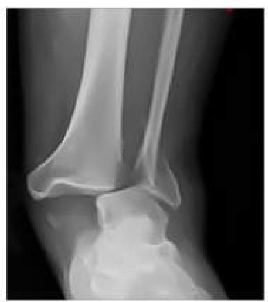
An RTA patient presented to the emergency department with severe pain in the ankle. An X-ray was performed, given below. What is the best next step in management?

A ballet dancer presents with chronic anterolateral ankle pain. Most likely diagnosis?
The operative procedure known as "microfracture" is done for the
Immediate treatment of compound fracture of tibia includes:
Commonest ligament injured in ankle injury ?
All of the following are indications for open reduction and internal fixation (ORIF) of fractures EXCEPT:
The rephrased question is:What is the most common complication of a fractured talus?
A 10-year-old obese boy was referred to the emergency department with a history of hip pain. He was observed to be limping and complained of severe pain. Which of the following investigations is least appropriate for this condition?
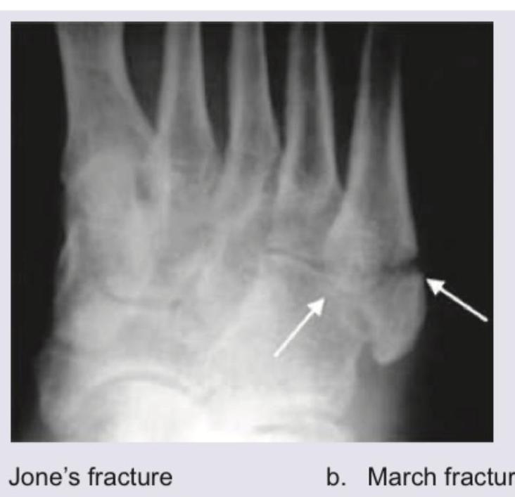
Comment on the diagnosis:

Match List-I with List-II and select the correct answer using the code given below the Lists: (Refer to the image below for the lists)
Explanation: ***Neurovascular Assessment and Closed reduction with slab application*** - The X-ray shows an **ankle dislocation without an obvious fracture**, making **closed reduction** the appropriate initial treatment. - A **slab (splint)** is preferred over a full cast initially for acute injuries to accommodate for swelling, reducing the risk of compartment syndrome, and allowing for serial neurovascular checks. *Neurovascular Assessment and Closed reduction with cast application* - While closed reduction is correct, applying a **full cast** immediately after an acute injury carries a risk of **compartment syndrome** due to potential swelling that cannot be accommodated by a rigid cast. - A cast would typically be applied after the initial swelling has subsided, usually a few days to a week after initial reduction and splinting. *Neurovascular Assessment and Immediate surgery* - **Immediate surgery** is generally reserved for **open fractures/dislocations**, dislocations that cannot be reduced closed (irreducible dislocations), or those with significant associated fractures that require surgical fixation to stabilize the joint. - In this case, the dislocation appears to be isolated and amenable to closed reduction, making surgery not the immediate next step. *Neurovascular Assessment and Immediate open reduction* - **Open reduction** is performed when closed reduction fails or is contraindicated, for example, due to soft tissue interposition or highly unstable fracture patterns. - Since closed reduction has not yet been attempted, immediate open reduction is premature and unnecessary for an apparently simple dislocation.
Explanation: **Lateral Ankle Sprain** - **Chronic anterolateral ankle pain** in a ballet dancer is highly suggestive of a **lateral ankle sprain**, often due to repetitive strain and instability. - Sprains commonly involve the **anterior talofibular ligament (ATFL)** and **calcaneofibular ligament (CFL)**, leading to persistent discomfort and potential functional deficits. *Calcaneal Stress Fracture* - A **calcaneal stress fracture** typically presents with **heel pain** that is worse with weight-bearing activities, rather than primarily anterolateral pain. - While common in athletes, the pain location is less consistent with the description in the question. *Tibialis Posterior Tendinitis* - **Tibialis posterior tendinitis** causes pain and tenderness along the **medial arch** and posterior aspect of the ankle, often associated with a **flatfoot deformity**. - The pain location described (anterolateral) does not align with the typical presentation of this condition. *Anterior Ankle Impingement Syndrome* - **Anterior ankle impingement syndrome** results from compression of soft tissues or bony spurs at the **anterior ankle joint**, typically causing pain with **dorsiflexion**. - While possible in a dancer, the presentation as chronic anterolateral pain without specific mention of dorsiflexion-related pain makes a lateral ankle sprain a more probable initial diagnosis.
Explanation: ***Osteochondral defect of femur*** - **Microfracture** is a surgical technique used to stimulate the growth of **fibrocartilage** in areas of damaged articular cartilage, such as an **osteochondral defect**. - It involves creating small holes in the **subchondral bone** to allow stem cells and growth factors from the bone marrow to form a new reparative tissue. *Delayed union of femur* - **Delayed union** typically involves an extended time for fracture healing, which is often managed through prolonged immobilization, **bone grafting**, or sometimes revision surgery. - Microfracture specifically targets cartilage repair, not the process of **bony union** after a fracture. *Non union of tibia* - **Non-union** refers to the failure of a fractured bone to heal within a reasonable timeframe, often requiring surgical intervention with **bone grafts** or **internal fixation**. - This condition involves bone healing problems, distinct from cartilage defects that microfracture addresses. *Loose bodies of ankle joint* - **Loose bodies** in a joint are typically removed surgically, often arthroscopically, to relieve pain and prevent joint damage. - This procedure does not involve the repair of cartilage defects, which is the primary goal of microfracture.
Explanation: ***Antibiotics, debridement, and splinting*** - **Antibiotics** are crucial to prevent infection in **compound (open) fractures** due to communication with the external environment. - **Debridement** removes contaminated and devitalized tissue, while **splinting** stabilizes the fracture and minimizes further soft tissue damage. *Antibiotics and debridement only* - While antibiotics and debridement are essential, **splinting** is also critical for immobilizing the fracture and preventing further injury. - Without stabilization, the fracture site can move, causing additional soft tissue damage and increasing the risk of infection and delayed healing. *Debridement and splinting only* - This option overlooks the critical need for **antibiotics** in compound fractures, which are at high risk of infection due to the exposure of bone and tissue to bacteria. - Infection can lead to serious complications such as **osteomyelitis**, significantly impacting recovery and patient outcomes. *Debridement, splinting, and blood transfusion* - While debridement and splinting are correct, a **blood transfusion** is generally not an immediate routine treatment for all compound tibial fractures unless there is significant hemorrhage leading to hypovolemic shock. - The primary immediate concerns are infection prevention and stabilization, not typically massive blood loss requiring transfusion in every case.
Explanation: ***Anterior talofibular ligament*** - The **anterior talofibular ligament (ATFL)** is the **most frequently injured ligament** in ankle sprains because it is the weakest and most commonly stretched during **inversion injuries**. - Its position makes it vulnerable during movements where the foot rolls inward, a common mechanism for ankle sprains. *Calcaneofibular ligament* - The **calcaneofibular ligament (CFL)** is stronger than the ATFL and is typically injured with more severe inversion forces, often in conjunction with ATFL rupture. - While it plays a crucial role in ankle stability, it is not the *most* commonly injured ligament. *Posterior talofibular ligament* - The **posterior talofibular ligament (PTFL)** is the strongest of the lateral ankle ligaments and is rarely injured in isolated ankle sprains. - Its injury usually signifies a **severe ankle sprain** with significant talar displacement or dislocation. *Spring ligament* - The **spring ligament**, also known as the **plantar calcaneonavicular ligament**, is located on the medial side of the foot and supports the medial longitudinal arch. - It is not directly involved in typical ankle sprains, which primarily affect the lateral collateral ligaments.
Explanation: ***Stable closed fracture*** - A **stable closed fracture** typically does not require surgical intervention with ORIF as it can usually be managed non-surgically with casting or bracing. - The goal of ORIF is to achieve **anatomic reduction and rigid fixation**, which is not necessary for stable fractures that maintain alignment. *Multiple trauma* - In patients with **multiple trauma**, early stabilization of long bone fractures using ORIF can help reduce pain, prevent further injury, and facilitate patient mobilization. - This approach aims to reduce the risk of complications such as **ARDS (acute respiratory distress syndrome)** and fat embolism for critically ill patients. *Compound fracture* - **Compound (open) fractures** involve a break in the skin, exposing the bone to the external environment, and are a classic indication for surgical management. - ORIF in these cases helps to achieve **stabilization** after debridement, crucial for preventing infection and promoting bone healing. *Intra-articular fracture* - **Intra-articular fractures** involve the joint surface, and accurate anatomical reduction is critical to prevent post-traumatic arthritis and preserve joint function. - ORIF provides the precise reduction and stable fixation needed to restore the **joint congruity**.
Explanation: ***Avascular necrosis (AVN)*** - The talus has a **precarious blood supply**, with arterial branches entering at multiple points but often centrally, making it vulnerable to **ischemia** after fracture. - Fractures, especially neck fractures, can disrupt these delicate vessels, leading to **osteonecrosis** and collapse of the bone. *Nonunion of the talus* - While possible, talar nonunion is **less common** than AVN due to the talus's dense cortical bone and limited muscle attachments. - Nonunion is more frequently seen with fractures of other bones, such as the **scaphoid**. *Osteoarthritis of the subtalar joint* - **Subtalar osteoarthritis** can occur post-talar fracture, often as a **secondary complication** of disrupted articular surfaces or AVN. - However, the **initial and most common direct complication** stemming from the blood supply disruption is AVN. *Osteoarthritis of the ankle joint* - **Ankle osteoarthritis** can also develop after certain talar fractures, particularly those involving the talar dome or leading to incongruity of the ankle joint. - Similar to subtalar arthritis, it is often a **later or secondary sequela**, rather than the immediate and most frequent direct complication like AVN.
Explanation: ***USG of hip*** - An **ultrasound (USG)** of the hip is generally not the primary imaging modality for diagnosing conditions like **slipped capital femoral epiphysis (SCFE)**, which is suggested by the patient's presentation. - While USG can detect effusions or synovitis, it provides poor visualization of bony structures and the physeal plate, which are crucial for diagnosing SCFE. *X-ray of the hip* - **X-rays** (AP and frog-leg lateral views) are the **initial and most important imaging study** for diagnosing SCFE. - They effectively visualize the **epiphyseal displacement** relative to the metaphysis and are sufficient for diagnosis in most cases. *MRI of the hip* - **MRI** is highly sensitive for detecting early or subtle SCFE, especially when X-rays are inconclusive. - It can evaluate the **physeal edema**, chondral changes, and avascular necrosis, providing more detailed information than X-rays. *CT scan of hip* - A **CT scan** provides excellent bony detail and can precisely assess the **degree of physeal slip** and femoral head deformity. - It may be used for surgical planning, especially in complex cases or when the slip is difficult to assess with X-rays.
Explanation: ***Jones fracture*** - The image indicates a fracture located at the **proximal metaphyseal-diaphyseal junction of the fifth metatarsal**, which is characteristic of a **Jones fracture**. - This fracture involves the **base of the fifth metatarsal** and is often associated with a higher risk of nonunion due to limited blood supply. *March fracture* - A **March fracture** is a type of stress fracture, typically affecting the **shaft of the second, third, or fourth metatarsals**, often seen in military recruits or those who engage in prolonged walking or running. - It results from repetitive stress rather than an acute injury, and its location is distinct from the proximal fifth metatarsal. *Shepherd's fracture* - A **Shepherd's fracture** refers to an avulsion fracture of the **posterolateral tubercle of the talus**, also known as an os trigonum fracture. - This fracture is located in the ankle region, distinct from the metatarsals. *Cotton's fracture* - A **Cotton's fracture** is a trimalleolar fracture of the ankle, involving the **medial malleolus**, **lateral malleolus**, and the **posterior malleolus** of the tibia. - This is a complex ankle injury, entirely unrelated to fractures of the metatarsals.
Explanation: ***A→4 B→3 C→2 D→1*** - **Atrial fibrillation** is characterized by **irregularly irregular rhythm** without distinct P waves, making the R-R interval highly variable. It is a supraventricular tachyarrhythmia, originating above the ventricles. - **Ventricular tachycardia** typically presents with a **wide QRS complex** (>0.12 s) and a **rapid, regular heart rate**, as it originates from the ventricles. - **Complete heart block** is characterized by complete dissociation between **P waves and QRS complexes**, meaning the atria and ventricles beat independently. This is reflected in an irregular P-P interval and a regular but slower R-R interval often due to an escape rhythm. - **Ventricular fibrillation** is an ECG emergency characterized by chaotic, **irregular electrical activity** and an absence of discernible P waves, QRS complexes, or T waves, leading to cardiac arrest. *A→4 B→3 C→1 D→2* - This option correctly matches A (Atrial fibrillation) with 4 (Irregular R-R interval without P waves) and B (Ventricular tachycardia) with 3 (Wide QRS complexes and regular rapid rate). However, it incorrectly matches C (Complete heart block) with 1 (Chaotic rhythm) and D (Ventricular fibrillation) with 2 (Dissociation of P and QRS waves). - **Complete heart block** involves **dissociation of P and QRS waves**, and **Ventricular fibrillation** is defined by a **chaotic rhythm**, not the other way around as suggested by C→1 and D→2. *A→3 B→2 C→4 D→1* - This option incorrectly matches A (Atrial fibrillation) with 3 (Wide QRS complexes and regular rapid rate), which describes ventricular tachycardia. - It also incorrectly matches C (Complete heart block) with 4 (Irregular R-R interval without P waves) and D (Ventricular fibrillation) with 1 (Chaotic rhythm), instead of the correct associations. *A→3 B→2 C→1 D→4* - This option incorrectly matches A (Atrial fibrillation) with 3 (Wide QRS complexes and regular rapid rate) which is characteristic of ventricular tachycardia. - It also incorrectly matches B (Ventricular tachycardia) with 2 (Dissociation of P and QRS waves), which is a characteristic of complete heart block, not ventricular tachycardia.
Get full access to all questions, explanations, and performance tracking.
Start For Free