What is the best imaging modality for detecting early osteomyelitis?
What is the condition commonly known as jumper's knee?
Who is often referred to as the father of joint replacement surgery?
Gold standard investigation for breast carcinoma screening in a patient with silicone breast implants
In which of the following situations is breast conservation surgery not indicated?
All of the following are indications for open reduction and internal fixation (ORIF) of fractures EXCEPT:
Which technique is considered the best for evaluating bone regeneration after periodontal surgery?
Best investigation to detect rupture of silicone breast implants is-
Vacuum assisted closure is contraindicated in which of the following conditions -
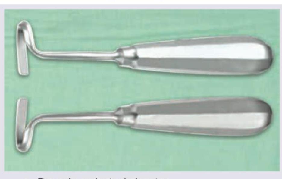
What does the given image show?

Explanation: ***MRI*** - **Magnetic Resonance Imaging (MRI)** is considered the **gold standard** for detecting early osteomyelitis due to its excellent soft tissue contrast and ability to visualize **bone marrow edema**, which is an early sign of infection. - It can identify changes within **3-5 days** of infection onset, much earlier than other modalities. *CT scan* - While useful for showing **bone destruction**, cortical integrity, and sequestra, **CT scans** are less sensitive than MRI for detecting early marrow edema. - Its ability to diagnose osteomyelitis is usually delayed until significant **bony changes** have occurred, typically around 1-2 weeks. *X-ray* - **Plain radiographs** are often the initial imaging study but are **insensitive** for early osteomyelitis, showing changes only after 10-14 days or more. - Early findings on X-rays can be subtle, such as **periosteal elevation** or **soft tissue swelling**, but frank bone destruction is a late finding. *Bone scintigraphy* - **Bone scintigraphy** (e.g., technetium-99m) is sensitive for detecting increased bone turnover associated with infection but lacks **specificity**, as it can be positive in other conditions like trauma or tumors. - While it can detect changes earlier than X-rays, typically within 2-3 days, it cannot clearly differentiate infection from other processes, and its spatial resolution is poor compared to MRI.
Explanation: ***Patellar tendonitis due to overuse of the patellar tendon.*** - **Jumper's knee** is the common term for **patellar tendonitis**, which specifically refers to inflammation of the patellar tendon. - This condition is frequently caused by **overuse**, especially in activities involving repetitive jumping and landing. *Inflammation of the patellar tendon at its insertion on the patella.* - While jumper's knee does involve inflammation of the patellar tendon, it is more commonly at its insertion on the **tibial tubercle** or specifically its origin at the **inferior pole of the patella**, not necessarily at the patella itself. - This option is less precise as it describes only one aspect of the condition without mentioning the critical role of overuse. *Tendinopathy of the quadriceps tendon.* - **Tendinopathy of the quadriceps tendon** is a distinct condition affecting the tendon above the patella, known as **quadriceps tendinopathy**. - It presents with pain proximal to the patella, differentiating it from jumper's knee, which involves the tendon distal to the patella. *Injury to the hamstring tendon.* - An **injury to the hamstring tendon** would cause pain and symptoms on the posterior aspect of the knee or thigh. - This is completely unrelated to jumper's knee, which is characterized by anterior knee pain.
Explanation: ***Sir John Charnley*** - **Sir John Charnley** is widely recognized as the **father of modern hip replacement surgery** due to his pioneering work in developing low-friction arthroplasty in the 1960s. - He introduced concepts such as **bone cement fixation**, **aseptic operating environments**, and the use of **ultra-high molecular weight polyethylene** as a bearing surface. *Sir John Manning* - While significant in medical history, **Sir John Manning** is not associated with the development of joint replacement surgery. - His contributions do not primarily lie in orthopedic surgery innovations. *Dr. Ignacio Ponseti* - **Dr. Ignacio Ponseti** is renowned for developing the **Ponseti method** for the treatment of **clubfoot**, a non-surgical technique involving manipulation and casting. - His work focuses on pediatric orthopedics and congenital deformities rather than joint replacement. *Sir John Girdlestone* - **Sir John Girdlestone** is known for the **Girdlestone arthroplasty**, a salvage procedure involving the **excision of the femoral head** to treat severe hip infections or failed hip replacements. - This procedure aims to create a pseudoarthrosis rather than replacing the joint, distinguishing it from modern joint replacement.
Explanation: ***MRI*** - **MRI** is considered the **gold standard** for breast cancer screening in patients with silicone breast implants due to its superior ability to visualize breast tissue through the implant and detect subtle lesions. - It offers **high sensitivity** in detecting both implant rupture and early malignancies, often providing better clarity than mammography in augmented breasts where implants can obscure tissue. *Mammography* - While a standard screening tool, **mammography** can be limited in patients with silicone implants because the implants can **obscure adjacent breast tissue**, making detection of small masses challenging. - Special views (e.g., **Eklund views**) can be used, but sensitivity is still reduced compared to MRI in augmented breasts. *CT scan* - **CT scans** are not routinely used for primary breast cancer screening due to their use of **ionizing radiation** and lower sensitivity for detecting early breast lesions compared to MRI. - CT is more commonly used for **staging** advanced cancers or evaluating complex masses detected by other modalities. *USG* - **Ultrasound (USG)** is a valuable complementary tool, especially for evaluating palpable lumps or clarifying findings from mammography, but it is **operator-dependent** and has a lower overall sensitivity for general screening compared to MRI. - It is particularly useful for differentiating between **cystic and solid masses** and detecting implant ruptures but is not the gold standard for comprehensive screening in augmented breasts.
Explanation: ***All of the options*** - All listed scenarios—**large pendular breast**, **SLE**, and **diffuse microcalcification**—represent situations where breast conservation surgery is generally contraindicated or challenging. - Their presence often necessitates alternative treatment approaches, such as mastectomy, to achieve optimal oncologic and cosmetic outcomes. *Large pendular breast* - While not an absolute contraindication, a **very large or pendulous breast** can make it difficult to achieve a satisfactory cosmetic outcome after breast conservation surgery. - The disproportionate breast size post-lumpectomy may lead to significant **asymmetry**, requiring further reconstructive procedures. *SLE* - Patients with **Systemic Lupus Erythematosus (SLE)** are at an increased risk of complications from radiation therapy, a mandatory component of breast conservation surgery. - They tend to experience more severe and prolonged **acute and chronic skin reactions** to radiation, which can significantly impair healing and quality of life. *Diffuse microcalcification* - **Diffuse microcalcification** within the breast can indicate widespread in situ carcinoma (e.g., DCIS) or an invasive carcinoma with extensive intraductal component. - In such cases, achieving **clear surgical margins** with breast conservation surgery can be challenging and often leads to multiple re-excisions or an increased risk of local recurrence.
Explanation: ***Stable closed fracture*** - A **stable closed fracture** typically does not require surgical intervention with ORIF as it can usually be managed non-surgically with casting or bracing. - The goal of ORIF is to achieve **anatomic reduction and rigid fixation**, which is not necessary for stable fractures that maintain alignment. *Multiple trauma* - In patients with **multiple trauma**, early stabilization of long bone fractures using ORIF can help reduce pain, prevent further injury, and facilitate patient mobilization. - This approach aims to reduce the risk of complications such as **ARDS (acute respiratory distress syndrome)** and fat embolism for critically ill patients. *Compound fracture* - **Compound (open) fractures** involve a break in the skin, exposing the bone to the external environment, and are a classic indication for surgical management. - ORIF in these cases helps to achieve **stabilization** after debridement, crucial for preventing infection and promoting bone healing. *Intra-articular fracture* - **Intra-articular fractures** involve the joint surface, and accurate anatomical reduction is critical to prevent post-traumatic arthritis and preserve joint function. - ORIF provides the precise reduction and stable fixation needed to restore the **joint congruity**.
Explanation: ***Cone Beam Computed Tomography*** - **CBCT** offers a three-dimensional view, which is superior for assessing **bone density, volume**, and the precise morphology of bone regeneration. - It allows for detailed analysis of defects and regeneration without the superimposition of anatomical structures, which is a limitation of 2D radiographs. *Intraoral Periapical radiography* - **Periapical radiographs** provide only a two-dimensional image and are poor at differentiating between **bony infill** and soft tissue. - They are limited by projection geometry and cannot accurately represent **bone volume** or complex defects. *Orthopantomogram* - An **OPG** provides a panoramic view but suffers from significant **magnification, distortion**, and superimposition of structures. - It is not precise enough for the detailed assessment required to evaluate **bone regeneration** after periodontal surgery. *Digital subtraction radiography* - **Digital subtraction radiography** can detect subtle changes in bone mineral content by subtracting two images taken at different times. - While useful for showing small changes, it is still a **2D technique** and does not provide information on **bone volume** or the three-dimensional architecture of the regenerated bone.
Explanation: ***MRI*** - **Magnetic Resonance Imaging (MRI)** is considered the **gold standard** for detecting silicone breast implant ruptures due to its superior soft tissue contrast and ability to differentiate silicone from other tissues. - It can accurately identify both **intracapsular** (linguine sign) and **extracapsular** ruptures, as well as associated silicone granulomas. *Mammography* - While useful for breast cancer screening, **mammography** has limited sensitivity for detecting silicone implant ruptures, especially subtle ones. - It can show indirect signs like implant contour abnormalities or increased implant density but is often inconclusive for rupture diagnosis. *X-ray* - **X-rays** provide very little information regarding the integrity of silicone breast implants because silicone is radiolucent and does not show up clearly on standard radiographs. - Its utility is primarily for detecting calcifications or foreign bodies, not implant rupture. *USG* - **Ultrasound (USG)** can be a useful initial screening tool for detecting implant ruptures, showing signs like the **"stepladder sign"** for intracapsular rupture or anechoic collections (silicone outside the capsule). - However, its accuracy is highly operator-dependent, and it may miss subtle ruptures or be limited by poor visualization due to scar tissue, making MRI a more definitive choice.
Explanation: ***Large amount of necrotic tissue with eschar*** - The presence of a large amount of **necrotic tissue** and **eschar** is a contraindication for VAC therapy because it prevents effective contact between the foam and viable tissue, impairing wound healing. - Eschar acts as a physical barrier, trapping bacteria and hindering the proper function of negative pressure by preventing uniform pressure distribution and fluid removal from the wound bed. *Chronic osteomyelitis* - While chronic osteomyelitis can be challenging, VAC therapy can sometimes be used as an **adjunctive treatment** after surgical debridement to manage the wound and promote granulation tissue formation. - It helps in controlling infection and closing the wound by removing exudates, reducing edema, and improving blood flow. *Abdominal wound* - VAC therapy is commonly used for **abdominal wounds**, especially after damage control surgery or in cases of open abdomen management. - It facilitates closure by promoting granulation, reducing edema, and protecting the abdominal contents. *Surgical wound dehiscence* - **Surgical wound dehiscence** is a common indication for VAC therapy, as it helps to manage the open wound, promote granulation tissue, and prepare the wound for eventual secondary closure or grafting. - VAC therapy reduces surgical site infections, removes exudates, and enhances tissue perfusion, leading to better wound healing outcomes.
Explanation: ***Doyen's periosteal elevator*** - This instrument is characterized by its **curved, spoon-like working end** and solid handle, designed for **elevating the periosteum** from bone. - Doyen's elevator comes in various sizes and angles, but the distinct hook-like curve is a key identifying feature, distinguishing it from other elevators. *Cobb's spinal elevator* - Cobb's elevator typically has a **flat, broad, and slightly curved blade** with a rounded or tapered tip, suitable for dissecting along bony surfaces, particularly in spinal surgery. - Its design prioritizes broad, even separation of tissue layers, contrasting with the more acute curve of the Doyen's. *Leksell's rongeur* - A rongeur is a biting instrument used for **gnawing away bone or tough tissue**, characterized by hinged jaws with sharp, cup-shaped ends. - The image clearly shows a single-piece instrument without hinged jaws, ruling out a rongeur. *Key periosteal elevator* - The Key elevator typically features a **broader, more rounded or slightly angled tip** and a flat, somewhat spatula-like working end, used for general periosteal elevation. - While also a periosteal elevator, its working end does not exhibit the characteristic deep, hook-like curve seen in the Doyen's elevator.
Get full access to all questions, explanations, and performance tracking.
Start For Free