I/V contrast is not used in -
Recommended angle of root end resection is:
Best site for administering spinal anesthesia is the intervertebral space between.
Which of the following is NOT a contraindication for spinal anaesthesia?
Removal of vertebral disc can be done by all these approaches except:
Minimally invasive Percutaneous plate osteosynthesis (MIPPO technique) is of use in:
In an accident involving potential cervical spine damage, the first line of management is:
What is the first step to be taken in the management of a cervical spine injury?
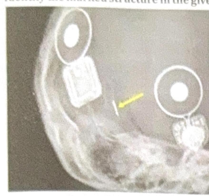
Identify the marked structure in the given image.

A surgeon experiences pin-site fracture during reference array fixation in computer-navigated TKA in an osteoporotic patient. Subsequently, three more cases develop similar complications. What systematic approach should be implemented to prevent this complication?
Explanation: ***Myelography*** - Myelography involves injecting contrast material directly into the **subarachnoid space** of the spinal canal to visualize nerve roots and the spinal cord, and therefore does not use intravenous contrast. - The contrast in myelography is typically **iodinated non-ionic contrast** injected intrathecally, not intravenously. *IVP* - **Intravenous Pyelogram (IVP)** is a radiological procedure that specifically uses **intravenous iodinated contrast** to visualize the kidneys, ureters, and bladder. - The contrast is excreted by the kidneys, highlighting the urinary tract structures on X-ray images. *MRI* - While many MRI scans do not require contrast, **intravenous gadolinium-based contrast agents** are commonly used to enhance visualization of certain pathologies like tumors, inflammation, or vascular anomalies. - The contrast is administered intravenously to accumulate in areas with increased vascularity or disrupted blood-brain barrier. *CT scan* - **CT scans** frequently utilize **intravenous iodinated contrast** to improve the visibility of blood vessels, organs, and various lesions like tumors or inflammatory processes. - The contrast enhances density differences between tissues, making pathologies more conspicuous.
Explanation: ***0 degrees*** - A **0-degree** resection angle is recommended to minimize the number of exposed **dentinal tubules** and therefore potential **leakage channels**, fostering better apical sealing. - This approach aims for a **flat** or perpendicular cut to the long axis of the tooth, preserving as much root structure as possible. *30 degrees* - A **30-degree** resection angle would expose a significantly larger number of **dentinal tubules** and increase the risk of **apical leakage**. - It would also unnecessarily remove more **root structure**, which could weaken the tooth. *15 degrees* - While less severe than 30 or 45 degrees, a **15-degree** angle still exposes more **dentinal tubules** and creates a larger surface area for potential **leakage** compared to a 0-degree resection. - This angle is not considered ideal for maximizing the **seal** and preserving root integrity. *45 degrees* - A **45-degree** resection angle is associated with the **greatest exposure** of **dentinal tubules** and the highest risk of **microleakage**. - This aggressive angle also leads to the removal of the most **root structure**, potentially compromising the **tooth's stability**.
Explanation: ***L3 - L4*** - The **spinal cord** typically ends at the level of **L1-L2** in adults, making the L3-L4 intervertebral space a safe choice to avoid inadvertent cord injury. - This interspace is easily identified by drawing an imaginary line between the highest points of the **iliac crests**, which usually intersects the L4 vertebra or the L3-L4 interspace. *L1 - L2* - This interspace is generally considered too high for routine spinal anesthesia due to the risk of directly puncturing the **spinal cord**, which often extends to this level in adults. - Puncturing the spinal cord can lead to severe neurological complications, so it is usually avoided. *L2 - L3* - While safer than L1-L2, the **L2-L3 interspace** is still relatively high and carries a slightly increased risk of spinal cord injury compared to lower levels. - The **L3-L4** or **L4-L5** interspaces are generally preferred as they offer a wider margin of safety. *L5 - S1* - The **L5-S1 interspace** is often difficult to access due to the angulation of the **vertebrae** and the presence of the **iliac crests**, making needle insertion challenging. - While anatomically safe in terms of spinal cord termination, the technical difficulty makes it a less preferred site for routine lumbar punctures or spinal anesthesia.
Explanation: ***Hypertension*** - While **severe uncontrolled hypertension** may necessitate blood pressure stabilization before surgery, **mild to moderate hypertension** is not an absolute contraindication for spinal anesthesia. - In fact, spinal anesthesia can sometimes be beneficial in hypertensive patients due to its **vasodilatory effects**, which may help lower blood pressure. *Bleeding disorder* - A **bleeding disorder** (e.g., thrombocytopenia, coagulopathy) is a **major contraindication** due to the high risk of **epidural or spinal hematoma** formation. - A hematoma can lead to **spinal cord compression** and irreversible neurological damage. *Raised intracranial tension* - **Raised intracranial tension (ICT)** is a **strict contraindication** because the drop in cerebrospinal fluid (CSF) pressure during spinal anesthesia can worsen the pressure gradient across the foramen magnum. - This can precipitate **herniation of the brainstem** and lead to catastrophic neurological injury or death. *Infection at injection site* - The presence of an **infection at the injection site** is an absolute contraindication as it poses a significant risk of introducing bacteria into the **subarachnoid space**. - This can lead to serious complications such as **meningitis** or a **spinal abscess**.
Explanation: ***Laminoplasty*** - **Laminoplasty** is a procedure that *expands the spinal canal* by reshaping and repositioning the lamina, rather than removing it, to relieve pressure on the spinal cord. - Unlike disc removal techniques, it aims to *preserve the posterior spinal elements* and maintain spinal stability. *Hemilaminectomy* - A **hemilaminectomy** involves the *partial removal of a lamina on one side* of the vertebra. - This approach allows access to the spinal canal to remove disc material or decompress nerve roots. *Laminotomy* - **Laminotomy** is a procedure where a *small opening is made in the lamina* to access the spinal canal. - This minimal removal of bone is often sufficient for **microdiscectomy**, allowing for the removal of herniated disc fragments. *Laminectomy* - A **laminectomy** involves the *complete removal of the lamina* of one or more vertebrae. - This wider exposure is used for more extensive decompression, such as for **spinal stenosis** or larger disc herniations.
Explanation: ***Fracture with metaphyseal comminution*** - The **MIPPO technique** is particularly useful for achieving stability in fractures with **metaphyseal comminution** by bridging the comminuted zone with a plate applied percutaneously. - This approach minimizes soft tissue dissection, preserving **periosteal blood supply**, which is crucial for healing in these complex fractures. *Segmental fracture* - While MIPPO can be used, **segmental fractures** often require more direct reduction and stabilization of both fracture segments, which might be challenging with a purely percutaneous approach alone. - The primary concern in segmental fractures is often maintaining length and alignment across two distinct fracture lines. *Spiral fracture* - **Spiral fractures** are typically inherently stable after reduction and are often amenable to intramedullary nailing or less invasive plate fixation, as the fracture pattern allows for good interfragmentary compression. - The main advantage of MIPPO (minimizing soft tissue stripping around comminution) is less critical in these stable, non-comminuted patterns. *Oblique fracture* - Similar to spiral fractures, **oblique fractures** are often amenable to primary screw fixation or conventional plating techniques due to their stable nature after reduction and good contact between fracture fragments. - The specific advantages of MIPPO for comminuted fractures are less relevant for simple oblique patterns.
Explanation: ***Correct: Maintain airway*** - In trauma management, the **ATLS protocol** follows the **A-B-C-D-E** approach where **Airway is the first priority** - In suspected cervical spine injury, airway management is performed **with concurrent cervical spine protection** (using jaw thrust maneuver instead of head tilt-chin lift) - A compromised airway leads to death within minutes, making it the **immediate first-line intervention** - **Cervical spine stabilization is performed simultaneously** during airway assessment and management, not as a separate preceding step - The correct approach: **"Airway with cervical spine protection"** - both are done together, but airway assessment/management takes priority *Incorrect: Stabilize the cervical spine* - While **cervical spine stabilization** is critical and must be maintained throughout trauma management, it is **not performed before airway assessment** - Manual inline stabilization and cervical collar application are done **during** airway management, not before it - ATLS teaches that C-spine protection is **integrated into** airway management, not a separate first step *Incorrect: X-ray* - **X-ray** is a diagnostic tool performed after initial stabilization and resuscitation - Imaging is part of the **secondary survey**, not primary trauma management - Never delay life-saving interventions for diagnostic studies *Incorrect: Turn head to side* - **Turning the head** is absolutely contraindicated in suspected cervical spine injury - Any movement can convert an unstable fracture into a **complete spinal cord injury** - If airway management is needed, use **jaw thrust** or **chin lift without head tilt**
Explanation: ***Immobilization of spine*** - In the context of **isolated cervical spine injury management**, **spinal immobilization** is the primary intervention to prevent further neurological damage. - This is typically achieved using a **cervical collar** and **backboard** to maintain in-line spinal stabilization. - **Note**: In actual trauma scenarios following **ATLS protocols**, airway management and cervical spine immobilization occur **simultaneously** as the first priority (Airway with C-spine protection). *Turn head* - **Turning the head** is absolutely contraindicated as it can exacerbate a cervical spine injury, leading to further compression or damage to the **spinal cord**. - Maintaining a **neutral, in-line position** is critical to avoid neurological deterioration. *Maintain airway* - In comprehensive trauma management per **ATLS guidelines**, **airway management with simultaneous cervical spine protection** is the first priority in the ABC sequence. - Airway is maintained using methods that do not compromise spinal stability, such as a **jaw thrust maneuver** or **endotracheal intubation with manual in-line stabilization**. - The distinction here is that this question focuses on the specific step for **spinal injury management** rather than overall trauma priorities. *None of the options* - This option is incorrect because **immobilization of the spine** is a definitive priority in managing a suspected cervical spine injury. - Both spinal immobilization and airway management are critical interventions that should occur together in actual practice.
Explanation: ***Coil*** - The marked structure appears to be a **cochlear implant's internal coil**, which is common in X-ray imaging of these devices. - The **cochlear implant internal coil** is crucial for transmitting processed sound signals via electromagnetic induction to the electrode array within the cochlea. *Electrode* - An **electrode array** is typically a thin, flexible wire with multiple contacts inserted into the cochlea, which is not what the arrow is pointing to directly. - While electrodes are part of a cochlear implant, the marked structure's shape and position are more consistent with the **internal coil** that connects to the electrode array. *Magnet* - A **magnet** is present in a cochlear implant system, typically in both the external processor and internal receiver, to hold these two components together through the skin. - Magnets usually appear as dense, circular structures in X-rays, often seen more anteriorly or superiorly to the coil for external component alignment. *Processor* - The **processor** for a cochlear implant is an external device worn behind the ear, not an implanted component visible on an X-ray. It processes sound and sends it to the internal coil. - The structures seen in the X-ray are **implanted components** of the cochlear implant, not the external sound processor.
Explanation: ***Use unicortical pins instead of bicortical pins with reduced insertion torque protocol*** - **Pin-site fractures** are a known complication in navigated TKA, especially in **osteoporotic bone**, and can be mitigated by reducing the **stress risers** created by drilling. - Using **unicortical pins** and avoiding power drivers to limit **insertion torque** provides sufficient stability for reference arrays while minimizing the risk of cortical failure. *Switch to electromagnetic navigation system* - **Electromagnetic navigation** aims to resolve line-of-sight issues but does not inherently eliminate the need for stable skeletal fixation of reference sensors. - Switching systems is a costly equipment change that does not directly address the underlying **biomechanical failure** of the bone-pin interface in osteoporosis. *Abandon navigation in all osteoporotic patients* - Abandoning navigation denies the patient population the benefits of **precise alignment** and component positioning where it is often most needed due to poor bone quality. - Systematic technical modifications are preferred over total abandonment of a beneficial **surgical technology**. *Increase pin diameter for better fixation* - Increasing the **pin diameter** is counterproductive as larger holes create larger **stress concentrators**, significantly increasing the risk of **periprosthetic fracture** in brittle bone. - A thicker pin displaces more cortical volume, which reduces the **structural integrity** of the femur or tibia in osteoporotic patients.
Get full access to all questions, explanations, and performance tracking.
Start For Free