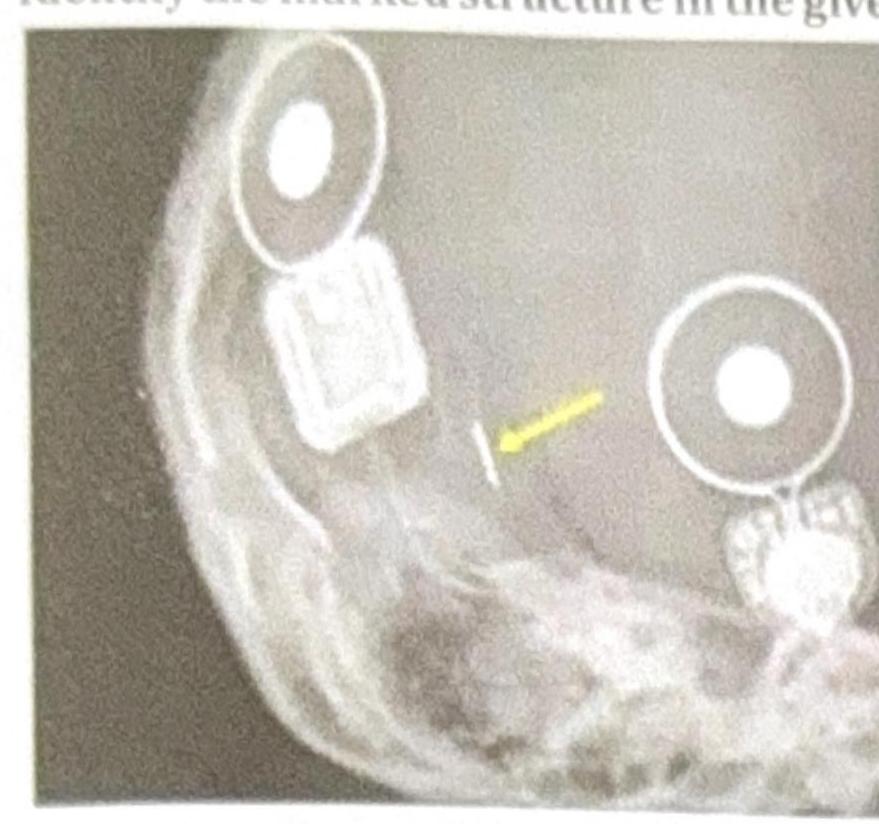
Identify the marked structure in the given image.

Recommended angle of root end resection is:
Which prosthesis is shown below in the X-ray?

A 75-year-old man presents with a fracture of the intracapsular neck of the femur. What is the most common management option for this patient?
A surgeon experiences pin-site fracture during reference array fixation in computer-navigated TKA in an osteoporotic patient. Subsequently, three more cases develop similar complications. What systematic approach should be implemented to prevent this complication?
A tertiary care center is planning to implement computer-assisted surgery program for joint replacement. They have limited budget and expertise. Which factor should be prioritized when selecting a navigation system?
A study compares outcomes of computer-navigated versus conventional total knee arthroplasty. Navigation group shows 95% implants within 3 degrees of neutral mechanical axis versus 80% in conventional group (p<0.05). However, 5-year functional outcomes and survival rates are similar. What is the most appropriate interpretation?
During computer-navigated total hip arthroplasty, the navigation system shows 38 degrees of cup abduction and 18 degrees of anteversion. However, the surgeon's visual assessment suggests more abduction. Intraoperative fluoroscopy confirms navigation data. What is the most likely cause of this discrepancy?
A 55-year-old patient is scheduled for computer-assisted pedicle screw placement in lumbar spine. During registration, the navigation system shows a registration error of 3.5 mm. What should be the surgeon's action?
A 68-year-old patient with severe varus deformity (15 degrees) undergoes computer-navigated total knee arthroplasty. During surgery, the navigation system shows 3 degrees residual varus after bone cuts but before trial implant insertion. What is the most appropriate next step?
Explanation: ***Coil*** - The marked structure appears to be a **cochlear implant's internal coil**, which is common in X-ray imaging of these devices. - The **cochlear implant internal coil** is crucial for transmitting processed sound signals via electromagnetic induction to the electrode array within the cochlea. *Electrode* - An **electrode array** is typically a thin, flexible wire with multiple contacts inserted into the cochlea, which is not what the arrow is pointing to directly. - While electrodes are part of a cochlear implant, the marked structure's shape and position are more consistent with the **internal coil** that connects to the electrode array. *Magnet* - A **magnet** is present in a cochlear implant system, typically in both the external processor and internal receiver, to hold these two components together through the skin. - Magnets usually appear as dense, circular structures in X-rays, often seen more anteriorly or superiorly to the coil for external component alignment. *Processor* - The **processor** for a cochlear implant is an external device worn behind the ear, not an implanted component visible on an X-ray. It processes sound and sends it to the internal coil. - The structures seen in the X-ray are **implanted components** of the cochlear implant, not the external sound processor.
Explanation: ***0 degrees*** - A **0-degree** resection angle is recommended to minimize the number of exposed **dentinal tubules** and therefore potential **leakage channels**, fostering better apical sealing. - This approach aims for a **flat** or perpendicular cut to the long axis of the tooth, preserving as much root structure as possible. *30 degrees* - A **30-degree** resection angle would expose a significantly larger number of **dentinal tubules** and increase the risk of **apical leakage**. - It would also unnecessarily remove more **root structure**, which could weaken the tooth. *15 degrees* - While less severe than 30 or 45 degrees, a **15-degree** angle still exposes more **dentinal tubules** and creates a larger surface area for potential **leakage** compared to a 0-degree resection. - This angle is not considered ideal for maximizing the **seal** and preserving root integrity. *45 degrees* - A **45-degree** resection angle is associated with the **greatest exposure** of **dentinal tubules** and the highest risk of **microleakage**. - This aggressive angle also leads to the removal of the most **root structure**, potentially compromising the **tooth's stability**.
Explanation: ***Austin Moore's prosthesis*** - The image clearly shows a **femoral stem with a long intramedullary component** and an **integrated prosthetic head** that articulates directly with the native acetabulum. This is characteristic of a hemiarthroplasty design, specifically resembling an Austin Moore prosthesis. - This type of prosthesis is commonly used for **femoral neck fractures** in older patients, replacing only the femoral head and neck rather than the entire hip joint. *Articular resurfacing* - **Articular resurfacing** involves capping the femoral head and lining the acetabulum with metallic implants, preserving more bone than a traditional total hip replacement. - The X-ray image does not show a cap on the femoral head or a separate acetabular component, which are features of resurfacing. *Thompson prosthesis* - The **Thompson prosthesis** is another type of hemiarthroplasty, but it typically has a **shorter, bulkier femoral stem** and a **relatively smaller head** compared to the Austin Moore prosthesis shown. - While both Thompson and Austin Moore prostheses are hemiarthroplasties, the specific shape and length of the stem in the X-ray are more consistent with an Austin Moore design. *Birmingham hip replacement* - The **Birmingham hip replacement** is a type of **hip resurfacing arthroplasty**, which, as explained earlier, involves capping the femoral head and is not depicted in this image. - It maintains more of the patient's original bone structure compared to conventional total hip replacement but still requires both femoral and acetabular components.
Explanation: **Hemiarthroplasty** - **Hemiarthroplasty** is the most common management for **intracapsular neck of femur fractures** in elderly patients, especially those who are frail or have substantial comorbidities. - This procedure replaces the **femoral head** with a prosthesis, preserving the native acetabulum, which is sufficient given the higher risk of complications with a full replacement in this age group. *Total Hip Replacement* - **Total hip replacement (THR)** is typically reserved for more active elderly patients with **pre-existing osteoarthritis** or for those requiring revision surgery, due to better functional outcomes but higher surgical risks. - It involves replacing both the **femoral head** and the **acetabulum**, *Dynamic Hip Screw* - A **dynamic hip screw (DHS)** is primarily used for **extracapsular femur fractures** (e.g., trochanteric fractures), where the blood supply to the femoral head is largely preserved. - It involves fixation, which is not suitable for most **intracapsular fractures** due to the disruption of blood supply, increasing the risk of **avascular necrosis** and non-union. *Conservative Management* - **Conservative management** (e.g., bed rest, pain control) is generally not recommended for **intracapsular neck of femur fractures** in mobile elderly patients due to high rates of complications such as **deep vein thrombosis**, **pressure ulcers**, and **avascular necrosis**. - It may be considered only in patients who are **non-ambulatory** or have severe contraindications to surgery.
Explanation: ***Use unicortical pins instead of bicortical pins with reduced insertion torque protocol*** - **Pin-site fractures** are a known complication in navigated TKA, especially in **osteoporotic bone**, and can be mitigated by reducing the **stress risers** created by drilling. - Using **unicortical pins** and avoiding power drivers to limit **insertion torque** provides sufficient stability for reference arrays while minimizing the risk of cortical failure. *Switch to electromagnetic navigation system* - **Electromagnetic navigation** aims to resolve line-of-sight issues but does not inherently eliminate the need for stable skeletal fixation of reference sensors. - Switching systems is a costly equipment change that does not directly address the underlying **biomechanical failure** of the bone-pin interface in osteoporosis. *Abandon navigation in all osteoporotic patients* - Abandoning navigation denies the patient population the benefits of **precise alignment** and component positioning where it is often most needed due to poor bone quality. - Systematic technical modifications are preferred over total abandonment of a beneficial **surgical technology**. *Increase pin diameter for better fixation* - Increasing the **pin diameter** is counterproductive as larger holes create larger **stress concentrators**, significantly increasing the risk of **periprosthetic fracture** in brittle bone. - A thicker pin displaces more cortical volume, which reduces the **structural integrity** of the femur or tibia in osteoporotic patients.
Explanation: ***Imageless navigation with good technical support and training program*** - **Imageless navigation** is the most cost-effective choice for limited budgets as it avoids the preoperative costs and infrastructure associated with **CT scans** or dedicated imaging. - Prioritizing **technical support** and **training programs** is essential for overcoming the **learning curve** (typically 15-20 cases) in a center with limited initial expertise. *Image-based system requiring dedicated CT scanner* - These systems increase the **per-case cost** and require significant capital investment in preoperative **CT imaging** infrastructure. - They introduce additional **radiation exposure** to the patient and complex logistics that may not suit a center with limited resources. *Most expensive system with all features available* - Purchasing the most expensive system is counterproductive for a facility with a **limited budget**, as many advanced features may not be utilized during the initial phase. - High-cost systems often have higher **maintenance contracts** and operational costs that can lead to program failure in resource-constrained environments. *System with steepest learning curve to ensure only expert surgeons use it* - A **steep learning curve** is a major barrier to the adoption of new technology and can lead to increased **operation time** and operative complications. - The goal of implementing a new program should be **sustainable integration** and broad clinical utility rather than restricting access through technical difficulty.
Explanation: ***Improved radiographic alignment may not translate to short-term functional improvement but could affect long-term survival*** - Navigation significantly improves **radiographic alignment** consistency (p<0.05), but high-quality evidence shows this does not always create immediate **functional differences** within 5 years. - While **short-term functional outcomes** are similar, reducing **outliers** to within 3 degrees of the neutral axis is theoretically linked to better **implant survival** beyond 10-15 years. *Navigation is inferior due to longer operative time without functional benefit* - Increased **operative time** is a known drawback, but it does not make the technique **inferior** if it achieves superior technical precision. - The term "inferior" is clinically inaccurate here as the study demonstrates **statistically significant** improvements in surgical accuracy. *Conventional technique should be abandoned* - **Conventional techniques** are still the gold standard because they provide identical **short-term survival** and clinical scores at a lower cost. - Abandonment is unjustified as long as the **clinical outcome** parity exists and long-term superiority of navigation is not yet definitively proven by this data. *The study proves navigation provides no clinical benefit* - Improved **mechanical axis** alignment is itself a technical clinical benefit that reduces mechanical stress on the polyethene liner. - A lack of difference in **5-year scores** does not prove a lack of benefit; it only indicates that **short-term follow-up** may be insufficient to detect long-term wear advantages.
Explanation: ***Incorrect pelvic tilt registration affecting surgeon's visual perception*** - Visual assessment in total hip arthroplasty is highly susceptible to **pelvic tilt**; a change in tilt can dramatically alter the surgeon’s perception of **cup abduction** and **anteversion**. - Since **fluoroscopy** (an objective imaging tool) confirms the **navigation data**, the discrepancy must originate from a subjective error in visual judgment caused by the patient's physical orientation. *Navigation system calibration error* - While calibration errors can occur, they would not be consistent with **intraoperative fluoroscopy** confirmation of the navigation data. - Modern systems use rigid **registration points** that usually trigger an alert if the tracking integrity or calibration is lost. *Electromagnetic interference from surgical equipment* - Electromagnetic interference typically causes **tracking jitter** or signal loss rather than a stable, plausible-looking but incorrect numerical reading. - If interference were present, the navigation data would likely appear inconsistent or fail to correlate with the **fluoroscopic images**. *Fluoroscopy machine malfunction* - It is highly unlikely for a fluoroscopy machine to malfunction in a way that perfectly correlates with an incorrect navigation reading; instead, it serves as the **gold standard** for confirming implant position. - A malfunction would usually involve **imaging artifacts** or failure to capture images rather than providing a misleadingly precise anatomical visualization.
Explanation: ***Re-register the anatomy to reduce error*** - In **computer-assisted navigation**, a registration error exceeding **2.0 mm** is considered clinically unacceptable and unsafe for screw placement. - The surgeon must perform **re-registration** to identify potential issues such as loose **reference frames**, patient movement, or poorly chosen anatomical landmarks. *Switch to freehand technique* - While the freehand technique is an alternative, the primary goal of using navigation is to enhance **accuracy**; one should first attempt to troubleshoot the system before abandoning it. - Reverting to freehand without attempting to fix the navigation error ignores the potential benefits of **intraoperative imaging** already obtained. *Proceed with surgery as error is acceptable* - A **3.5 mm error** is significantly above the safety threshold and increases the risk of **pedicle breach**, nerve root injury, or vascular complications. - Proceeding with such high **deviation** defeats the purpose of precision-guided surgery and compromises patient safety. *Increase the screw diameter to compensate* - Increasing the **screw diameter** is dangerous and counterintuitive, as it would likely lead to a structural **pedicle wall blowout** if the trajectory is slightly off. - Accuracy must be maintained through **spatial alignment**, not by altering hardware size to cover up for navigation inaccuracies.
Explanation: ***Perform additional medial soft tissue release*** - In **total knee arthroplasty (TKA)**, achieving neutral alignment often requires both accurate **bone cuts** and meticulous **soft tissue balancing** to correct deformity. - Residual **varus alignment** after navigation-validated bone cuts indicates that the **medial collateral ligament** and associated medial structures are still too tight and require release to balance the joint. *Accept the alignment as navigation has 5-degree error margin* - **Computer-assisted navigation** is highly precise, typically having an error margin significantly smaller (around **1-2 degrees**) than traditional manual instrumentation. - Leaving a **3-degree residual varus** is clinically significant and increases the risk of **asymmetric polyethylene wear** and early implant failure. *Recut the tibial surface with more valgus angle* - Recutting the bone after a navigation-verified cut may lead to **excessive bone loss** and potentially compromise the **joint line** or stability. - Since the navigation system confirms the cuts were made as planned, the persistent deformity is a **soft tissue issue** rather than a bony one. *Proceed with cementing the final components* - Cementing components before achieving **neutral mechanical alignment** and balanced gaps leads to poor postoperative kinematics and **instability**. - Final implantation should only occur once the **navigation system** confirms that the alignment and stability are optimized during trials.
Get full access to all questions, explanations, and performance tracking.
Start For Free