
Bone Tumors — MCQs

On this page
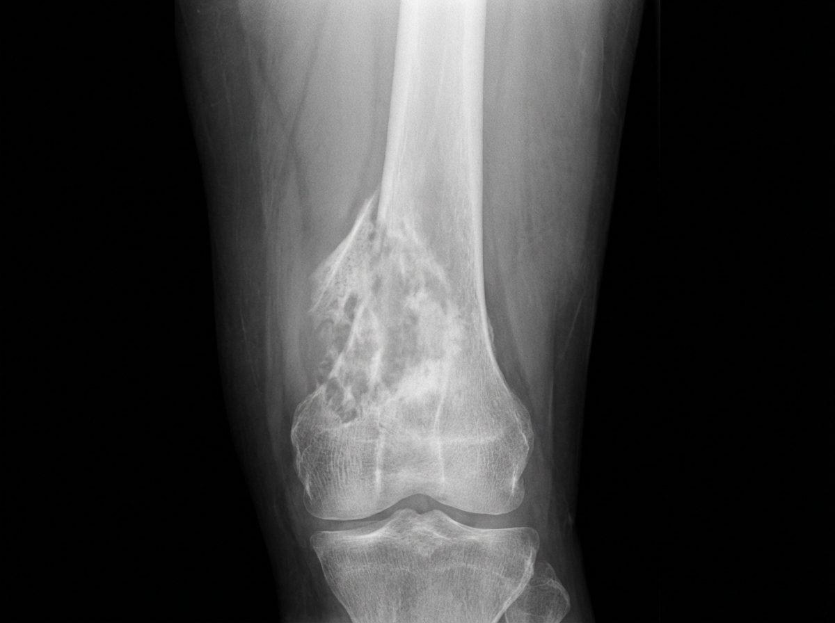
A 12-year-old boy presents with chronic knee pain for 2 years. The X-ray shows significant pathology. What is the probable diagnosis?
An Aneurysmal Bone Cyst (ABC) has a striking radiographic resemblance to which of the following conditions?
Which of the following is true about non-ossifying fibroma of bone?
What is true about osteoclastoma?
Which tumour most commonly metastasizes to bone in females?
A 20-year-old male presented with pain in the right shoulder region which aggravated with activity. On examination, a mass was felt in the right shoulder region which was warm and tender, had increased surface vascularity and a bruit was discernible over the mass. There was decreased range of motion of the right shoulder joint. X-ray and radionuclide scan were performed. The tumour was resected and sent for HPE examination. Which is the most likely diagnosis in the above scenario?
According to the Enneking system, which of the following is NOT true regarding an active benign tumor?
Which bone tumor arises from the area around the epiphyseal plate?
Which of the following is true for chondrosarcoma?
Which of the following bone tumors most commonly presents with lung secondaries and pneumothorax?
Practice by Chapter
Classification of Bone Tumors
Practice Questions
Benign Bone Tumors
Practice Questions
Malignant Primary Bone Tumors
Practice Questions
Metastatic Bone Disease
Practice Questions
Tumor-Like Lesions of Bone
Practice Questions
Soft Tissue Tumors
Practice Questions
Evaluation and Staging of Bone Tumors
Practice Questions
Biopsy Principles
Practice Questions
Limb Salvage Surgery
Practice Questions
Amputation for Bone Tumors
Practice Questions
Adjuvant Therapies
Practice Questions
Surveillance and Follow-up
Practice Questions
Want unlimited practice?
Get full access to all questions, explanations, and performance tracking.
Scan to download app