
Bone Tumors — MCQs

On this page
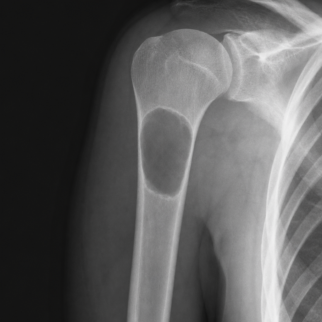
A patient presents with a lytic lesion in an X-ray of the upper end of the humerus. What is the most likely diagnosis?
Sun burst appearance is seen in which of the following conditions?
A 20-year-old boy complains of pain in his leg at a particular site, worst at night and relieved by salicylates. What is the most likely diagnosis?
What is the most common site of metastases of osteosarcoma?
Which of the following is NOT true about diaphyseal aclasia?
Which of the following conditions is least likely to present as an acentric osteolytic lesion?
What is the commonest true benign tumor of bone?
Osteochondroma is a disease of which part of the bone?
A bone tumor seen in children presents with a characteristic lamellated periosteal reaction and a mottled appearance, with extension into soft tissue. Which of the following is the most likely diagnosis?
Which of the following is NOT a malignant bone tumor?
Practice by Chapter
Classification of Bone Tumors
Practice Questions
Benign Bone Tumors
Practice Questions
Malignant Primary Bone Tumors
Practice Questions
Metastatic Bone Disease
Practice Questions
Tumor-Like Lesions of Bone
Practice Questions
Soft Tissue Tumors
Practice Questions
Evaluation and Staging of Bone Tumors
Practice Questions
Biopsy Principles
Practice Questions
Limb Salvage Surgery
Practice Questions
Amputation for Bone Tumors
Practice Questions
Adjuvant Therapies
Practice Questions
Surveillance and Follow-up
Practice Questions
Want unlimited practice?
Get full access to all questions, explanations, and performance tracking.
Scan to download app