Identify the lesion in the X-ray shown below:

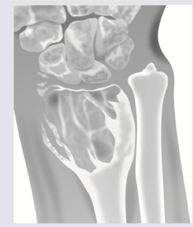
The most likely diagnosis for the lesion shown in the image is:

The following hallmarks characterise Diaphyseal aclasis except
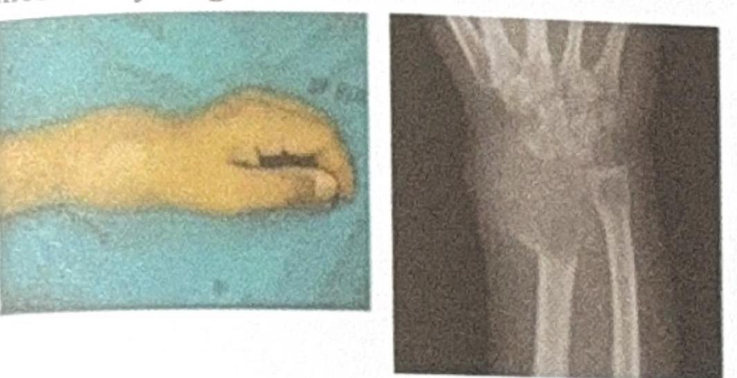
The image shows a wrist deformity and an X-ray of a bone lesion near the distal radius. Based on the clinical and radiological features, what is the most likely diagnosis?

Adamantinoma affects
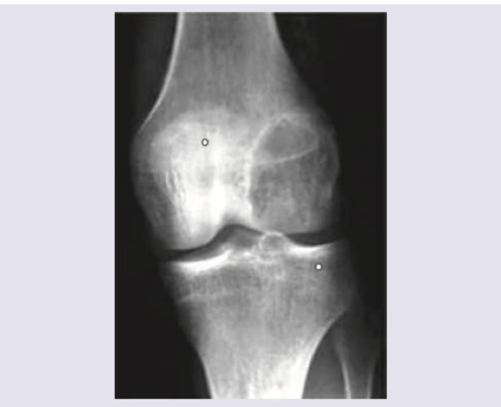
A patient presents with pain in the thigh, relieved by aspirin. X-ray shows a radiolucent mass surrounded by sclerosis. Diagnosis is ?
Which of the following is the most frequent tumor of the bone in the hand:
Classification system of bone tumors is -
All of the following are the causes of sudden increase in pain in osteochondroma, except:
Most common benign tumor of bone?
Explanation: ***Aneurysmal bone cyst*** - The image shows a **lytic, expansile lesion** in the distal radius with a **"soap bubble" appearance**, characteristic of an aneurysmal bone cyst. - These are typically **benign, blood-filled lesions** that cause bone remodeling and expansion. *Giant cell tumour* - While also a **lytic lesion**, giant cell tumors are usually more aggressive and often show **cortical destruction** and less distinct margins without the typical septations seen here. - They tend to occur in the **epiphysis** of long bones after epiphyseal closure. *Osteoma* - An osteoma is a **benign, dense, sclerotic bone growth**, appearing as a very bright, compact lesion on X-ray, which is clearly not depicted in the image. - These are typically **asymptomatic** and found incidentally. *Osteosarcoma* - This is a **highly malignant primary bone tumor** that typically presents with a **blastic or mixed lytic-blastic appearance**, often with a **"sunburst" pattern** or **Codman's triangle**, characteristics not seen in this image. - It usually causes significant **cortical destruction** and a more aggressive soft tissue mass.
Explanation: ***Giant cell tumour*** - The image shows an **epiphyseal lytic lesion** in the distal femur that extends to the subchondral bone, which is characteristic of a giant cell tumour. - Giant cell tumours are usually **benign but locally aggressive**, often presenting in patients aged 20-40 years and typically involve the **epiphysis** of long bones, most commonly around the knee. *Unicameral bone cyst* - These are typically **metaphyseal lesions**, often seen in children and adolescents, and are usually **lucent** with a thin sclerotic rim. - They often present with a **"fallen fragment sign"** if there's a pathological fracture, which is not visible here. *Aneurysmal bone cyst* - An aneurysmal bone cyst is usually a **multiloculated, expansile, lytic lesion** that can be metaphyseal or diaphyseal, most commonly found in patients under 20 years old. - While it can be destructive, it typically has a **fluid-fluid level** on MRI due to blood products, which is not appreciable on this plain radiograph, and its common location differs. *Osteoid osteoma* - An osteoid osteoma is a small, benign tumour characterized by a **small radiolucent nidus** (typically less than 1.5 cm) surrounded by a large area of **sclerosis**. - It usually presents with **pain that improves with NSAIDs**, and its radiographic appearance is distinctly different from the large lytic lesion shown.
Explanation: ***Chondrosarcoma may occur in less than 1% cases*** - This statement is incorrect as the risk of malignant transformation to **chondrosarcoma** in Diaphyseal aclasis (hereditary multiple exostoses) is commonly reported to be significantly higher, ranging from **5-25%**. - Therefore, stating it occurs in "less than 1% cases" is factually inaccurate, making this the correct answer as to what does *not* characterize the condition. *Pseudoarthrosis is common* - **Pseudoarthrosis** (false joint formation) can occur as a complication of **bony deformities** and the presence of osteochondromas, particularly in regions where they interfere with bone growth or cause fractures. - The abnormal growth of osteochondromas can predispose to fractures, which may heal poorly and result in pseudoarthrosis. *It is inherited as an autosomal dominant dysplasia* - Diaphyseal aclasis, also known as **hereditary multiple exostoses (HME)**, is indeed an **autosomal dominant** skeletal dysplasia. - It is caused by mutations in the **EXT1 or EXT2 genes**, which are involved in heparan sulfate proteoglycan synthesis, leading to abnormal bone growth. *Genu valgum can be found* - **Genu valgum** (knock-knees) is a common **skeletal deformity** observed in individuals with Diaphyseal aclasis. - The multiple osteochondromas can disturb the normal growth and alignment of long bones, frequently affecting the distal femur and proximal tibia leading to this angular deformity.
Explanation: ***Giant Cell Tumor (GCT)*** * The image shows a **lytic (lucid) lesion** located in the **epiphysis/metaphysis of the distal radius**, which is a classic presentation site for GCT. * GCTs are typically seen in individuals aged 20-40, often present with **pain, swelling, and reduced range of motion**, and can show a **soap bubble appearance** on X-ray even with cortical erosion as seen in the image. *Ewing's Sarcoma* * Ewing's Sarcoma commonly affects the **diaphysis of long bones** and may present with an **onion skin periosteal reaction**, none of which are clearly depicted. * It primarily affects children and young adults (5-20 years old), which does not align with the assumed adult presentation given the fused epiphysis. *Osteochondroma* * Osteochondromas are **bony prominences covered by cartilage** and grow **outward from the bone surface**, often away from the joint, unlike the intraosseous lytic lesion seen. * They typically appear as **pedunculated or sessile exostoses** and are benign growth plate abnormalities, not lytic lesions of the marrow cavity. *Osteoid Osteoma* * Osteoid osteomas are characterized by a **small lucent nidus** (usually <1.5 cm) surrounded by a significant margin of **sclerotic bone**, which is not seen here. * They classically cause **nocturnal pain** relieved by NSAIDs and are typically smaller than the lesion depicted, which appears expansile.
Explanation: ***Tibia*** - **Adamantinoma** is a rare, malignant bone tumor that almost exclusively affects the **tibia**, accounting for over 90% of cases. - It often presents as a **slow-growing mass** associated with pain and swelling in the shin. *Humerus* - While other primary bone tumors can affect the humerus, adamantinoma is **extremely rare** in this location. - The humerus is more commonly affected by tumors like **osteosarcoma** or **Ewing sarcoma**. *Femur* - The femur is a common site for various bone tumors, but **adamantinoma is not typically found** here. - Tumors like **osteosarcoma** and **chondrosarcoma** are much more prevalent in the femur. *Radius* - Similar to the humerus and femur, the radius is **not a characteristic location** for adamantinoma. - Tumors of the radius are generally less common than in the major long bones.
Explanation: ***Osteoid osteoma*** - Characterized by **pain that is classically relieved by aspirin or NSAIDs**, due to high prostaglandin production within the lesion. - Radiographically, it appears as a **small radiolucent nidus (lesion) less than 2 cm**, surrounded by a dense sclerotic bone reaction. *Osteoma* - This is a **benign tumor of mature bone**, most commonly found on the skull and facial bones. - It typically presents as a **hard, solitary, immobile mass** and is usually asymptomatic, not causing pain relieved by aspirin. *Osteoclastoma* - Also known as a **giant cell tumor of bone**, it is a locally aggressive tumor. - It typically affects the **epiphysis and metaphysis of long bones** (e.g., around the knee) and is not characteristically relieved by aspirin. *Osteoblastoma* - This is a **larger variant of osteoid osteoma** (>2 cm), also bone-forming, but the pain is generally less responsive to aspirin, and it tends to be more aggressive. - It is more commonly found in the **spine and sacrum**, unlike the thigh in this case.
Explanation: ***Enchondroma*** - **Enchondromas** are the **most frequent benign tumors** of the bone in the hand, commonly arising from persistent hyaline cartilage rests within the medullary cavity. - They are typically discovered incidentally on radiographs and can present with pain if there's a **pathologic fracture**. *Hemangioma* - **Hemangiomas** are benign vascular tumors more commonly found in the **skull and vertebrae**, and less frequently in the hands. - While they can occur in bone, they are not the most common bone tumor in the hand. *Ganglion cyst of the bone* - An **intraosseous ganglion cyst** is a benign cystic lesion within the bone, typically communicating with a joint or tendon sheath. - While they can occur in the wrist and hand, they are less common than enchondromas as primary bone lesions within the digits. *Synovioma* - **Synovioma** (more accurately called **synovial sarcoma**) is a rare, malignant soft tissue tumor that typically arises near joints, bursae, or tendon sheaths, not primarily within the bone itself. - It is a highly aggressive tumor and not a frequent benign bone tumor of the hand.
Explanation: ***Enneking*** - The **Enneking staging system** is widely used for primary **bone tumors**, particularly sarcomas. - It classifies tumors based on their histological grade, local extension, and presence of metastases, which guides surgical planning and prognosis. *Edmonton* - The **Edmonton classification** is primarily used for **periprosthetic fractures** around hip and knee replacements. - It does not classify primary bone tumors but rather describes fracture patterns related to prosthetic implants. *TNM* - The **TNM (Tumor, Node, Metastasis)** classification is a general staging system used for many types of cancer, but it's not the primary system for bone tumors. - While applicable for some bone cancers, the **Enneking system** provides a more specific functional and anatomical assessment for limb-sparing surgery in bone sarcomas. *Manchester* - The **Manchester staging system** is primarily used for **lymphoma**, particularly Hodgkin lymphoma. - It describes the extent of lymph node involvement and extralymphatic disease, completely unrelated to bone tumors.
Explanation: ***Degenerative changes*** - While other conditions listed can cause acute pain in an osteochondroma, **degenerative changes** typically manifest as a slow, progressive increase in pain rather than a sudden exacerbation. - This option is the least likely cause of a **sudden** increase in pain among the choices provided. *Bursitis* - An osteochondroma can irritate adjacent soft tissues, leading to the formation of a **bursa** overlying the lesion, which can become inflamed (**bursitis**) and cause sudden pain. - The inflammation of the bursa can cause acute, localized pain due to pressure and friction. *Sarcomatous change* - The malignant transformation of an osteochondroma into a **chondrosarcoma** or osteosarcoma can cause a rapid increase in pain, size, and in some cases, lead to pathological fracture. - This is a serious complication and a significant cause of sudden pain. *Fracture* - The bony stalk or the cartilaginous cap of an osteochondroma can fracture, especially with trauma or repetitive stress. - A **fracture** of the lesion will typically result in a sudden onset of severe pain.
Explanation: ***Osteochondroma*** - This is the **most common benign bone tumor**, characterized by a bony spur with a cartilaginous cap. - It typically arises from the **metaphysis of long bones**, especially around the knee. *Osteoma* - Osteomas are **benign, slow-growing tumors** composed of mature compact or cancellous bone. - They are most commonly found in the **skull and facial bones**, not typically in long bones. *Simple bone cyst* - This is a **fluid-filled lesion** of bone, not a true neoplasm, frequently found in the metaphysis of long bones in children. - It is often discovered incidentally or after a **pathological fracture**. *Osteoid osteoma* - Characterized by a **small, benign bone tumor** with a central nidus of osteoid and trabecular bone, surrounded by reactive sclerotic bone. - It classically causes **nocturnal pain** that is relieved by NSAIDs.
Classification of Bone Tumors
Practice Questions
Benign Bone Tumors
Practice Questions
Malignant Primary Bone Tumors
Practice Questions
Metastatic Bone Disease
Practice Questions
Tumor-Like Lesions of Bone
Practice Questions
Soft Tissue Tumors
Practice Questions
Evaluation and Staging of Bone Tumors
Practice Questions
Biopsy Principles
Practice Questions
Limb Salvage Surgery
Practice Questions
Amputation for Bone Tumors
Practice Questions
Adjuvant Therapies
Practice Questions
Surveillance and Follow-up
Practice Questions
Get full access to all questions, explanations, and performance tracking.
Start For Free