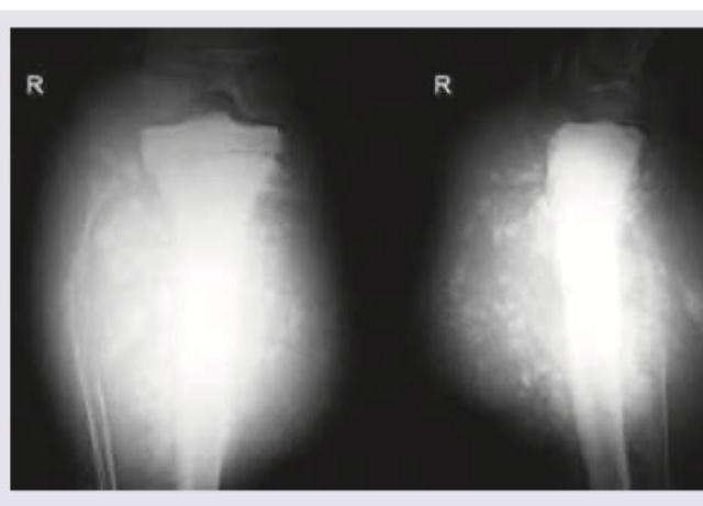
A 17-year-old boy presents with a progressively increasing swelling over the tibia along with fever. Radiological examination reveals a Codman triangle and sunburst appearance. What is the most likely diagnosis?
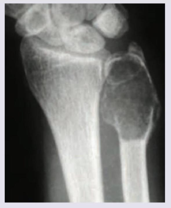
99. A 20-year-old male patient presents with a lesion in the wrist joint. The X-ray appearance is given below. What is the likely diagnosis?

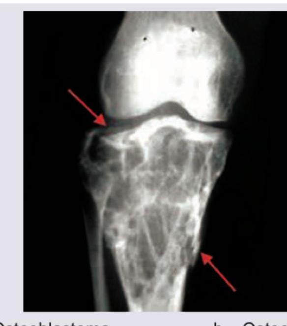
101. Identify the bone tumor:

96. Spot the diagnosis: (Recent NEET Pattern 2016-17)

97. Spot the diagnosis based on X-ray:

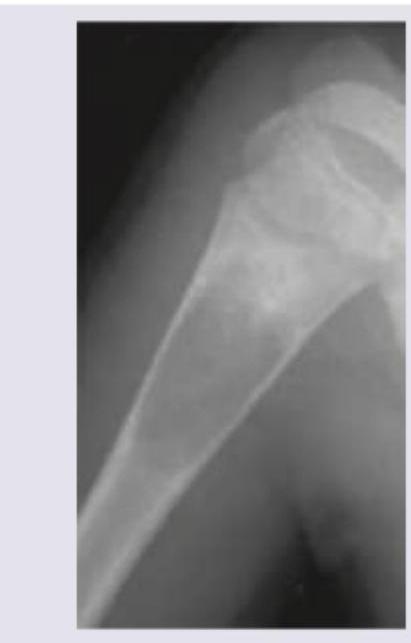
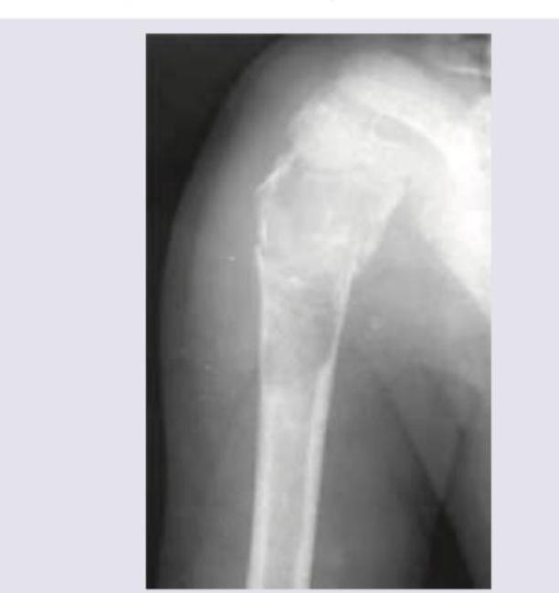
A 15-year-old boy presents with pain in the right upper arm and stiffness after playing cricket in the school. Since the complaints of the child were persisting, the family physician performed X-ray of right upper arm. The X-ray humerus shows presence of:

Spot the diagnosis based on the given X-ray of hand.

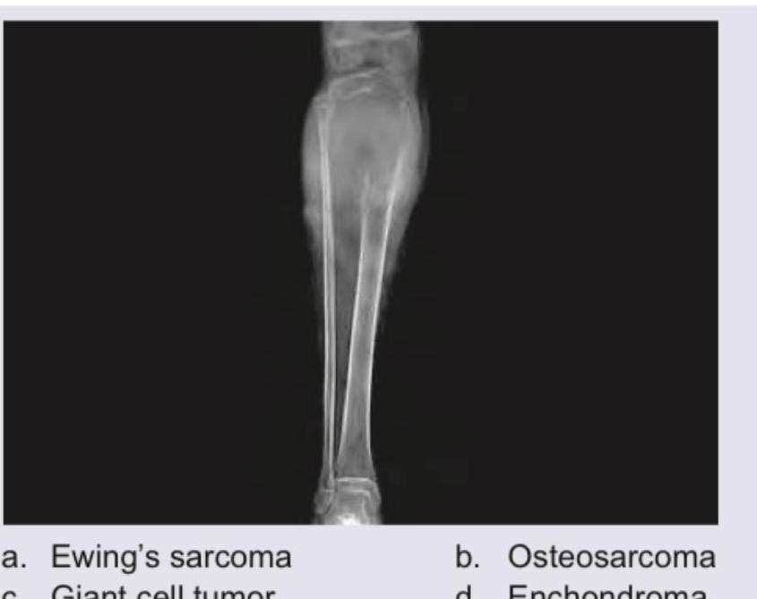
A 20-year-old college girl presents with pain in the upper part of tibia especially after dance classes. The pain has increased to a level that she cannot practice for forthcoming college festival. X-ray of the lower leg shows:

Comment on the diagnosis of the presentation shown below:

Comment on the test being performed in the patient.

Explanation: ***Osteosarcoma*** - This is the most common primary malignant bone tumor in adolescents, often presenting with pain and swelling, typically affecting the **metaphysis** of long bones (like the tibia). - The presence of the **Codman triangle** (periosteal elevation) and the **sunburst appearance** (spicules of bone radiating outwards) are pathognomonic radiological signs due to aggressive bone formation. *Ewing sarcoma* - While also affecting adolescents and associated with systemic features like **fever** (mimicking infection), its classic radiological sign is the **'onion-peel' (laminated) periosteal reaction**. - It typically involves the **diaphysis** of long bones or flat bones, unlike the metaphyseal involvement seen here. *Giant cell tumour* - This tumor usually affects slightly older adults (20-40 years) and predominantly involves the **epiphysis** of long bones. - Radiographically, GCT exhibits a non-sclerotic, **'soap bubble' appearance** (multiloculated lytic lesion) but does not feature the aggressive Codman triangle or sunburst pattern. *Chondrosarcoma* - This diagnosis is unlikely in a 17-year-old, as it typically presents in older adults (40-70 years). - The radiologic hallmark of chondrosarcoma is the presence of **ring-and-arc** or **'popcorn' calcifications** within the cartilaginous matrix, not the ossifying reactions seen in this case.
Explanation: ***Simple bone cyst*** - The X-ray image shows a **lucent, well-defined lesion** in the diaphysis/metaphysis of the ulna, often seen in the proximal humerus or femur, but can occur in other long bones. - The lesion has a **thin, sclerotic rim** without significant periosteal reaction or cortical destruction, suggesting a benign, fluid-filled lesion like a simple bone cyst. *Osteoclastoma* - This typically presents as an **eccentric, expansile lytic lesion** in the epiphysis or metaphysis of long bones, often with a "soap bubble" appearance. - It usually occurs near the **knee or distal radius** but the lesion in the image lacks the characteristic aggressive features and epiphyseal involvement of osteoclastoma. *Adamantinoma* - This is a rare, malignant bone tumor that almost exclusively occurs in the **tibia**. - It presents as a **lytic, often multifocal lesion** with central sclerosis or cystic changes, which does not match the appearance or location of the lesion in the ulna. *Multiple myeloma* - This is a systemic malignancy causing **punched-out lytic lesions** in the axial skeleton (skull, spine, pelvis) and proximal long bones. - The appearance in the image is of a single, well-defined cyst, not the widespread, multiple lucencies typical of multiple myeloma.
Explanation: *Ewing's sarcoma* - The image displays a **large lytic lesion** in the diaphysis/metaphysis of the tibia, with a **"onion skin" periosteal reaction** and possible soft tissue extension, which are characteristic radiological features of Ewing's sarcoma. - Ewing's sarcoma typically affects **children and young adults** and is known for its aggressive nature and diaphyseal involvement of long bones. *Osteosarcoma* - Osteosarcoma usually presents with a **sclerotic (bone-forming) lesion** and a **"sunburst" periosteal reaction** or Codman's triangle, not primarily a lytic lesion with onion-skinning. - While it also affects metaphyses of long bones and is common in adolescents, its radiological appearance often differs significantly from what is shown. *Giant cell tumor* - Giant cell tumors are typically **lytic lesions** found in the **epiphysis/metaphysis** of long bones, often described as a "soap bubble" appearance. - They do not typically show significant periosteal reaction or the widespread diaphyseal involvement seen in the image. *Enchondroma* - Enchondromas are **benign cartilaginous tumors** usually found in the medullary cavity of small bones of the hands and feet, or less commonly in long bones. - They appear as **well-circumscribed, lytic lesions** with chondroid matrix calcifications and do not typically cause aggressive periosteal reactions or extensive bone destruction.
Explanation: ***Aneurysmal bone cyst*** - The image shows an **expansile, lytic lesion** in the metaphysis of the tibia, with a **thin sclerotic rim** and **soap bubble appearance**, which is characteristic of an aneurysmal bone cyst. - The lesion extends close to the **epiphyseal plate** but generally respects it, and the presence of internal trabeculations often gives it a multiloculated appearance. *Osteoblastoma* - This lesion is typically a **well-circumscribed, lytic lesion** often associated with a **sclerotic rim**, but it usually presents as a nidus greater than 2 cm, frequently in the spine or long bones, and is less expansile than what is seen here. - While it can be destructive, the **multiloculated, expansile appearance** with distinct fluid levels (not directly visible on plain X-ray but implied by "soap bubble") is not typical for osteoblastoma. *Osteoclastoma* - Also known as **Giant Cell Tumor of Bone**, this tumor is typically an **epiphyseal lesion**, extending to the subarticular bone, often described as an **expansile osteolytic lesion** without a sclerotic rim. - The lesion in the image is more metaphyseal and shows a well-defined sclerotic border with internal septations, not the typical "soap bubble" appearance of ABC or the purely lytic destructive nature of an osteoclastoma. *Chondrosarcoma* - Chondrosarcomas are malignant tumors characterized by the formation of cartilage, often presenting as **lytic lesions with calcifications** (popcorn or ring-and-arc patterns) and cortical destruction, but typically lack the multiloculated, expansile, "soap bubble" appearance of an aneurysmal bone cyst. - While they can be expansile, the **radiographic features** in the image, particularly the internal architecture and clear margination, are not typical for chondrosarcoma.
Explanation: ***Osteoid osteoma*** - The X-ray image shows a small, well-defined **radiolucent nidus** (indicated by the arrow) surrounded by a significant amount of **sclerotic bone reaction**. This appearance is characteristic of an osteoid osteoma. - The nidus, which contains osteoid tissue, is typically less than 1.5 cm in diameter and is the source of the pain, often relieved by NSAIDs. *Osteochondroma/exostosis* - An osteochondroma is a **bony projection** covered with a cartilaginous cap that arises from the external surface of a bone, usually near a growth plate. - It would appear as a clearly defined, sessile or pedunculated outgrowth from the cortex, which is not what is seen here. *Ewing's sarcoma* - Ewing's sarcoma is an aggressive malignant tumor characterized by a **permeative or moth-eaten osteolytic pattern** and often an **"onion-skin" periosteal reaction**. - It does not typically present with a small, central nidus surrounded by dense sclerosis like an osteoid osteoma. *Osteosarcoma* - Osteosarcoma is also a highly malignant bone tumor, often characterized by **sclerotic and lytic areas**, a **Codman triangle**, or a **sunburst appearance** due to aggressive periosteal reaction. - The image does not show these features; instead, it presents with a focal nidus and organized sclerosis, typical of a benign process.
Explanation: ***Unicameral bone cyst*** - The X-ray image shows a **lucent lesion** in the metaphysis of the humerus, consistent with a **unicameral bone cyst (UBC)**. These cysts are common in children and adolescents, typically located in the proximal humerus or femur, and can present with pain after minor trauma due to **pathological fracture**. - The age of the patient (15-year-old boy), location (right upper arm/humerus), and presentation (pain after playing cricket indicating a potential pathological fracture) all align with the characteristics of a UBC. *Aneurysmal bone cyst* - While aneurysmal bone cysts (ABCs) can occur in similar age groups and locations, they typically present as **multiloculated, expansile, eccentric lesions** with thin bony septa, often described as a "blown-out" appearance. - The image provided shows a more uniform lytic lesion without prominent septations or extensive cortical expansion, making ABC less likely. *Osteoblastoma* - **Osteoblastoma** is a benign bone tumor that often presents with localized pain, but it is typically a **sclerotic or mixed lytic-sclerotic lesion**, sometimes with a nidus, and not a purely lucent, cystic lesion like the one seen. - It most commonly affects the spine and long bones, but its radiographic appearance would differ significantly from the image. *Osteoclastoma* - **Osteoclastoma**, also known as **giant cell tumor of bone**, typically occurs in individuals in their 20s to 40s, often at the **epiphysis** of long bones (e.g., distal femur, proximal tibia, distal radius) after epiphyseal plate closure. - While it is a lytic lesion, its characteristic location and patient age range make it a less likely diagnosis for a 15-year-old with a metaphyseal lesion in the humerus.
Explanation: ***Enchondroma*** - The X-ray image shows a **lucent, well-defined lesion** within the shaft of the 5th metacarpal bone, accompanied by some **cortical thinning** but largely preserved cortex. This appearance, particularly in the small bones of the hand, is classic for an enchondroma. - Enchondromas are benign cartilaginous tumors, often found incidentally, and their radiographic features include a **radiolucent geographic lesion** with occasional internal calcifications, though not clearly visible here. *Osteoid osteoma* - An osteoid osteoma typically presents as a small **lucid nidus less than 1.5 cm** surrounded by a prominent rim of **sclerotic bone**. - While it can occur in the hand, the lesion in the image is larger and lacks the characteristic central nidus with reactive sclerosis. *Simple bone cysts* - Simple bone cysts (also known as unicameral bone cysts) are typically **lucent, centrally located lesions** often found in the **metaphysis of long bones**. - They tend to be large and usually lack internal calcifications or the characteristic lobulated appearance seen in this lesion. *Chloroma* - A chloroma (also known as granulocytic sarcoma) is a solid tumor of **myeloid blasts**, typically associated with leukemia. - On X-ray, they usually present as **lytic bone lesions** that can cause bone destruction and soft tissue involvement, which is not consistent with the well-defined, lytic but contained lesion shown.
Explanation: ***Osteoblastoma*** - On X-ray, osteoblastomas commonly appear as a **lytic lesion** with a **sclerotic rim**, often larger than 2 cm, which can be seen in the provided image. The pain in the **tibia** that worsens with activity and affects a young individual is also consistent with the presentation of osteoblastoma. - This benign bone tumor is known to cause significant localized pain and can be found in the **metaphysis or diaphysis** of long bones like the tibia. *Osteosarcoma* - Osteosarcoma is a **highly aggressive malignant bone tumor** that typically presents with a **"sunburst" or "Codman's triangle"** appearance and areas of amorphous calcification (osteoid matrix) on X-ray, which are not clearly evidenced here. - While it can occur in young adults and cause pain, the radiographic findings are more indicative of a benign lytic lesion. *Chondrosarcoma* - Chondrosarcoma is a **malignant cartilaginous tumor** often characterized by **"rings and arcs"** or **popcorn-like calcifications** within a lytic lesion on X-ray, typical of cartilage matrix. - While it can cause pain, its radiographic features are distinct from those observed. *Pigmented villonodular synovitis* - This is a **benign proliferative disorder of the synovium** affecting joints or bursae, not primarily the bone itself. - It would typically demonstrate **erosions** and **soft tissue masses** within a joint on imaging, not a lytic bone lesion as shown.
Explanation: ***Unicameral bone cyst*** - This X-ray shows a **well-defined, lytic lesion** in the proximal humerus, consistent with a unicameral bone cyst, often presenting in the **metaphysis** of long bones in children and adolescents. - The lesion has a **thin, sclerotic rim** and shows **no significant periosteal reaction** or soft tissue mass, which are classic features. *Aneurysmal bone cyst* - While also a lytic lesion, an aneurysmal bone cyst typically appears more **expansile** and **multiloculated**, often with a **"soap bubble" appearance**. - It usually has a **thicker, more irregular rim** and can present with faster growth or more aggressive features than a unicameral bone cyst. *Osteoblastoma* - Osteoblastomas are typically **osteoblastic (bone-forming)** tumors, which would appear more **sclerotic** or mixed lytic-sclerotic on X-ray, rather than a purely lytic lesion like the one shown. - They commonly affect the **spine or long bones**, but their radiographic appearance of predominant bone formation is not seen here. *Osteoclastoma* - Osteoclastomas, also known as **giant cell tumors of bone**, are usually found in the **epiphysis** of long bones in mature skeletons, which is a different location than observed. - They are typically **aggressive lytic lesions** with poorly defined margins and can extend to the articular surface, which is not characteristic of the well-demarcated lesion in the image.
Explanation: ***Hoover sign*** - The image depicts the Hoover sign, which tests for **non-organic weakness** in a lower limb. - The examiner asks the patient to lift one leg while feeling for contralateral heel pressure; absence of pressure indicates an inconsistent effort. *McMurray* - The McMurray test evaluates for a **meniscal tear** in the knee. - It involves flexing and rotating the knee while extending it to elicit a click or pain. *Apley's grinding test* - Apley's grinding test is used to assess for **meniscal or ligamentous injury** in the knee. - The patient lies prone, and the examiner applies downward pressure with rotation to the flexed knee. *Gordon sign* - The Gordon sign is a pathological reflex used to identify **upper motor neuron lesions**. - It involves squeezing the calf muscle, which results in **dorsiflexion of the great toe** and fanning of the other toes, similar to a Babinski sign.
Classification of Bone Tumors
Practice Questions
Benign Bone Tumors
Practice Questions
Malignant Primary Bone Tumors
Practice Questions
Metastatic Bone Disease
Practice Questions
Tumor-Like Lesions of Bone
Practice Questions
Soft Tissue Tumors
Practice Questions
Evaluation and Staging of Bone Tumors
Practice Questions
Biopsy Principles
Practice Questions
Limb Salvage Surgery
Practice Questions
Amputation for Bone Tumors
Practice Questions
Adjuvant Therapies
Practice Questions
Surveillance and Follow-up
Practice Questions
Get full access to all questions, explanations, and performance tracking.
Start For Free