
Bone Tumors — MCQs

On this page
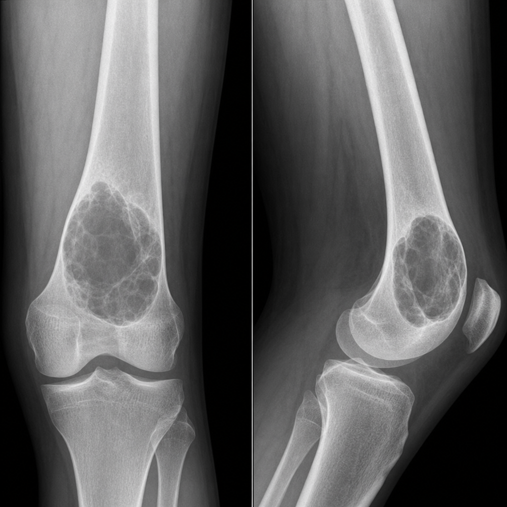
The most probable diagnosis of the lesion depicted in the X-ray is?
Which of the following conditions is least likely to present as an eccentric osteolytic lesion?
What is the most appropriate treatment for a soap bubble appearance at the lower end of the radius?
Pediatric patient with an upper humerus lytic lesion and cortical thinning, which among the following is not a treatment modality?
Most common site of Ewing's sarcoma?
Expansile lytic lesion with fluid-fluid levels involving the proximal metaphysis of fibula in an early adolescent female is typical of?
Practice by Chapter
Classification of Bone Tumors
Practice Questions
Benign Bone Tumors
Practice Questions
Malignant Primary Bone Tumors
Practice Questions
Metastatic Bone Disease
Practice Questions
Tumor-Like Lesions of Bone
Practice Questions
Soft Tissue Tumors
Practice Questions
Evaluation and Staging of Bone Tumors
Practice Questions
Biopsy Principles
Practice Questions
Limb Salvage Surgery
Practice Questions
Amputation for Bone Tumors
Practice Questions
Adjuvant Therapies
Practice Questions
Surveillance and Follow-up
Practice Questions
Want unlimited practice?
Get full access to all questions, explanations, and performance tracking.
Scan to download app