
Bone Tumors — MCQs

On this page
Expansile pulsating secondary metastasis is a feature of which of the following?
A 22-year-old male presents with pain and swelling over the distal radius. X-ray shows an expansile lytic lesion in the metaphysis. Fine needle aspiration (FNA) reveals a bloody aspirate with hemosiderin-laden macrophages. What is the most likely diagnosis?
A 20-year-old male patient presented with localized pain, which is gradual in onset and worsened over time. X-ray showed the following finding. What is the diagnosis?
A 20-year-old patient presents with painful swelling in the middle finger around the proximal phalanx. An X-ray was done, and it is shown below. What is the most likely diagnosis? 
A 17-year-old boy presents to the clinic complaining of a painless lump on the lateral aspect of his left knee. The radiograph of the patient is shown below. Which of the following is the most likely diagnosis?
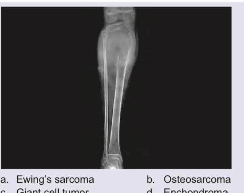
A 17-year-old boy presents with a progressively increasing swelling over the tibia along with fever. Radiological examination reveals a Codman triangle and sunburst appearance. What is the most likely diagnosis?
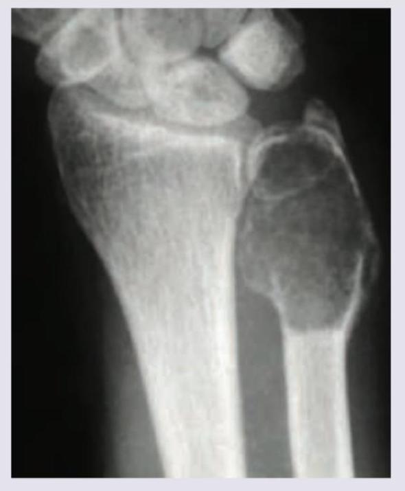
99. A 20-year-old male patient presents with a lesion in the wrist joint. The X-ray appearance is given below. What is the likely diagnosis?
101. Identify the bone tumor:
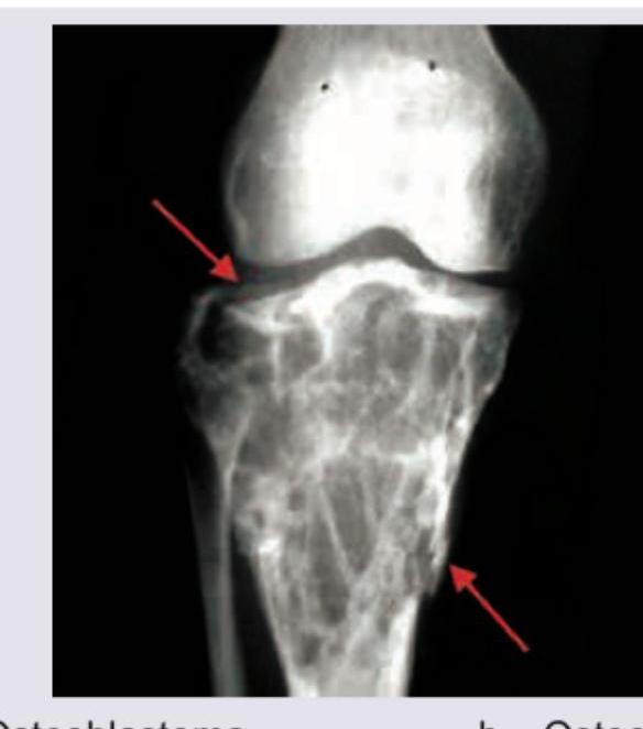
A 16-year-old boy presents with pain and swelling around the knee for 3 months. X-ray of the affected region is shown below. What is the most likely diagnosis?
96. Spot the diagnosis: (Recent NEET Pattern 2016-17)
Practice by Chapter
Classification of Bone Tumors
Practice Questions
Benign Bone Tumors
Practice Questions
Malignant Primary Bone Tumors
Practice Questions
Metastatic Bone Disease
Practice Questions
Tumor-Like Lesions of Bone
Practice Questions
Soft Tissue Tumors
Practice Questions
Evaluation and Staging of Bone Tumors
Practice Questions
Biopsy Principles
Practice Questions
Limb Salvage Surgery
Practice Questions
Amputation for Bone Tumors
Practice Questions
Adjuvant Therapies
Practice Questions
Surveillance and Follow-up
Practice Questions
Want unlimited practice?
Get full access to all questions, explanations, and performance tracking.
Scan to download app