A patient presents with pain in the thigh, relieved by aspirin. X-ray shows a radiolucent mass surrounded by sclerosis. Diagnosis is ?
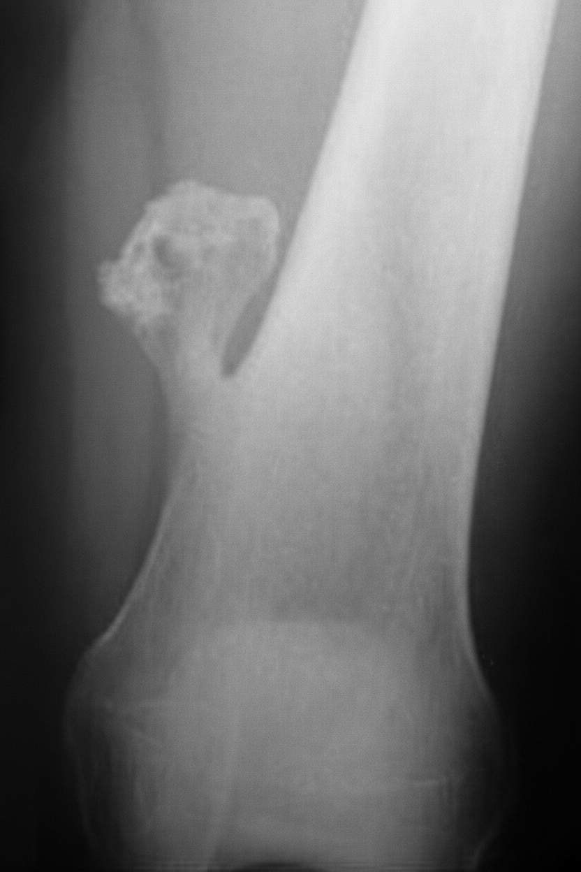
Which of the following is an epiphyseal tumor?
Which bone tumor involves the epiphysis?
Characteristic radiological feature of fibrous dysplasia is:
Which of the following is a bone-forming malignant tumor?
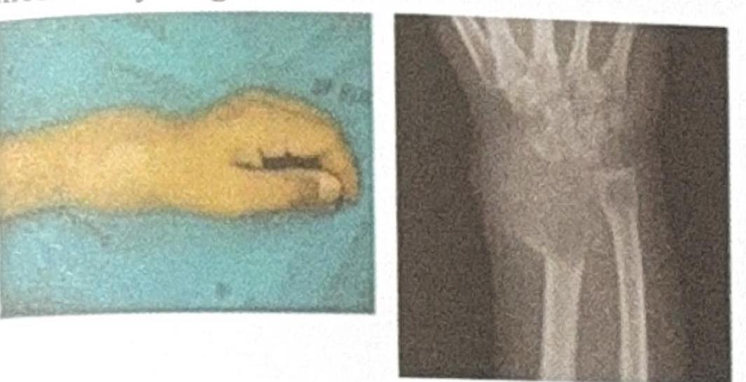
The image shows a wrist deformity and an X-ray of a bone lesion near the distal radius. Based on the clinical and radiological features, what is the most likely diagnosis?

A patient with GCT, which of the following is false?
Most common benign tumor of bone?
Most common site of adamantinoma of the long bones is -
Which of the following is an epiphyseal lesion?
Explanation: ***Osteoid osteoma*** - Characterized by **pain that is classically relieved by aspirin or NSAIDs**, due to high prostaglandin production within the lesion. - Radiographically, it appears as a **small radiolucent nidus (lesion) less than 2 cm**, surrounded by a dense sclerotic bone reaction. *Osteoma* - This is a **benign tumor of mature bone**, most commonly found on the skull and facial bones. - It typically presents as a **hard, solitary, immobile mass** and is usually asymptomatic, not causing pain relieved by aspirin. *Osteoclastoma* - Also known as a **giant cell tumor of bone**, it is a locally aggressive tumor. - It typically affects the **epiphysis and metaphysis of long bones** (e.g., around the knee) and is not characteristically relieved by aspirin. *Osteoblastoma* - This is a **larger variant of osteoid osteoma** (>2 cm), also bone-forming, but the pain is generally less responsive to aspirin, and it tends to be more aggressive. - It is more commonly found in the **spine and sacrum**, unlike the thigh in this case.
Explanation: ***Chondroblastoma*** - **Chondroblastoma** is a rare, benign cartilaginous tumor that typically originates in the **epiphysis** of long bones before the closure of growth plates. - It is histologically characterized by chondroblast-like cells, multinucleated giant cells, and chondroid matrix, and radiographically appears as a lytic lesion with a sclerotic rim in the epiphysis. *Osteosarcoma* - **Osteosarcoma** is the most common primary malignant bone tumor and typically originates in the **metaphysis** of long bones, particularly around the knee. - It invades the surrounding bone and soft tissues, often presenting with a **Codman triangle** or **sunburst pattern** on imaging studies. *Osteoid osteoma* - **Osteoid osteoma** is a benign bone-forming tumor primarily found in the **cortex** of long bones, although it can occur in other locations, presenting with nocturnal pain relieved by NSAIDs. - It is characterized by a central radiolucent nidus surrounded by reactive sclerotic bone. *Adamantinoma* - **Adamantinoma** is an extremely rare, low-grade malignant tumor that almost exclusively occurs in the **tibia diaphysis**. - It is thought to originate from epithelial cells and presents as a lytic lesion within the cortical bone, often with a polycystic appearance.
Explanation: ***Giant cell tumor*** - **Giant cell tumor (GCT)**, also known as osteoclastoma, characteristically arises in the **epiphysis** [1] or **metaphysis** of long bones in adults. - It is a benign but locally aggressive tumor that often presents with pain, swelling, and reduced range of motion in the affected joint [1]. *Osteosarcoma* - **Osteosarcoma** typically arises in the **metaphysis** of long bones, particularly around the knee (distal femur, proximal tibia). - It is a highly malignant primary bone tumor characterized by the production of **osteoid** by tumor cells [2]. *Ewing's sarcoma* - **Ewing's sarcoma** most commonly affects the **diaphysis** of long bones or flat bones (e.g., pelvis, scapula, ribs). - It is characterized by small, round, blue cells and often presents with pain, swelling, and systemic symptoms like fever. *Multiple myeloma* - **Multiple myeloma** is a malignancy of **plasma cells** that primarily affects the **bone marrow** and can cause widespread osteolytic lesions. - It typically presents in older adults and affects bones with active marrow, such as the vertebrae, ribs, skull, and pelvis, rather than being localized to the epiphysis as a primary bone tumor. **References:** [1] Kumar V, Abbas AK, et al.. Robbins and Cotran Pathologic Basis of Disease. 9th ed. Bones, Joints, and Soft Tissue Tumors, pp. 1205-1206. [2] Cross SS. Underwood's Pathology: A Clinical Approach. 6th ed. Common Clinical Problems From Osteoarticular And Connective Tissue Disease, pp. 673-674.
Explanation: ***Ground glass appearance*** - The "ground glass" appearance on **radiography** is a classic and highly characteristic feature of **fibrous dysplasia**, reflecting the immature, woven bone and fibrous tissue within the lesion. - This specific pattern results from the uniform, finely trabeculated osseous tissue that is less dense than normal bone, creating a hazy, frosted appearance. *Cortical erosion* - While significant **tumor growth** in any bone lesion can lead to cortical erosion, it is not a specific or characteristic primary feature of **fibrous dysplasia**. - **Cortical erosion** is more commonly associated with aggressive or rapidly expanding lesions, such as malignant tumors or infections. *Bone enlargement* - **Bone enlargement** can occur in fibrous dysplasia due to the expansion of the abnormal fibrous and osseous tissue, but it is a non-specific finding. - Many conditions, including various **benign** and **malignant tumors**, can cause bone enlargement, making it less diagnostic on its own. *Thickened bone matrix* - Fibrous dysplasia involves a disorganized and immature bone matrix, characterized by **woven bone**, not a thickened, well-organized normal bone matrix. - **Thickened bone matrix** may be seen in conditions like osteopetrosis or Paget's disease, which involve different pathological bone remodeling processes.
Explanation: ***Osteosarcoma*** - **Osteosarcoma** is the most common **primary malignant bone tumor**, characterized by the production of **osteoid** (immature bone) by malignant osteoblasts [1], [2]. - It typically arises in the **metaphysis of long bones** (e.g., distal femur, proximal tibia, proximal humerus) and often presents with pain and swelling [1], [2]. *Osteoid osteoma* - **Osteoid osteoma** is a benign bone tumor characterized by a small nidus of **osteoid-forming tissue** surrounded by sclerotic bone. - While it involves osteoid formation, it is **not malignant** and does not metastasize. *Chondroblastoma* - **Chondroblastoma** is a rare, **benign cartilage-forming tumor** that typically occurs in the **epiphysis of long bones** in skeletally immature individuals. - It is composed of chondroblast-like cells and does not produce osteoid or show malignant features. *Giant cell tumor* - **Giant cell tumor** of bone is an **aggressive, locally destructive benign tumor** characterized by numerous osteoclast-like giant cells and spindle-shaped stromal cells. - It arises in the **epiphysis/metaphysis** of long bones (e.g., distal femur, proximal tibia) but is not a bone-forming tumor and is typically not malignant, though it has a high recurrence rate. **References:** [1] Cross SS. Underwood's Pathology: A Clinical Approach. 6th ed. Common Clinical Problems From Osteoarticular And Connective Tissue Disease, pp. 671-674. [2] Kumar V, Abbas AK, et al.. Robbins and Cotran Pathologic Basis of Disease. 9th ed. Bones, Joints, and Soft Tissue Tumors, pp. 1200-1202.
Explanation: ***Giant Cell Tumor (GCT)*** * The image shows a **lytic (lucid) lesion** located in the **epiphysis/metaphysis of the distal radius**, which is a classic presentation site for GCT. * GCTs are typically seen in individuals aged 20-40, often present with **pain, swelling, and reduced range of motion**, and can show a **soap bubble appearance** on X-ray even with cortical erosion as seen in the image. *Ewing's Sarcoma* * Ewing's Sarcoma commonly affects the **diaphysis of long bones** and may present with an **onion skin periosteal reaction**, none of which are clearly depicted. * It primarily affects children and young adults (5-20 years old), which does not align with the assumed adult presentation given the fused epiphysis. *Osteochondroma* * Osteochondromas are **bony prominences covered by cartilage** and grow **outward from the bone surface**, often away from the joint, unlike the intraosseous lytic lesion seen. * They typically appear as **pedunculated or sessile exostoses** and are benign growth plate abnormalities, not lytic lesions of the marrow cavity. *Osteoid Osteoma* * Osteoid osteomas are characterized by a **small lucent nidus** (usually <1.5 cm) surrounded by a significant margin of **sclerotic bone**, which is not seen here. * They classically cause **nocturnal pain** relieved by NSAIDs and are typically smaller than the lesion depicted, which appears expansile.
Explanation: ***Chemotherapy is the mainstay of treatment*** - This statement is **false** because **Giant Cell Tumor of Bone (GCT)** therapy primarily involves **surgical resection**, with or without adjuvant therapies like **denosumab**. - **Chemotherapy** is generally *not* the first-line treatment for GCT, as these tumors respond poorly to it; it's usually reserved for cases of **metastatic GCT** or when other treatments fail. *Defined margins* - GCTs often present radiographically with **well-defined, non-sclerotic margins**, which indicates a lytic lesion that is often locally aggressive but typically doesn't invade widely. - While they are locally destructive, their borders are usually visible, helping distinguish them from other bone tumors. *Epiphyseo-metaphyseal location* - GCTs commonly originate in the **metaphysis** of long bones and **extend into the epiphysis** after the growth plate has closed. - This characteristic location near a joint is a classic diagnostic feature of GCT, especially in adults. *Eccentric* - GCTs typically arise **eccentrically** within the bone, meaning they originate off-center in the bone marrow cavity before expanding and thinning the cortex. - This eccentric growth pattern is a distinguishing feature, particularly in contrast to other bone tumors which might be centrally located.
Explanation: ***Osteochondroma*** - This is the **most common benign bone tumor**, characterized by a bony spur with a cartilaginous cap. - It typically arises from the **metaphysis of long bones**, especially around the knee. *Osteoma* - Osteomas are **benign, slow-growing tumors** composed of mature compact or cancellous bone. - They are most commonly found in the **skull and facial bones**, not typically in long bones. *Simple bone cyst* - This is a **fluid-filled lesion** of bone, not a true neoplasm, frequently found in the metaphysis of long bones in children. - It is often discovered incidentally or after a **pathological fracture**. *Osteoid osteoma* - Characterized by a **small, benign bone tumor** with a central nidus of osteoid and trabecular bone, surrounded by reactive sclerotic bone. - It classically causes **nocturnal pain** that is relieved by NSAIDs.
Explanation: ***Tibia*** - Adamantinoma is a rare, malignant bone tumor that has a strong predilection for the **tibia**, accounting for approximately 85-90% of cases in long bones. - It most commonly occurs in the **diaphysis of the tibia** but can also be found in the metaphysis. *Ulna* - While adamantinoma can rarely affect other long bones, the **ulna is not a common site** for its occurrence. - The incidence of adamantinoma in the upper extremities, including the ulna, is significantly lower compared to the tibia. *Fibula* - The **fibula is an uncommon site** for adamantinoma, with only a small percentage of cases reported in this bone. - When it does occur in the fibula, it usually involves the mid-diaphysis. *Femur* - The **femur is also a rare location** for adamantinoma, with only a handful of cases documented in medical literature. - The vast majority of adamantinomas consistently originate in the lower leg, particularly the tibia.
Explanation: **Chondroblastoma** - **Chondroblastoma** is a rare, benign bone tumor that typically arises in the **epiphysis** of long bones before epiphyseal fusion. - It specifically originates from **chondroblasts** within the epiphyseal growth plate region. *Fibrosarcoma* - **Fibrosarcoma** is a malignant tumor of fibrous connective tissue origin, typically found in the **metaphysis** or **diaphysis** of long bones. - It rarely affects the **epiphyseal** region and is characterized by aggressive local invasion and metastases. *Chondrosarcoma* - **Chondrosarcoma** is a malignant tumor of cartilage, commonly arising in the **metaphysis** or **diaphysis** of long bones, particularly the femur, humerus, and pelvis. - While it involves cartilage, its typical location is not primarily **epiphyseal** and it is characterized by malignant cartilaginous matrix. *Non-ossifying fibroma* - A **non-ossifying fibroma** (NOF), also known as a fibrous cortical defect, is a common benign fibrous lesion typically found in the **metaphysis** of long bones. - These lesions are usually asymptomatic and self-limiting, often resolving spontaneously, and do not originate in the **epiphysis**.
Get full access to all questions, explanations, and performance tracking.
Start For Free