
Adjuvant Therapies — MCQs

Which statement is incorrect about the pathology of the bone tumor?
Classification system of bone tumors is -
A patient with GCT, which of the following is false?
Most radiosensitive tumor among the following is
Which of the following tumours is radiosensitive?
What is the most appropriate treatment for a soap bubble appearance at the lower end of the radius?
A 28-year-old female presents to the clinic with a complaint of dull, aching pain in her left hand after accidentally striking her finger against a door frame. On physical examination, there is mild tenderness over the affected area, but the range of m otion is preserved. An X -ray of the hand is shown below. Based on the clinical presentation and radiographic findings, what is the most likely diagnosis?
A 9-year-old child presents with pain and swelling over the mid -shaft of the tibia, associated with low -grade fever and an elevated erythrocyte sedimentation rate (ESR). An X-ray of the affected limb is shown below. Based on the clinical features and radiographic findings, what is the most likely diagnosis?
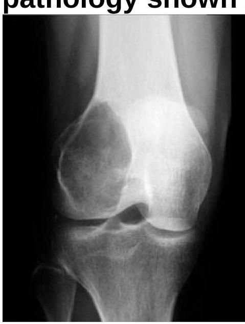
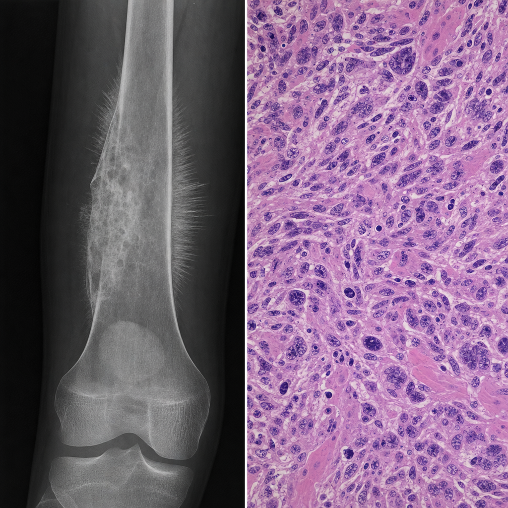
A 16-year-old boy presents with a 3-month history of progressively worsening pain and swelling around the distal right femur. Plain radiograph shows a permeative lytic lesion with Codman's triangle and a sunburst periosteal reaction. A core needle biopsy is performed and the histopathological specimen is shown in Image 1. Following staging workup, imaging confirms the lesion is confined to the bone with no skip lesions and no distant metastases. What is the most appropriate next step in management?
In the treatment of osteosarcoma, all of the following chemotherapy agents are used EXCEPT:
Want unlimited practice?
Get full access to all questions, explanations, and performance tracking.
Scan to download app