What is the preferred site for a first-order bone graft?
Which site is the most common source for autologous bone grafts?
Clamshell technique is required for which type of bone graft?
Cancellous bone graft can be taken from which of the following anatomical locations?
What is the standard site for a primary bone graft?
Which of the following bone grafts possesses osteogenic properties?
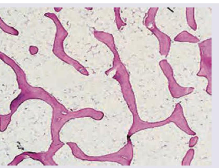
All are correct about the bone tissue shown except:

All are correct about the X-ray film shown below except:

Which of the following bone defects offers the best chance for bone fill?
The graft with the maximum osteogenic potential is:
Explanation: The **Pelvis (specifically the Iliac Crest)** is the gold standard and preferred site for a first-order bone graft because it provides the highest concentration of **cancellous bone**. Cancellous bone is superior for grafting because it is highly vascularized and contains a large population of osteoblasts and mesenchymal stem cells, facilitating rapid **osteogenesis, osteoconduction, and osteoinduction**. ### Evaluation of Options: * **A. Pelvis (Correct):** The iliac crest (anterior or posterior) is the most common site for autologous bone grafting. It offers a large volume of high-quality marrow and trabecular bone with minimal structural morbidity. * **B. Tibial metaphysis:** While used for smaller grafts (e.g., in foot and ankle surgery), it provides a significantly lower volume of cancellous bone compared to the pelvis. * **C. Medial malleolus:** This is a site for very small, localized bone grafts. It lacks the volume and cellular density required for major reconstructive procedures. * **D. Femoral condyle:** Though it contains cancellous bone, the surgical morbidity of entering a major weight-bearing joint makes it a secondary choice compared to the iliac crest. ### High-Yield Clinical Pearls for NEET-PG: * **Gold Standard:** Autologous bone graft (Autograft) remains the gold standard because it possesses all three properties: **Osteogenesis** (living cells), **Osteoinduction** (growth factors like BMP), and **Osteoconduction** (scaffold). * **Posterior vs. Anterior Iliac Crest:** The posterior iliac crest yields a higher volume of bone graft than the anterior crest. * **Cortical vs. Cancellous:** Cortical grafts (e.g., Fibula) provide immediate **structural support** but have poor osteogenic potential. Cancellous grafts (e.g., Pelvis) provide **rapid revascularization** and healing but no structural strength. * **Creeping Substitution:** The process by which the host replaces the grafted bone with new living bone.
Explanation: **Explanation:** The **Iliac Crest** is the "gold standard" and most common donor site for autologous bone grafting. This is because it provides a rich supply of both **corticocancellous bone** and osteoprogenitor cells. The posterior iliac crest is preferred when a large volume of bone is needed, while the anterior crest is used for smaller grafts due to easier surgical access. **Why the other options are incorrect:** * **Tibial tuberosity:** While it can be used for small amounts of cancellous bone (e.g., in foot and ankle surgeries), the volume available is significantly less than the ilium, and there is a risk of weakening the extensor mechanism. * **Greater trochanter:** This site provides a limited amount of cancellous bone. It is rarely the primary choice unless the surgery is already being performed in the hip region. * **Fibula:** The fibula is the most common source for **vascularized cortical bone grafts**. It is used for structural support (e.g., bridging large bony defects) rather than as a general source of cancellous bone. **High-Yield Clinical Pearls for NEET-PG:** * **Osteogenesis, Osteoinduction, and Osteoconduction:** Autografts are unique because they possess all three properties. * **Complications:** The most common complication of iliac crest harvesting is **chronic donor site pain**. Injury to the lateral femoral cutaneous nerve (Meralgia paresthetica) can occur during anterior harvesting. * **Bone Morphogenetic Proteins (BMPs):** These are the most potent osteoinductive growth factors found within the bone matrix.
Explanation: **Explanation:** The **Clamshell technique** (also known as the Trapdoor or Tessier technique) is a specific surgical approach used to harvest large amounts of **ilio-cancellous bone graft** from the iliac crest while preserving the overall shape and structural integrity of the pelvis. **1. Why the Correct Answer is Right:** In this technique, the iliac crest is split longitudinally along its superior border. The inner and outer tables of the ilium are then pried apart like a "clamshell" or "trapdoor." This provides direct access to the rich **cancellous bone** (medulla) located between the cortical plates. After the cancellous bone is scooped out, the cortical plates are repositioned and sutured back together. This minimizes donor site morbidity and maintains the aesthetic contour of the hip. **2. Why Other Options are Wrong:** * **Ilio-cortical graft:** These grafts involve taking the full thickness of the bone (cortex). The clamshell technique specifically aims to *spare* the cortex while harvesting the marrow. * **Posterior/Anterior iliac graft:** These refer to the *anatomical site* of harvest rather than the *technique* used to extract the bone. While the clamshell technique can be performed on either the anterior or posterior crest, it specifically describes the method of accessing cancellous bone, not the location. **3. High-Yield Clinical Pearls for NEET-PG:** * **Gold Standard:** Autologous cancellous bone graft is the gold standard for promoting osteogenesis, osteoinduction, and osteoconduction. * **Donor Site:** The **Posterior Iliac Crest** is the richest source of cancellous bone in the body (provides more volume than the anterior crest). * **Complication:** The most common complication of iliac crest harvest is **chronic donor site pain** and injury to the **lateral femoral cutaneous nerve** (in anterior approaches). * **Creeping Substitution:** This is the process by which the host bone replaces the graft with new bone.
Explanation: **Explanation:** Cancellous bone grafts (also known as "spongy" or "trabecular" bone) are highly valued in orthopaedic surgery for their superior **osteogenic** and **osteoinductive** properties. Unlike cortical bone, which provides structural stability, cancellous bone has a large surface area and contains viable osteoblasts and growth factors that facilitate rapid revascularization and incorporation (creeping substitution). **Anatomical Sources:** Cancellous bone is primarily harvested from the metaphyses of long bones and flat bones where the marrow space is abundant. * **Pelvis (Iliac Crest):** This is the **gold standard** and most common site for harvesting autologous cancellous bone. Both the anterior and posterior iliac crests provide large volumes of high-quality graft. * **Greater Trochanter:** A common local site used during hip surgeries. It contains a significant amount of cancellous bone accessible through a simple cortical window. * **Femoral Condyles:** The distal femur (metaphyseal region) is a rich source often utilized when the primary surgical site is around the knee. **Why "All of the above" is correct:** All three listed sites—the pelvis, greater trochanter, and femoral condyles—are classic anatomical reservoirs of cancellous bone. Other common sites include the proximal tibia (Gerdy’s tubercle) and the distal radius. **High-Yield Clinical Pearls for NEET-PG:** * **Creeping Substitution:** The process by which the host replaces the grafted bone with new bone. This is faster in cancellous grafts compared to cortical grafts. * **Cortical Bone Graft:** Best for **structural support** (e.g., Fibula). * **Cancellous Bone Graft:** Best for **osteogenesis** and filling voids. * **Most common complication of Iliac Crest harvest:** Chronic donor site pain and injury to the lateral femoral cutaneous nerve (Meralgia Paraesthetica).
Explanation: ### Explanation The **lateral malleolus** is considered the standard site for a primary bone graft, specifically when a **cortical bone graft** is required. In orthopaedic surgery, the distal fibula (lateral malleolus) is frequently utilized because it provides a strong, straight piece of cortical bone that can be harvested with minimal donor site morbidity, as the fibula is a non-weight-bearing bone in its mid and proximal portions. #### Analysis of Options: * **D. Lateral Malleolus (Correct):** It is the classic donor site for cortical grafts (e.g., for bridging defects or providing structural stability). The distal portion (malleolus) is a landmark, though the graft is typically harvested from the shaft just above it. * **A. Pelvis:** While the **Iliac Crest** (part of the pelvis) is the "Gold Standard" for **cancellous** bone grafts due to its high osteogenic potential, the question asks for the "standard site" in a general context where the lateral malleolus/fibula is the traditional answer for structural cortical needs in classic textbooks. * **B. Greater Trochanter:** This site is occasionally used for small amounts of local cancellous bone during hip surgeries, but it is not a "standard" primary donor site for general grafting. * **C. Medial Malleolus:** This is a critical weight-bearing component of the ankle joint. Harvesting bone from here would compromise joint stability and is avoided. #### NEET-PG High-Yield Pearls: * **Gold Standard for Cancellous Graft:** Iliac Crest (highest concentration of osteoblasts and growth factors). * **Standard for Cortical Graft:** Fibula (Lateral malleolus area). * **Creeping Substitution:** The process by which the host bone replaces the necrotic bone of the graft with new viable bone. * **Osteoconduction:** The graft acts as a scaffold (all grafts). * **Osteoinduction:** The graft stimulates stem cells to differentiate into osteoblasts (BMPs). * **Osteogenesis:** The graft provides living cells that directly form bone (only fresh autografts).
Explanation: To understand bone grafting, it is essential to distinguish between the three fundamental properties of graft materials: **Osteogenesis** (contains living cells that form bone), **Osteoinduction** (contains growth factors like BMPs that recruit stem cells), and **Osteoconduction** (provides a physical scaffold for bone growth). ### **Why Bone Marrow Aspirate (BMA) is Correct** **Bone Marrow Aspirate** is the only option listed that possesses **Osteogenic** properties. It contains viable **Mesenchymal Stem Cells (MSCs)** and osteoprogenitor cells that can directly differentiate into osteoblasts to produce new bone. While it lacks a structural scaffold (not osteoconductive), it is a potent source of living cells and growth factors. ### **Analysis of Incorrect Options** * **A & B. Calcium Sulfate and Calcium Triphosphate:** These are synthetic bone substitutes. They are purely **Osteoconductive**. They provide a mineral framework for native bone to grow into but contain no living cells or inductive proteins. * **C. Demineralized Bone Matrix (DBM):** DBM is prepared by removing the mineral content of allograft bone, leaving behind Type I collagen and non-collagenous proteins, including **Bone Morphogenetic Proteins (BMPs)**. Therefore, DBM is primarily **Osteoinductive** and weakly osteoconductive, but it is not osteogenic because the processing kills all living cells. ### **High-Yield NEET-PG Pearls** * **The "Gold Standard":** Autologous Bone Graft (Autograft) is the only material that is **Osteogenic, Osteoinductive, and Osteoconductive.** * **Iliac Crest:** The most common site for harvesting autograft. * **Creeping Substitution:** The process by which the host replaces the grafted bone with new viable bone. * **BMP-2 and BMP-7:** The specific growth factors responsible for the osteoinductive property of DBM and autografts.
Explanation: ***Provides tensile strength*** - Compact bone, not the cancellous bone shown, **primarily provides tensile strength** and resistance to bending forces due to its dense, organized structure. - While cancellous bone contributes to the overall strength of bone, its primary role is in **withstanding compressive forces** and providing space for bone marrow. *Cancellous bone* - The image displays a network of **bone trabeculae** interspersed with large spaces, which is characteristic of **cancellous (spongy) bone**. - This type of bone is typically found in the **ends of long bones**, vertebrae, and flat bones. *Preferred in bone grafting* - **Cancellous bone** is often preferred for bone grafting due to its high **porosity**, which allows for better vascularization and integration with host bone. - It also contains a higher proportion of **osteogenic cells**, promoting faster bone regeneration. *Metabolically less active* - **Compact bone** is metabolically less active than cancellous bone due to its denser structure and **lower surface area-to-volume ratio**. - **Cancellous bone** has a much larger surface area and is therefore **more metabolically active**, playing a significant role in calcium homeostasis.
Explanation: ***Antibiotic weight should be less than 10 % of bone cement weight*** - This statement is incorrect because the antibiotic weight should ideally be between **10% and 20%** of the bone cement (PMMA) weight to ensure effective local antibiotic release without compromising the mechanical integrity of the cement. - Adding less than 10% may result in **sub-therapeutic antibiotic concentrations**, while significantly more than 20% can weaken the cement. *Antibiotic bead implantation* - The X-ray image clearly shows multiple radiopaque **beads connected by a wire**, which is characteristic of antibiotic bead implantation, often used in the treatment of osteomyelitis. - These beads, typically made of **PMMA (polymethylmethacrylate)**, are designed for local, sustained release of high concentrations of antibiotics at the infection site. *Preferred antibiotic impregnated in the beads is aminoglycosides* - **Aminoglycosides** like gentamicin and tobramycin are frequently incorporated into PMMA beads due to their **broad-spectrum activity** against common bone infection pathogens and their **heat stability** during cement polymerization. - Vancomycin is also commonly used, especially for methicillin-resistant infections, and can be combined with aminoglycosides. *PMMA beads used prophylactically to limit SSI* - While primarily used for treating existing infections, **antibiotic-impregnated PMMA cement or beads** can be used prophylactically, particularly in patients at high risk for surgical site infections (SSI) in orthopedic procedures. - This local delivery method helps achieve high local antibiotic concentrations that **exceed systemic levels**, reducing the risk of bacterial colonization and infection around implants.
Explanation: ***3 Walled defect*** - A **3-walled defect** provides the best prognosis for bone fill because it retains the most natural bone structure, enhancing the ability to contain bone graft material effectively. - The presence of three bony walls offers **excellent support and blood supply** for graft survival and successful bone regeneration. *Hemisepta* - A **hemisepta** refers to a one-walled defect, which offers very limited containment for graft materials. - It has a **poor prognosis** for bone fill due to insufficient support and rapid loss of grafting material. *Osseous crater* - An **osseous crater** is a two-walled defect where the buccal and lingual walls are present, but the interproximal walls are missing. - While better than a one-walled defect, it still presents challenges in graft containment and has a **less predictable outcome** compared to a 3-walled defect. *2 Walled defect* - A **2-walled defect** offers less containment and support for bone graft materials compared to a 3-walled defect. - The reduced number of walls means there is a **higher chance of graft material displacement** and a slower healing process.
Explanation: ***autocancellus*** - **Autocancellous** bone grafts are considered the gold standard due to their high osteogenic potential as they contain **viable osteoblasts** and **bone marrow stromal cells**. - The porous structure of cancellous bone also provides a **scaffold** for new bone formation and excellent revascularization. *Allograft* - **Allografts** are grafts transferred between genetically dissimilar individuals of the same species and are **osteoconductive** and **osteoinductive** but lack viable cells and thus have lower osteogenic potential than autografts. - They undergo sterilization processes that can further reduce their osteoinductive properties and may elicit an **immune response** from the recipient. *xenograft* - **Xenografts** are grafts transferred between different species and primarily serve as an **osteoconductive scaffold**. - They lack live osteogenic cells and have the **lowest osteogenic potential** due to significant immunological barriers and species-specific biochemical differences. *Autocortical* - **Autocortical** grafts are structural and provide mechanical support, but their **osteogenic potential is lower** than cancellous bone. - Cortical bone has a denser structure with fewer viable cells and a slower revascularization rate compared to cancellous bone.
Explanation: ***Marrow- cancellous graft*** - **Cancellous bone** contains a high concentration of **bone marrow**, which is rich in **osteogenic stem cells** and growth factors essential for bone formation. - These cells contribute significantly to **osteogenesis**, making cancellous grafts potent for bone healing and fusion. *Marrow- cortical graft* - While cortical grafts provide structural support, the **bone marrow** within them is less abundant and less readily accessible compared to cancellous grafts. - The primary contribution of a cortical graft with marrow is **mechanical strength**, with less emphasis on osteogenic cell concentration. *Costochondral graft* - A costochondral graft includes both **cartilage** and bone, making it useful for specific reconstructions, such as mandibular condyle regeneration. - However, its primary osteogenic potential comes from the osseous component, which typically has a lower concentration of osteogenic cells compared to a cancellous bone graft. *Cortical graft* - **Cortical bone** is dense and provides significant **structural support**, but it contains very few **osteogenic cells** and has limited intrinsic capacity for new bone formation. - Its main roles are providing **load-bearing strength** and acting as a scaffold, rather than contributing a high concentration of osteogenic cells.
Biology of Bone Grafting
Practice Questions
Autografts: Harvesting and Applications
Practice Questions
Allografts: Processing and Applications
Practice Questions
Bone Graft Substitutes
Practice Questions
Vascularized Bone Grafts
Practice Questions
Growth Factors in Bone Healing
Practice Questions
Demineralized Bone Matrix
Practice Questions
Ceramic Materials
Practice Questions
Composite Grafts
Practice Questions
Bone Graft Immunology
Practice Questions
Bone Morphogenetic Proteins
Practice Questions
Tissue Engineering in Orthopaedics
Practice Questions
Get full access to all questions, explanations, and performance tracking.
Start For Free