Osteoclasts have all of the following functions except -
Estrogen administration in a menopausal woman increases the:
What is the primary organic component of bone?
The most important regulator of serum 1,25(OH)2 vitamin D concentration is:
A patient is on a low calcium diet for 8 weeks. Which of the following increases to maintain serum calcium levels?
Most metabolically active part in bone is
The success of estrogen and estrogen-like drugs in combating osteoporosis in postmenopausal women may indicate that estrogen:
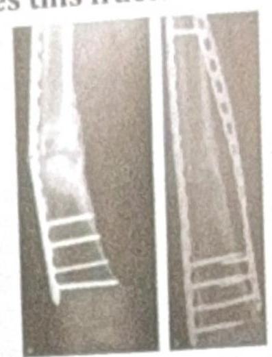
The X-ray shows plating done for a fracture. How does this fracture heal?

The compression fracture is commonest in
Fracture at which site affects the longitudinal growth of a bone?
Explanation: ***Receptor for parathormone*** - **Osteoclasts** do not directly have receptors for **parathormone (PTH)**; instead, **osteoblasts** have PTH receptors. - When PTH binds to osteoblasts, they release factors (like **RANKL**) that stimulate osteoclast activity, thus indirectly regulating bone resorption. *Bone resorption* - **Osteoclasts** are specialized cells primarily responsible for **resorbing bone matrix**, a critical process in bone remodeling. - They secrete **acids and enzymes** to break down the mineral and organic components of bone. *Ruffled border* - The **ruffled border** is a characteristic morphological feature of active osteoclasts, representing a highly folded plasma membrane. - This specialized structure increases the surface area for the secretion of **protons and lysosomal enzymes** into the bone-resorbing compartment. *RANK ligand production* - **Osteoclasts** do not produce **RANK ligand (RANKL)**; rather, they have **RANK receptors** that bind to RANKL produced by **osteoblasts and stromal cells**. - The binding of RANKL to RANK is essential for the **differentiation, activation, and survival** of osteoclasts.
Explanation: ***Bone mass*** - Estrogen plays a crucial role in maintaining **bone density** by inhibiting osteoclast activity and promoting osteoblast function. - In menopausal women, estrogen administration counteracts bone loss and thus **increases bone mass**, reducing the risk of osteoporosis. *Gonadotropin secretion* - In menopausal women, **gonadotropin-releasing hormone (GnRH)** and subsequent **FSH and LH levels are elevated** due to the absence of ovarian estrogen feedback. - Estrogen administration would exert a **negative feedback** on the hypothalamus and pituitary, thereby **decreasing**, not increasing, gonadotropin secretion. *Muscle mass* - While estrogen has some anabolic effects, **androgens** (like testosterone) are the primary hormones responsible for significantly increasing muscle mass. - Estrogen administration to menopausal women is not a primary intervention for increasing muscle mass; its effects on this parameter are generally **modest or negligible**. *LDL cholesterol* - Estrogen generally has a **favorable effect on lipid profiles**, typically leading to a **decrease in LDL cholesterol** and an increase in HDL cholesterol. - Therefore, estrogen administration would generally **reduce**, not increase, LDL cholesterol levels.
Explanation: ***90% collagen protein*** - **Type I collagen** constitutes around 90% of the organic matrix of bone, providing its tensile strength and flexibility [1]. - This extensive collagen network forms the framework upon which **mineral crystals** (hydroxyapatite) are deposited [1]. *10% collagen* - This percentage is significantly lower than the actual proportion of collagen in the organic matrix of bone. - If collagen only represented 10%, bone would lack its characteristic **tensile strength** and elasticity [2]. *10% noncollagenous protein* - While noncollagenous proteins like **osteocalcin** and **osteonectin** are important for bone mineralization and cell signaling, they only constitute about 10% of the *organic matrix*, not the entire bone, and are not the *primary organic component* [1]. - The dominant organic component is collagen, which provides the structural scaffold [1]. *20% noncollagenous protein* - This percentage is inaccurate; **noncollagenous proteins** typically make up about 10% of the bone's organic matrix [1]. - A higher proportion of noncollagenous proteins would alter the bone's mechanical properties, potentially making it more brittle.
Explanation: ***Parathyroid hormone*** - **Parathyroid hormone (PTH)** directly stimulates the **kidney's 1-alpha hydroxylase** enzyme, which converts **25(OH)D** to its active form, **1,25(OH)2D (calcitriol)**. - This regulation is critical for maintaining **calcium and phosphate homeostasis**, with PTH levels increasing when serum calcium is low, thereby boosting 1,25(OH)2D production. *Calcium levels in serum* - While **low serum calcium** indirectly stimulates **PTH** release, which then regulates 1,25(OH)2 vitamin D, calcium itself is not the direct or most important regulator. - The direct regulatory action on the conversion enzyme is mediated by PTH. *Magnesium levels in serum* - **Magnesium** plays a cofactor role in various enzymatic reactions, including those involving vitamin D metabolism, but it is not a direct or primary regulator of **1,25(OH)2 vitamin D concentration**. - Severe **hypomagnesemia** can sometimes impair PTH secretion and action, indirectly affecting vitamin D, but this is a secondary effect. *25-hydroxyvitamin D in serum* - **25-hydroxyvitamin D** is the precursor to **1,25(OH)2 vitamin D**, and its availability limits the maximum potential production of the active form. - However, the *rate* of conversion into the active form and thus the *concentration* of 1,25(OH)2D is primarily dictated by PTH, not the precursor itself.
Explanation: ***PTH*** - **Parathyroid hormone (PTH)** is the primary regulator of calcium homeostasis and the key hormone that **increases in response to hypocalcemia** (low serum calcium). - In a patient on a low calcium diet for 8 weeks, **PTH secretion increases** to maintain normal serum calcium levels. - PTH acts through three main mechanisms: increasing **bone resorption** (releasing calcium from bone), enhancing renal **calcium reabsorption** in the distal tubule, and stimulating the production of **active vitamin D (1,25-dihydroxycholecalciferol)** which increases intestinal calcium absorption. *Active 24,25 dihydroxy cholecalciferol* - **24,25-dihydroxycholecalciferol** is a relatively **inactive metabolite** of vitamin D and represents a pathway of vitamin D catabolism, not activation. - The **active form** of vitamin D that increases calcium absorption is **1,25-dihydroxycholecalciferol (calcitriol)**, whose production is stimulated by PTH. - This metabolite does **not increase** in response to hypocalcemia as a compensatory mechanism. *Serum phosphate level* - A low calcium diet would **not directly lead to an increase in serum phosphate levels**. - In fact, PTH (which increases in response to low calcium) typically causes a **decrease in serum phosphate** by promoting renal phosphate excretion (phosphaturic effect). - High phosphate levels can actually exacerbate hypocalcemia by forming insoluble calcium-phosphate complexes. *Calcitonin* - **Calcitonin** is released from the thyroid parafollicular cells (C cells) in response to **high serum calcium levels** (hypercalcemia). - It acts to **lower** calcium by inhibiting osteoclast activity and reducing renal calcium reabsorption. - In hypocalcemia (low calcium diet), calcitonin secretion would **decrease, not increase**, making this the opposite of what occurs to maintain calcium homeostasis.
Explanation: ***Cancellous bone*** - **Cancellous bone** (trabecular/spongy bone) is the **most metabolically active** part of bone due to its **large surface area-to-volume ratio** (approximately 10 times greater than cortical bone). - It has a **high rate of bone turnover** and remodeling, being 8-10 times more active than cortical bone. - Contains abundant **osteoblasts** and **osteoclasts** on trabecular surfaces, making it the primary site for **calcium homeostasis** and rapid response to metabolic demands. - Metabolic bone diseases like **osteoporosis** and **hyperparathyroidism** predominantly affect cancellous bone first due to its high metabolic activity. *Endosteal surface* - The **endosteal surface** (inner lining of cortical bone and trabecular surfaces) is metabolically active with osteoblasts and osteoclasts. - While technically the trabecular surfaces are endosteal surfaces, in clinical teaching, **cancellous bone as a whole** is recognized as the most metabolically active component. - This option represents an anatomical subdivision rather than the structural answer expected in standard physiology. *Cortical bone* - **Cortical bone** (compact bone) is dense and provides structural strength but has **lower metabolic activity** due to its compact structure and smaller surface area. - Remodeling rate is significantly slower (about 1/10th) compared to cancellous bone. - Makes up 80% of skeletal mass but contributes less to metabolic bone turnover. *Periosteal surface* - The **periosteal surface** (outer bone covering) is involved in bone growth in width and fracture repair. - Has osteoblasts and osteoclasts but accounts for a **smaller proportion** of total bone remodeling compared to the extensive trabecular surfaces. - Less metabolically active than cancellous bone overall.
Explanation: ***Decreases the activity of bone-resorbing cells*** - **Estrogen** plays a crucial role in maintaining **bone density** by inhibiting the activity of **osteoclasts**, which are the cells responsible for **bone resorption**. - In postmenopausal women, the decline in estrogen levels leads to increased osteoclast activity and accelerated **bone loss**, hence the effectiveness of estrogen therapy. *Increases the activity of bone-resorbing cells* - An increase in **bone-resorbing cell** (osteoclast) activity would lead to further **bone loss** and exacerbate osteoporosis, contrary to the observed therapeutic effect of estrogen. - Estrogen's protective role in **bone health** is primarily through its inhibitory effect on osteoclasts. *Prevents the mineralization of bone during turnover* - This option describes a process that would lead to **osteomalacia** or **rickets**, where new bone matrix fails to mineralize adequately. - Estrogen's action is not primarily on mineralization but on the **balance between bone formation and resorption**. *Reduces the activity of bone-forming cells* - Reducing the activity of **bone-forming cells** (osteoblasts) would also lead to reduced bone density and worsen osteoporosis. - While estrogen has complex effects, its main therapeutic benefit in osteoporosis is to **slow down bone breakdown**, not to reduce bone formation.
Explanation: **Primary healing** - **Plating of a fracture** aims to achieve **absolute stability** at the fracture site, which facilitates primary bone healing. - In primary healing, there is **direct bone formation** across the fracture gap without the formation of a significant callus. *Secondary healing* - Secondary healing involves the formation of a **callus** (fibrous tissue, cartilage, and immature bone) to bridge the fracture gap. - This type of healing occurs in situations with **relative stability** and some micromotion at the fracture site, such as with casting or intramedullary nailing. *Tertiary healing* - **Tertiary healing** is not a recognized term in the context of fracture healing. - Bone healing typically involves either primary or secondary mechanisms depending on the stability achieved. *Distraction histiogenesis* - **Distraction histiogenesis** is the process by which new bone is formed between bone surfaces that are gradually pulled apart using an external fixator (**distraction osteogenesis**). - This is used in procedures like **limb lengthening** and is distinct from the direct healing of a fracture fixed with a plate.
Explanation: ***Lower thoracic spine*** - The **thoracolumbar junction (T11-L2)** is the most common site for compression fractures due to its high biomechanical stress, transitioning from stiff thoracic spine to more flexible lumbar spine. - This area is particularly vulnerable to axial loading and flexion injuries because it's a zone of increased mobility and stress concentration. *Upper thoracic spine* - The upper thoracic spine has **rib cage support** and less mobility, making fractures here less common without significant traumatic force. - Fractures in this region often indicate a **high-energy injury** due to its inherent stability. *Cervical spine* - While cervical fractures can be serious, they typically result from **high-energy trauma** and are less commonly simple compression fractures compared to the thoracolumbar region. - The **cervical spine** is more prone to **burst fractures** or **dislocations** from flexion-distraction or extension injuries. *Lumbosacral region* - The **sacrum and coccyx** are relatively stable bone structures and are less prone to common compression fractures unless there is severe trauma or significant bone weakening (e.g., severe osteoporosis). - While lumbar compression fractures do occur, the **junctional region** between the thoracic and lumbar spine (lower thoracic/upper lumbar) is statistically more frequent.
Explanation: ***Epiphyseal plate*** - The **epiphyseal plate**, also known as the **growth plate**, is a cartilaginous disc responsible for the **longitudinal growth** of long bones. - A fracture in this region can damage the **chondrocytes** and disrupt the normal ossification process, potentially leading to **growth arrest** or limb length discrepancies. *Diaphysis* - The **diaphysis** is the **shaft** or central part of a long bone. - While a fracture here can cause pain and instability, it typically does not directly affect the **longitudinal growth** potential of the bone. *Epiphysis* - The **epiphysis** is the end part of a long bone, initially separated from the main bone by cartilage but later fused with it. - Although it contains the epiphyseal plate in growing individuals, a fracture to the epiphysis itself (excluding the growth plate) primarily affects the **joint surface** and stability, rather than longitudinal growth directly. *Metaphysis* - The **metaphysis** is the transitional zone between the diaphysis and the epiphysis, adjacent to the growth plate. - While fractures in this area can be close to the growth plate, a metaphyseal fracture generally does not directly damage the **growth plate cartilage** to the same extent as a fracture through the plate itself, making its impact on longitudinal growth less direct or severe.
Get full access to all questions, explanations, and performance tracking.
Start For Free