
Basic Science in Orthopaedics — MCQs

On this page
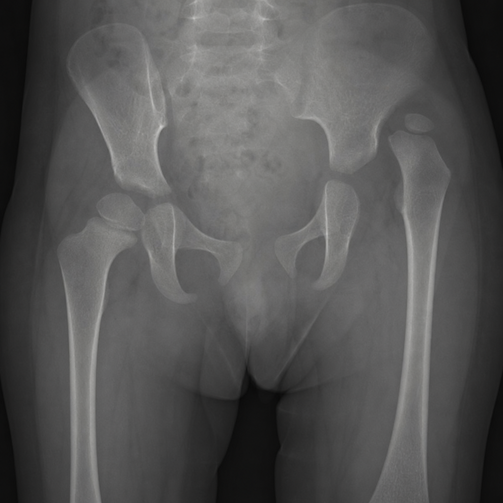
A 9-month-old girl is brought by her mother who noticed asymmetric skin folds on the thighs and difficulty abducting the left hip during diaper changes. She was a firstborn, delivered in breech presentation. Examination reveals limited abduction of the left hip to 30 degrees. The pelvis radiograph is shown (Image 1). Which of the following is the most appropriate next step in management?
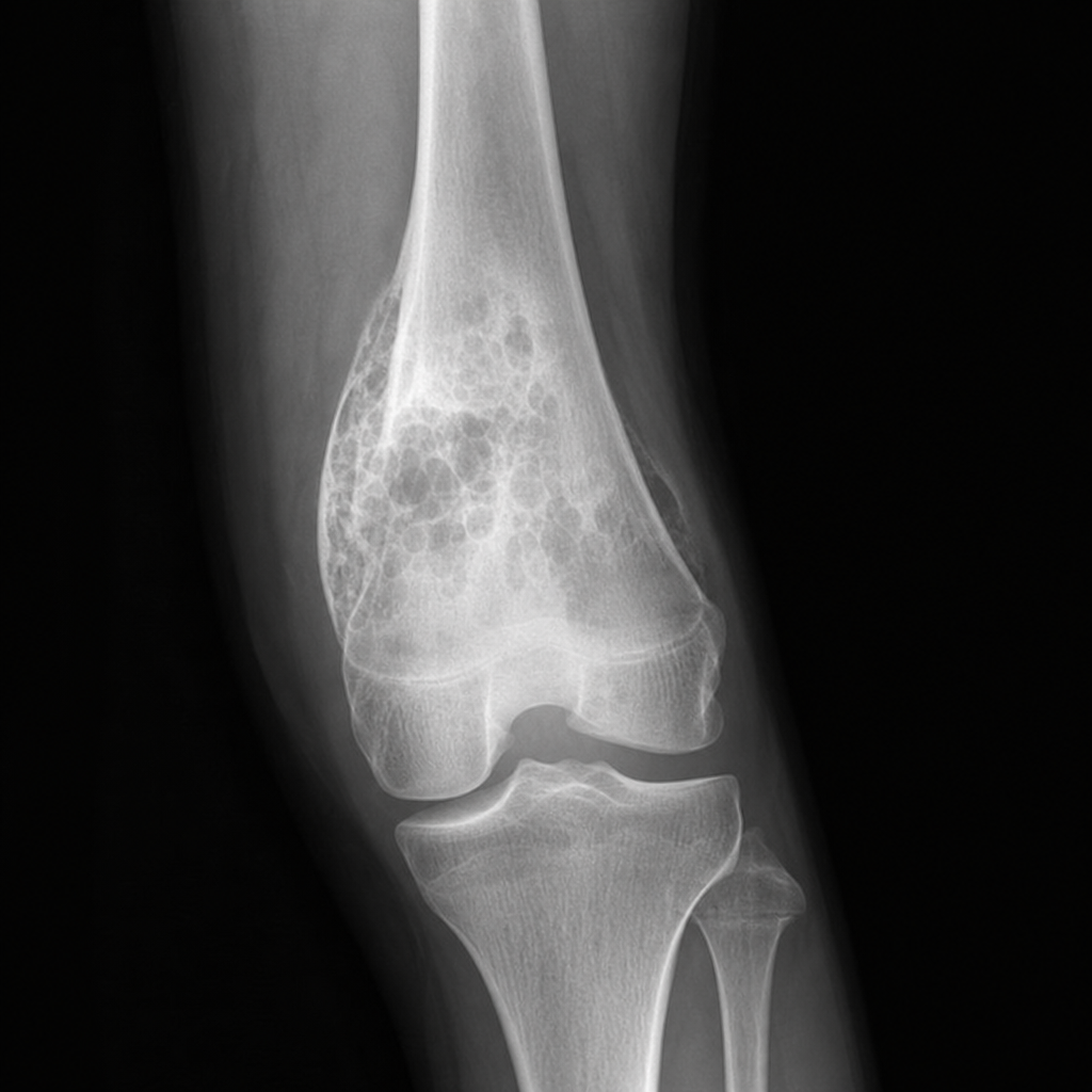
A 16-year-old boy presents with a 6-week history of progressively worsening pain and swelling around the right knee. He denies any trauma. On examination there is a firm, tender, warm swelling over the distal femur. Serum alkaline phosphatase is markedly elevated. The plain radiograph is shown in Image 1. Which of the following is the most appropriate next step in management?
Which of the following diagnostic studies is NOT useful in the evaluation of upper-extremity pain?
What is the most important factor in fracture healing?
A patient operated for a forearm fracture under general anesthesia with a tourniquet was unable to move his fingers and had sensory loss over the entire hand postoperatively. What is the most common type of nerve injury in this scenario?
What is Neurapraxia?
Tinel sign is indicative of?
What is the latent period in distraction osteogenesis?
Disruption of the myelin sheath without interruption in the axons is called:
Pseudarthrosis may be seen in all of the following conditions except?
Practice by Chapter
Bone Structure and Function
Practice Questions
Cartilage Biology and Physiology
Practice Questions
Muscle and Tendon Physiology
Practice Questions
Joint Biomechanics
Practice Questions
Fracture Healing Process
Practice Questions
Bone Metabolism and Turnover
Practice Questions
Orthopaedic Biomaterials
Practice Questions
Tribology in Orthopaedics
Practice Questions
Gait Analysis
Practice Questions
Biomechanics of Spine
Practice Questions
Applied Surgical Anatomy
Practice Questions
Bone Banking and Grafting
Practice Questions
Want unlimited practice?
Get full access to all questions, explanations, and performance tracking.
Scan to download app