A healthcare worker develops fever, night sweats, and cough. Sputum shows acid-fast bacilli. What is the next diagnostic test?
According to DOTS-PLUS guidelines 2013, which of the following statements about the treatment of multidrug-resistant TB is incorrect?
A patient with pulmonary tuberculosis, who is receiving anti-tuberculosis therapy consisting of rifampicin, isoniazid, ethambutol, and pyrazinamide, should be advised to take which of the following supplements?
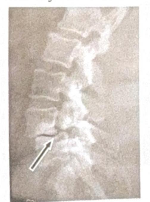
Identify the condition shown in the image:

In tuberculosis, a 'case' is
A 60-year-old male presents with chronic arthritis. Which of the following is the most likely cause?
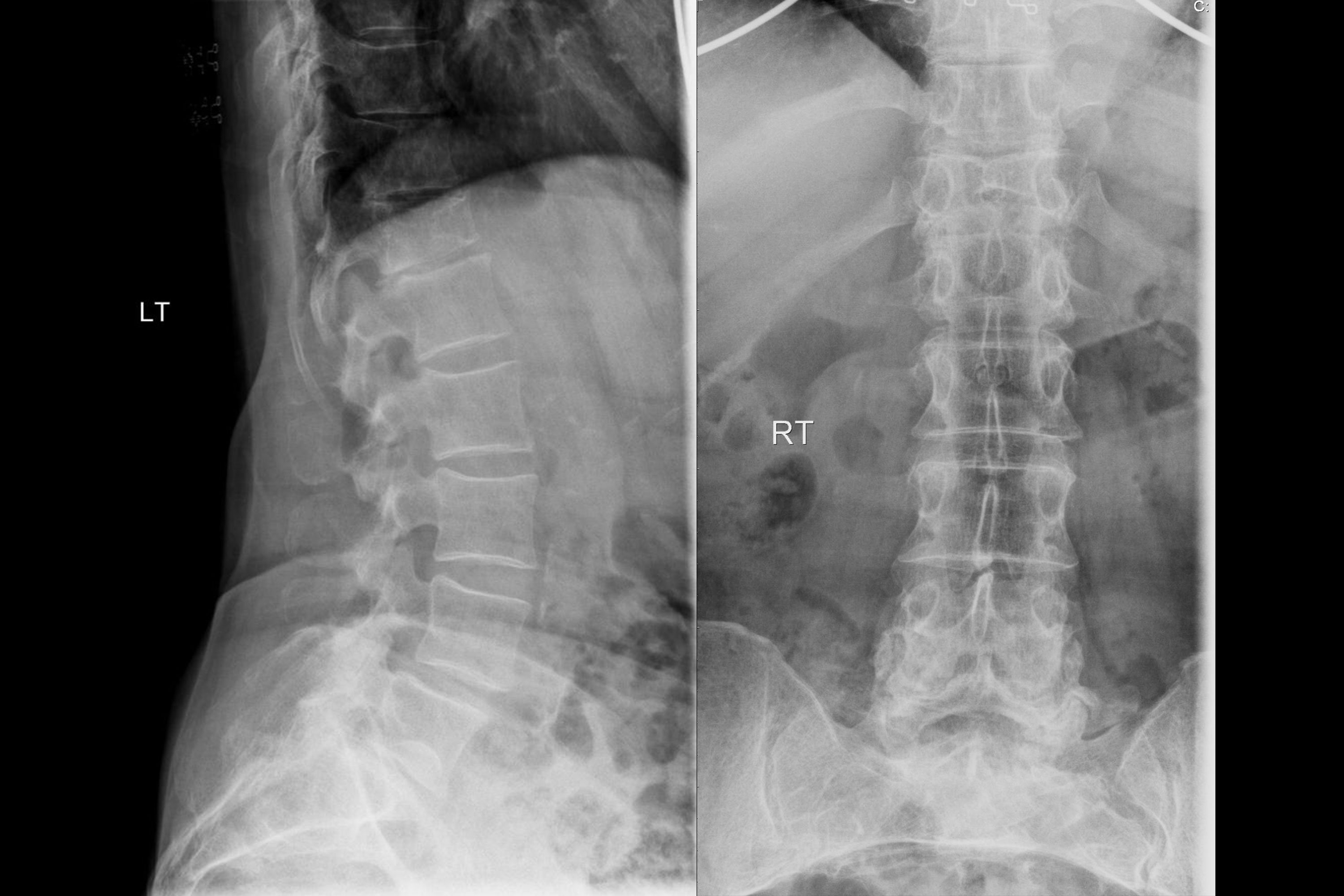
A 75-year-old female has chronic backache. X-ray of the spine is shown. What is the most likely diagnosis?

What is considered a poor prognostic indicator in Pott's paraplegia?
Tuberculosis of the spine; what is the most common site affected?
What is the earliest X-ray sign observed in spinal tuberculosis?
Explanation: ***NAAT for TB*** - Nucleic Acid Amplification Tests (**NAAT**) rapidly confirm the presence of **Mycobacterium tuberculosis** DNA or RNA, crucial after an **acid-fast bacilli (AFB) smear** is positive [1]. - This test offers high sensitivity and specificity and can also detect **drug resistance**, guiding immediate treatment decisions [1]. *Gram stain* - A **Gram stain** is not appropriate for **Mycobacterium tuberculosis** because these bacteria have a unique cell wall that makes them **acid-fast**, not readily stained by the Gram method. - The initial finding of **acid-fast bacilli** already indicates a general type of organism, making a Gram stain redundant and uninformative for TB. *Serology for TB* - **Serological tests for TB** (detecting antibodies to M. tuberculosis) are generally **not recommended** for the diagnosis of active pulmonary TB due to their **poor sensitivity and specificity**. - They have limited utility in diagnosing active disease and are not endorsed by major health organizations for this purpose. *Sputum culture* - **Sputum culture** is the **gold standard** for confirming TB diagnosis and for **drug susceptibility testing**, but it is a **slow process** (taking several weeks) [2]. - While essential for definitive diagnosis and resistance profiling, it is not the **"next" rapid diagnostic test** required given the positive AFB smear.
Explanation: ***Continuation phase - 2 drugs*** - According to DOTS-PLUS guidelines (2013), the continuation phase for multidrug-resistant TB (MDR-TB) should include at least **three to four effective drugs**, not two. - Using only two drugs in the continuation phase would be grossly inadequate and would likely lead to treatment failure and the development of extensively drug-resistant TB (XDR-TB). - This statement is **clearly incorrect** and represents a major deviation from standard treatment protocols. *Total duration 24-27 months* - According to DOTS-PLUS 2013 guidelines, the total treatment duration for MDR-TB is typically **18-24 months** (at least 18 months after culture conversion). - In complex cases, treatment may be extended beyond 24 months, though 24-27 months falls within acceptable parameters for difficult cases. - This statement is essentially correct for the upper range of treatment duration. *Intensive phase 6-9 months* - The intensive phase for MDR-TB treatment is indeed typically **6-9 months** or until culture conversion is documented. - This phase includes daily injectable agents and multiple oral drugs to rapidly reduce bacterial load. - This statement is **correct**. *Intensive phase - 6 drugs* - The 2013 DOTS-PLUS guidelines recommend an intensive phase regimen comprising **at least 4 effective drugs including an injectable agent**. - A 5-6 drug regimen may be used in complex cases or when drug susceptibility is uncertain. - While not the minimum standard, using 6 drugs is within acceptable practice, making this statement **generally correct**.
Explanation: ***Pyridoxine*** - **Isoniazid** in the anti-tuberculosis regimen can cause **peripheral neuropathy** by interfering with **pyridoxine (vitamin B6)** metabolism. - Supplementation with **pyridoxine** is advised to prevent this neurotoxic side effect, especially in patients at higher risk such as those with diabetes, malnutrition, or alcoholism. *Niacin* - **Niacin (vitamin B3)** deficiency can lead to **pellagra**, characterized by dermatitis, diarrhea, and dementia. - While important for general health, niacin supplementation is not specifically required to counteract side effects of standard anti-tuberculosis drugs. *Riboflavin* - **Riboflavin (vitamin B2)** is essential for various metabolic processes and cellular energy production. - There is no direct significant depletion or interference with riboflavin metabolism caused by the common anti-tuberculosis drugs. *Thiamine* - **Thiamine (vitamin B1)** deficiency can lead to **beriberi** and neurological symptoms, particularly in those with chronic alcoholism. - While thiamine is crucial for neurological function, antitubercular drugs do not specifically deplete or interfere with its metabolism to the extent of requiring routine supplementation.
Explanation: ***Spondylolysis*** * The image shows a **break in the pars interarticularis** of a vertebra, indicated by the arrow, which is characteristic of spondylolysis. * This condition is a **stress fracture** or defect in the pars interarticularis, a bony segment connecting the superior and inferior articular facets. *Renal osteodystrophy* * Renal osteodystrophy refers to a spectrum of **bone abnormalities** that occur in chronic kidney disease, not a specific vertebral fracture pattern. * It typically involves features such as **osteomalacia**, **osteitis fibrosa cystica**, or **osteoporosis**, which are not directly depicted as a fracture in this image. *Spondylolisthesis* * Spondylolisthesis is the **anterior slippage** of one vertebral body over another, which can be caused by bilateral spondylolysis but is not directly shown as a slip in this specific image. * The image distinctly highlights the **fracture line** itself, rather than the displacement of the vertebral body. *Tuberculosis (TB)* * Spinal tuberculosis (Pott's disease) typically presents with **destruction of vertebral bodies**, disc space narrowing, and often a **paravertebral abscess**. * The image does not show these features; instead, it demonstrates a clear **bony defect** in the pars interarticularis.
Explanation: ***Sputum positive*** - In the context of **tuberculosis (TB)**, a 'case' is defined by **bacteriological confirmation**, most commonly through **sputum smear positivity** for acid-fast bacilli (AFB). - According to **RNTCP (Revised National Tuberculosis Control Programme)** guidelines, a sputum smear-positive case is one with at least **two sputum specimens positive for AFB**, or one sputum specimen positive for AFB plus radiographic abnormalities consistent with active pulmonary TB. - A positive sputum smear confirms the presence of **Mycobacterium tuberculosis** in the respiratory tract, indicating active, transmissible infection requiring immediate treatment. *Cough* - **Cough** is a common symptom of tuberculosis but is not sufficient on its own to define a 'case'. - Many respiratory conditions can cause a cough, and it does not confirm the presence of **Mycobacterium tuberculosis** or infectiousness. - Cough lasting more than 2-3 weeks is a screening criterion for TB suspects, not a case definition. *X-ray positive* - A **positive chest X-ray** can show abnormalities consistent with tuberculosis, such as infiltrates, cavities, or hilar lymphadenopathy. - However, radiological findings alone do not definitively confirm a TB diagnosis, as many other conditions can mimic TB on chest X-ray. - Chest X-ray is used for **clinically diagnosed TB cases** when bacteriological confirmation is not possible, but it does not establish active infectivity without microbiological confirmation. *Mantoux positive* - A **positive Mantoux test** (tuberculin skin test) indicates exposure to Mycobacterium tuberculosis and the presence of a cell-mediated immune response. - It signifies **latent TB infection (LTBI)** but does not indicate active, infectious disease. - Many people with positive Mantoux tests never develop active TB and are not considered 'cases'.
Explanation: Crystal-induced arthritis, such as gout or calcium pyrophosphate deposition disease (CPPD), can lead to chronic arthritis associated with progressive joint damage and functional limitations [1]. While acute presentations are common, such as 'pseudogout' involving the knee, the underlying pathophysiology involves the balance of crystal formation and tissue concentration within the joint [1]. Specific causes like metabolic diseases or previous trauma may also predispose individuals to early-onset osteoarthritis [2].
Explanation: ***Osteoporosis*** - The X-ray shows diffuse **osteopenia** (reduced bone density) and **vertebral compression fractures**, particularly visible in the lateral view, which are characteristic findings in elderly patients with osteoporosis and chronic backache. - The vertebral bodies appear **demineralized** and some exhibit a loss of height, suggesting collapse due to weakened bone structure. *Spondylodiscitis* - This condition involves **inflammation of the vertebral body and adjacent intervertebral disc**, typically showing **erosions** of the vertebral endplates and **narrowing of the disc space** on X-ray, which are not clearly evident here as the primary issue. - While it can cause back pain, the dominant finding on this X-ray is widespread bone density loss and fractures, rather than localized infection-related changes. *Pott's spine* - Pott's spine (**tuberculous spondylitis**) is a form of osteomyelitis that causes **destruction of vertebral bodies** and adjacent discs, often leading to a **gibbus deformity** (sharp posterior angulation of the spine). - The X-ray does not show extensive vertebral destruction, paraspinal abscess formation, or typical kyphotic deformity associated with Pott's spine. *Spondylolisthesis* - Spondylolisthesis is characterized by the **forward slippage of one vertebral body over another**, often due to a defect in the pars interarticularis. - While there may be some degenerative changes, there is no clear evidence of significant anterior translation of a vertebral body on the lateral X-ray that would indicate spondylolisthesis.
Explanation: ***Rapid progression of neurological deficits*** - **Rapid progression** implies severe spinal cord damage occurring quickly, which is less likely to fully recover even with treatment. - This indicates a more aggressive disease process or significant compression that can lead to irreversible neurological impairment. *Healed disease* - **Healed disease** (even if paraplegia existed previously) indicates that the infection is resolved and the destructive process has stopped, allowing for potential neurological recovery or stability. - While residual neurological deficits might remain, the absence of active inflammation improves the long-term prognosis compared to ongoing damage. *Chronic disease* - **Chronic disease** in the context of Pott's paraplegia often refers to established deficits after a prolonged course, but it doesn't necessarily imply ongoing active deterioration. - The chronicity itself, without rapid progression, suggests a more stable state where the damage has already occurred, and further deterioration might be slow or absent. *Active disease* - **Active disease** means the infection is still present and causing bone destruction, which is a concern. - However, if the neurological deficits are not rapidly progressing, there is still a window for treatment to stop the disease and potentially allow for some recovery, distinguishing it from an acute, rapidly deteriorating situation.
Explanation: ***94ed055d-c7da-4d18-a2fd-52720dfe8b6e*** - The **dorsolumbar (thoracolumbar)** region is the most common site of **spinal tuberculosis (Pott's disease)** [1] due to its high vascularity, facilitating hematogenous spread. - **Spinal tuberculosis** typically affects the vertebral bodies, leading to their destruction, kyphosis (angular deformity), and potentially neurological deficits [1]. *aebdfe6c-98dc-4073-892f-bb24d047bab4* - The **sacral** region can be affected by **tuberculosis**, but it is considerably less common than the thoracolumbar region. - Involvement of the sacrum is often associated with **direct extension** from adjacent structures, such as the sacroiliac joint, rather than primary vertebral involvement. *15c1feef-e3ca-496f-a180-127d52b77bfa* - **Cervical spine tuberculosis** is relatively rare, accounting for a small percentage of all spinal tuberculosis cases. - While possible, it presents with specific challenges due to the proximity of vital neurological and vascular structures. *d05d4d13-bb83-4f26-aa2d-c9c0203d299c* - The **lumbosacral region** (L5-S1) can be involved in **tuberculosis**, but it is less frequently affected than the thoracolumbar region. - While the lumbar spine is a common site, the entire lumbosacral region as a single entity is not the most common spot for spinal TB.
Explanation: ***Narrowing of disc space*** - This is the **earliest radiographic finding** in spinal tuberculosis on plain X-ray, typically appearing within the first few weeks to months of infection. - Tuberculous spondylitis begins in the **anterior subchondral region** near the vertebral endplates, leading to early involvement of the intervertebral disc. - The infection spreads from the vertebral body to the adjacent disc, causing **disc space narrowing** along with endplate irregularities as initial manifestations. - Unlike pyogenic spondylitis, TB shows **relatively preserved** disc space initially, but narrowing is still the earliest visible change. *Endplate erosion* - **Endplate erosion** occurs concurrently with or shortly after disc space narrowing as the tuberculous infection destroys the subchondral bone. - Both findings appear early in the disease course and are often seen together on X-ray. - This represents active bone destruction at the vertebral margins. *Paravertebral shadow* - The **paravertebral abscess** is a characteristic and important finding in spinal tuberculosis, but it develops **later** in the disease course. - It forms as a secondary phenomenon when the infection spreads beyond the vertebral body, with caseous material and pus collecting along the paravertebral ligaments. - While highly suggestive of TB when present, it is not the earliest radiographic sign. *Gibbus* - A **gibbus deformity** is a **late complication** resulting from vertebral body collapse and subsequent angular kyphosis. - It indicates extensive vertebral destruction and significant structural compromise. - This is a clinical and radiographic sign of advanced disease, not an early finding.
Get full access to all questions, explanations, and performance tracking.
Start For Free