Which laser is used in the management of posterior capsule opacification (PCO)?
Which of the following is not a relative contraindication for breast conservative surgery?
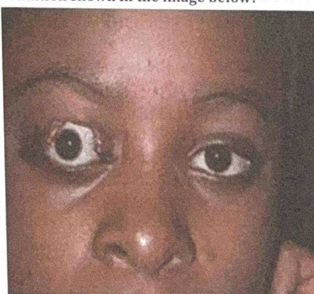
What is the most likely complication of the condition shown in the image below?

Which keratometry reading is most accurate in post-LASIK eyes for IOL power calculation?
Laser used in LASIK is:
The laser procedure, most often used for treating iris neovascularization is
A lady wants LASIK surgery for her daughter and asks for your opinion. All the following are suitable for performing LASIK surgery except:
A lady wants LASIK surgery for her daughter. All the following are indications for performing LASIK surgery, except:
Maximum correction of myopia can be done by?
What does a visual acuity test primarily assess?
Explanation: ***Nd:YAG*** - The **Nd:YAG laser** is the standard treatment for posterior capsule opacification (PCO) due to its ability to create a small opening in the **opacified posterior capsule** without damaging adjacent structures. - This laser works by **photodisruption**, using short pulses of high-energy light to create plasma and mechanically break apart the capsular tissue. *Krypton* - **Krypton lasers** are primarily used in ophthalmology for **retinal photocoagulation**, particularly for conditions like diabetic retinopathy and macular edema. - They are not used for treating PCO as their mechanism is based on thermal coagulation, which would be inappropriate for the delicate posterior capsule. *Argon* - **Argon lasers** are also used for **retinal photocoagulation** and for treating conditions like **glaucoma** (e.g., argon laser trabeculoplasty). - Similar to krypton lasers, their thermal effects are not suitable for the precise, non-thermal disruption required for PCO treatment. *Excimer* - The **excimer laser** is best known for its use in **refractive surgery** like LASIK and PRK to reshape the cornea. - Its mechanism involves **photoablation**, precisely removing tissue, but it is not used for creating an opening in the posterior capsule.
Explanation: ***Small tumor size (<3cm)*** ✓ - A small tumor size is **NOT a contraindication** for breast-conserving surgery; it is actually a **favorable condition** and an indication for breast conservation. - Small tumors allow for complete tumor removal with good cosmetic outcomes and adequate margins. - This is the **correct answer** as it is the only option that is NOT a relative contraindication. *Multicentric disease* - **Multicentric disease** refers to the presence of multiple tumor foci in **different quadrants** of the breast, making complete surgical removal challenging with breast-conserving surgery. - This is a **relative contraindication** as it increases the risk of **positive margins** and local recurrence, making mastectomy often a more appropriate option. *Previous radiation to breast* - Prior radiation therapy to the breast is a **contraindication** (often considered absolute) for subsequent breast radiation, which is an essential component of breast-conserving therapy. - Re-irradiation carries a high risk of severe **skin and tissue toxicity**, making further breast conservation unfeasible. *Large tumor size* - A large tumor size is a **relative contraindication** as it can make it difficult to achieve **clear surgical margins** while maintaining an acceptable cosmetic result. - However, **neoadjuvant chemotherapy** may downstage large tumors to make them suitable for breast-conserving surgery. - Without tumor reduction, it often requires **mastectomy**.
Explanation: ***Exposure Keratitis*** - The image shows **proptosis** (exophthalmos) of the right eye, where the eyeball protrudes forward. This condition often leads to incomplete eyelid closure (lagophthalmos). - **Exposure keratitis** occurs when the cornea is inadequately covered by the eyelids, leading to drying and damage due to constant exposure to air and environmental factors. *Difficulty in eye movement* - While **proptosis** can sometimes be associated with restricted eye movements (e.g., in severe Graves' ophthalmopathy due to muscle swelling), it is not the **most likely direct complication** of the exposure itself. - The image primarily depicts the physical displacement of the globe, which predisposes to corneal issues, not necessarily oculomotor dysfunction as the primary complication. *Cataract* - **Cataracts** are opacities of the lens and are typically associated with aging, trauma, or certain systemic conditions (e.g., diabetes, steroid use). - They are not a direct or common complication of **proptosis** or the resulting **exposure of the ocular surface**. *Glaucoma* - **Glaucoma** is a group of conditions characterized by damage to the optic nerve, often due to elevated intraocular pressure. - While severe **proptosis** leading to orbital congestion can theoretically increase intraocular pressure, it is not the most direct or prevalent complication compared to **exposure keratitis**, which is a direct consequence of inadequate globe protection.
Explanation: ***Total corneal power*** * After LASIK, the **anterior and posterior corneal curvatures** are altered, leading to discrepancies in standard keratometry readings. * **Total corneal power** methods, such as those derived from **corneal tomography** or **anterior segment OCT**, account for both surfaces, providing a more accurate estimation of the true refractive power. * *Topography-derived K* * While corneal topography is valuable for assessing the anterior corneal surface and identifying **irregular astigmatism**, it traditionally focuses on the anterior curvature and may not fully account for the altered **posterior corneal surface** after LASIK. * Standard topography-derived K values often rely on assumptions about the posterior-to-anterior corneal curvature ratio, which are invalid after refractive surgery. * *Manual keratometry* * Manual keratometry measures the **anterior corneal curvature** at a few discrete points and is highly susceptible to inaccuracies due to the post-LASIK changes in corneal shape. * It tends to **overestimate the corneal power** in eyes that have undergone myopic LASIK and **underestimate it** in hyperopic LASIK, leading to significant IOL power calculation errors. * *Automated keratometry* * Similar to manual keratometry, automated keratometry primarily measures the **anterior corneal surface** and relies on a fixed refractive index ratio that is no longer valid after corneal reshaping. * These devices generally provide **inaccurate keratometry readings** in post-refractive surgery eyes, contributing to refractive surprises after cataract surgery.
Explanation: ***Excimer*** - An **excimer laser** is used in LASIK procedures due to its ability to precisely ablate (remove) corneal tissue without causing thermal or collateral damage to surrounding tissues. - This laser operates in the **ultraviolet (UV) spectrum** and uses a mixture of inert and halogen gases (e.g., argon fluoride) to produce its beam. *Argon* - **Argon lasers** are primarily used for procedures involving the **retina and iris**, such as panretinal photocoagulation for diabetic retinopathy or iridotomy for narrow-angle glaucoma. - They emit light in the **visible blue-green spectrum**, which is absorbed by melanin and hemoglobin. *Holmium* - **Holmium lasers** are used in ophthalmology for procedures like **sclerostomy** (creating a drainage channel for glaucoma) or **transscleral cyclophotocoagulation**. - They operate in the **infrared spectrum** and are known for their strong tissue absorption and thermal effect. *Nd-yag* - A **Nd:YAG laser** is commonly used for **posterior capsulotomy** after cataract surgery to treat posterior capsular opacification, and for **peripheral iridotomy** in glaucoma. - It emits light in the **infrared spectrum** and works by creating photodisruption (plasma formation) to cut tissue without direct thermal effects.
Explanation: ***Panretinal photocoagulation (PRP)*** - **PRP** is the most effective laser procedure for **iris neovascularization** and **neovascular glaucoma**, as it ablates the ischemic retina, reducing the production of **vascular endothelial growth factor (VEGF)**. - By destroying the ischemic peripheral retina, PRP reduces the **angiogenic drive** that leads to new vessel formation on the iris and in the angle. *Laser iridoplasty* - This procedure involves applying laser energy to the peripheral iris to cause contraction and widen the **anterior chamber angle**, primarily used for **angle-closure glaucoma**. - While it can open a closed angle, it does not address the underlying **ischemic drive** causing neovascularization. *Laser trabeculoplasty* - This procedure targets the **trabecular meshwork** to improve aqueous humor outflow, commonly used for **open-angle glaucoma**. - It does not directly affect **iris neovascularization** or the ischemic factors driving its development. *Goniophotocoagulation* - This involves directly lasering new vessels in the **anterior chamber angle**, often as an adjunct to PRP, but it's not the primary treatment to prevent **iris neovascularization**. - It treats existing vessels but does not address the underlying cause of **retinal ischemia** that promotes new vessel growth.
Explanation: ***Age of 15 years*** - LASIK surgery is generally not recommended for individuals under the age of 18-21 years because their **refractive error** may still be changing. - **Refractive stability** is a critical prerequisite to ensure a lasting surgical outcome. *Myopia of 4 Diopters* - This level of **myopia** (nearsightedness) typically falls within the treatable range for LASIK, which can correct moderate to high degrees of refractive error. - LASIK is highly effective for reducing or eliminating dependence on **glasses or contact lenses** for this range. *Stable refraction for 1 year* - **Refractive stability** is an essential criterion for LASIK candidacy, indicating that the patient's prescription has not significantly changed over a specific period. - This ensures that the surgical correction will be durable and accurate, preventing the need for future enhancements due to progression of refractive error. *Corneal thickness of 600 microns* - A **corneal thickness** of 600 microns is considered adequate for LASIK, as it allows for sufficient residual stromal bed thickness after ablation. - The minimum required **residual stromal bed thickness** is typically around 250-300 microns to maintain corneal integrity and prevent ectasia.
Explanation: ***Age of 15 years*** - LASIK surgery is generally not recommended for individuals under the age of 18, as their eyes and **refractive error** are still developing and stabilizing. - Performing LASIK on a 15-year-old could lead to **regression** of the refractive correction as the eye continues to grow. *Myopia of 4 Diopters* - A **myopia** of 4 Diopters (D) falls within the treatable range for LASIK, which can effectively correct moderate degrees of nearsightedness. - This is a common indication for individuals seeking freedom from glasses or contact lenses. *Stable refraction for 1 year* - **Stable refraction** for at least one year is a crucial criterion for LASIK, ensuring that the patient's prescription is unlikely to change significantly post-surgery. - Unstable refraction could result in suboptimal visual outcomes and the need for further correction. *Corneal thickness of 600 microns* - A **corneal thickness** of 600 microns is considered well within the safe range for LASIK surgery, allowing sufficient residual stromal bed after flap creation and ablation. - Adequate corneal thickness is essential to prevent complications such as **corneal ectasia**.
Explanation: ***LASIK*** - **LASIK (Laser-Assisted In Situ Keratomileusis)** allows for significant correction of high myopia by reshaping the cornea with an excimer laser. - It involves creating a **corneal flap** and then ablating tissue underneath, offering precise and stable vision correction for a wide range of refractive errors. - Among the given corneal refractive procedures, LASIK can correct myopia up to **-10 to -12 D**. *Radial keratotomy* - **Radial keratotomy (RK)** involves making radial incisions in the cornea to flatten it, primarily used for low to moderate myopia (up to -3 to -4 D). - It has a higher risk of **unpredictable outcomes**, induced astigmatism, and glare compared to modern laser procedures. *Photorefractive keratectomy* - **Photorefractive keratectomy (PRK)** involves direct ablation of the corneal surface without creating a flap, which is suitable for moderate myopia (up to -8 to -10 D). - While effective, PRK typically has a **longer recovery period** and more post-operative pain than LASIK. *Orthokeratology* - **Orthokeratology (Ortho-K)** uses specially designed rigid contact lenses worn overnight to temporarily reshape the cornea and correct myopia. - The effect is **temporary**, requiring continuous lens wear to maintain vision correction, and is generally limited to low to moderate myopia (up to -4 to -6 D).
Explanation: ***Ability to recognize shapes and details*** - A visual acuity test, typically using a **Snellen chart**, measures the sharpness of vision, specifically the ability to discern letters or symbols at a given distance. - It assesses the eye's capacity to resolve fine **spatial detail**, which is crucial for tasks like reading and recognizing faces. - This is the fundamental definition of visual acuity and what these tests are specifically designed to measure. *Ability to perceive light* - This refers to **light perception (LP)**, the most basic form of vision, indicating whether a person can detect the presence or absence of light. - While essential for vision, it is a much simpler function than what visual acuity tests measure and is assessed separately. *Ability to differentiate colors* - This is assessed by **color vision tests**, such as the Ishihara plates, which evaluate the function of cone photoreceptors. - It specifically checks for **color blindness** (e.g., red-green or blue-yellow deficiencies) and is distinct from the sharpness of vision. *Ability to detect contrast* - This is measured by **contrast sensitivity tests**, which evaluate the ability to distinguish objects from their background at various contrast levels. - While related to overall visual quality, it is a different aspect of vision than the ability to recognize fine details at high contrast.
Get full access to all questions, explanations, and performance tracking.
Start For Free