
Pediatric Ophthalmology and Strabismus — MCQs

On this page
Which syndrome is characterized by the following ocular finding?
A patient presented with his head tilted towards the left. On examination, he was having left hypertropia which increased on looking towards the right or medially. Which muscle is most likely paralyzed?
Which of the following statements is FALSE regarding Duane's retraction syndrome?
Large angle Kappa gives rise to which type of squint?
All of the following are useful in the measurement of the angle of deviation in squint EXCEPT?
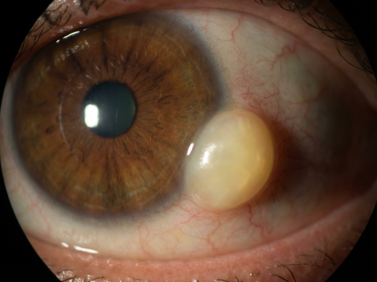
What is the primary treatment for traumatic cataract in children?
Ocular lesions of rubella include all of the following except?
Oculocardiac reflex is seen in which surgery?
Strabismic amblyopia is more common in patients with which type of squint?
How does retinoblastoma spread?
Practice by Chapter
Amblyopia
Practice Questions
Esotropia
Practice Questions
Exotropia
Practice Questions
Vertical Deviations
Practice Questions
Special Forms of Strabismus
Practice Questions
Nystagmus in Children
Practice Questions
Pediatric Cataract
Practice Questions
Retinopathy of Prematurity
Practice Questions
Pediatric Glaucoma
Practice Questions
Pediatric Neuro-ophthalmology
Practice Questions
Genetic Eye Diseases in Children
Practice Questions
Pediatric Ocular Trauma
Practice Questions
Want unlimited practice?
Get full access to all questions, explanations, and performance tracking.
Scan to download app