The cover-uncover test is performed to diagnose various eye conditions. Which of the following is not detected by this test?
A 5-month-old baby is brought by the mother with complaints of the left upper eyelid moving up and down during breastfeeding or thumb sucking, which disappears when the baby is not being fed. What is the most likely diagnosis?
A 3-year-old child has amblyopia in the right eye. What is the preferred treatment for this child?
All of the following are done in the treatment of amblyopia, except:
The essential foundational components of binocular single vision are:
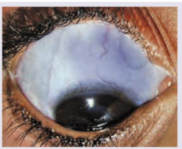
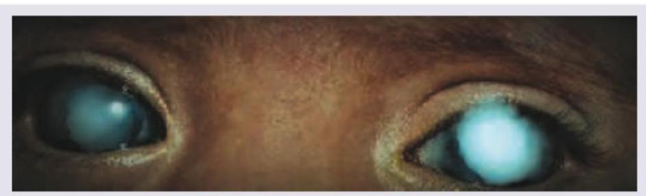
Identify the clinical sign shown in the picture:

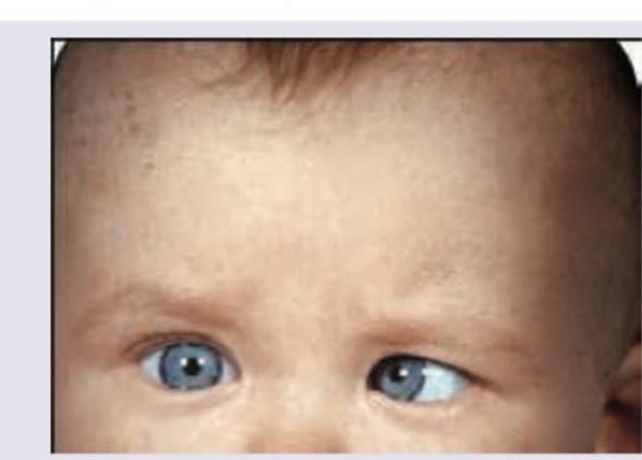
Based on the clinical photograph, identify the type of strabismus present:

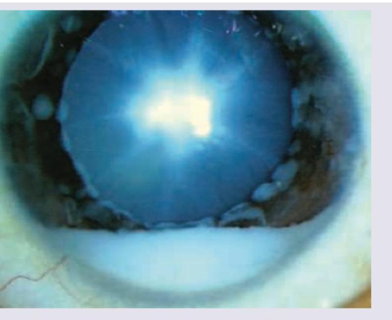
All are causes of the presentation shown below except:

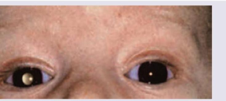
A 6-month-old child with retinoblastoma is brought with the following presentation in the right eye. The presentation shown is known as:

A 2-year-old child is brought with complaints of watering of eyes, photophobia and intermittently keeping eyes closed while watching TV. What may be the diagnosis? (AIIMS Nov 2018)

Explanation: ***Amblyopia*** - Amblyopia, often called a "lazy eye," is a neurodevelopmental disorder characterized by reduced **visual acuity** in one eye, which is not correctable by refractive means alone. - It is diagnosed by assessing vision with tools like a **Snellen chart**, whereas the cover-uncover test is designed to evaluate **ocular alignment** and motor fusion, not sensory function like visual acuity. *Latent Squint* - A latent squint, or **heterophoria**, is a tendency for the eyes to misalign when binocular vision is interrupted, such as when one eye is covered. - The **uncover** portion of the test reveals a phoria, as the eye that was covered will move to re-establish fixation once the occluder is removed. *Manifest Squint* - A manifest squint, or **heterotropia**, is a constant, observable misalignment of one eye. - The **cover test** component identifies a tropia by observing the movement of the uncovered eye; if it moves to take up fixation when the other eye is covered, a manifest squint is present. *Lateral Nystagmus* - **Nystagmus** is an involuntary, rhythmic movement of the eyes. Certain types, like latent nystagmus, become apparent or worsen when one eye is occluded. - The cover-uncover test, by breaking binocular fusion, can elicit or accentuate **latent nystagmus**, making it observable to the examiner.
Explanation: ***Marcus Gunn Jaw-Winking Syndrome***- This classic presentation involves **trigemino-oculomotor synkinesis**, where activation of the mandibular division of the **trigeminal nerve (V3)** during sucking or chewing causes involuntary co-contraction of the levator palpebrae superioris muscle (innervated by CN III).- The resulting unilateral ptosis is temporarily relieved (eyelid elevates or 'winks') only during jaw movement, confirming the mechanical synkinetic link.*Lagophthalmos*- This condition describes the **inability to close the eyelids completely**, leading to risk of corneal exposure and drying.- It is usually caused by **facial nerve palsy (CN VII)**, severe proptosis, or scarring, and does not involve intermittent, movement-related eyelid retraction.*Myasthenia Gravis*- Myasthenia gravis causes **fatigable ptosis** and diplopia that typically worsens with sustained muscle use or activity (e.g., end of the day or prolonged crying/sucking), distinguishing it from synkinesis.- Congenital myasthenia involves poor sucking effort and generalized or ocular muscle weakness, but the trigger mechanism is *fatigue*, not specific jaw movement.*3rd CN palsy*- A complete third cranial nerve palsy results in **severe ptosis** (paralysis of the levator palpebrae superioris) and limitations in eye movement (superior, inferior, medial recti, inferior oblique).- Although ptosis occurs, CN III palsy does not explain the *intermittent* and *synkinetic* nature of the eyelid movement linked specifically to mandibular division (V3) activity.
Explanation: ***Occlusion Therapy*** - This is the **cornerstone treatment** for functional amblyopia in children aged 3 to 7, as the visual system remains plastic and responsive to stimulation during this critical period. - It involves **patching the better-seeing eye** for designated periods to force the use and subsequent strengthening of vision in the amblyopic eye. - Occlusion therapy is most effective when combined with appropriate refractive correction if needed. *Observation* - Amblyopia is a **developmental disorder** requiring timely intervention during the critical period (before age 7-9) to prevent permanent visual loss. - Observation alone will result in **cortical suppression** becoming irreversible, leading to poor long-term visual acuity. - Active treatment is essential; the condition will not resolve spontaneously. *Spectacles with near addition* - **Refractive correction** (spectacles) is an essential adjunct when refractive errors (hyperopia, astigmatism, anisometropia) contribute to amblyopia, but **occlusion therapy remains the primary treatment** for established amblyopia. - Near addition (bifocals) is primarily used to treat **accommodative esotropia**, not as primary amblyopia therapy. - Spectacles alone are insufficient to reverse cortical suppression in functional amblyopia. *Radial keratotomy* - This is an **outdated refractive surgical technique** involving corneal incisions to correct myopia in adults. - It is **absolutely contraindicated** and medically inappropriate for treating amblyopia or refractive errors in a 3-year-old child. - Modern pediatric amblyopia management relies on non-surgical approaches during the critical visual development period.
Explanation: ***Strabismus surgery*** - Strabismus surgery is primarily performed to **correct ocular misalignment** (deviation) in strabismic amblyopia, improving cosmesis and binocular potential. - However, **surgery alone does NOT treat amblyopia** – it addresses the anatomical deviation but not the visual deficit itself. - Amblyopia treatment (patching, penalization, or vision therapy) must be performed **before and/or after surgery** to improve visual acuity in the amblyopic eye. - This makes it the exception as it's not a direct treatment modality for the amblyopic visual deficit. *Incorrect: Refractive error correction* - **Correcting refractive errors** with spectacles or contact lenses is the **absolute first step** in all amblyopia treatment. - This is especially crucial in **refractive amblyopia** (anisometropic or isoametropic). - Often, correction alone can lead to significant visual improvement in mild cases. *Incorrect: Video game therapy* - **Dichoptic video game therapy** (e.g., Luminopia) is an **emerging, evidence-based treatment** for amblyopia. - FDA-approved therapies use binocular games to promote visual development in the amblyopic eye. - Used as primary therapy or adjunct to patching, particularly in older children or cases with poor compliance. *Incorrect: Patching (occlusion therapy)* - **Occlusion therapy** (patching the better eye) is the **gold standard treatment** for amblyopia. - Forces the brain to use the amblyopic eye, promoting visual development. - Duration and regimen depend on severity and age, with part-time or full-time patching protocols.
Explanation: ***a. SMP b. Fusion*** - **Simultaneous Macular Perception (SMP)** is the foundational first grade of Binocular Single Vision (BSV), where both eyes perceive images simultaneously on corresponding retinal points. - **Fusion** is the second essential grade, where the brain actively merges these two simultaneously perceived images into a single unified percept. - These two components form the **essential foundation** for BSV. Stereopsis (Grade 3) is a higher function that depends on intact SMP and Fusion. - Together, SMP and Fusion represent the core sensory and motor mechanisms that enable binocular vision. *a. Stereopsis b. Fusion* - This option omits **SMP**, which is the primary prerequisite for all binocular functions. - Without SMP, the brain cannot perceive images from both eyes simultaneously, making fusion impossible. - Stereopsis cannot develop without the foundational presence of SMP. *a. SMP b. Stereopsis* - This option skips **Fusion** (Grade 2), which is the critical intermediate step between SMP and stereopsis. - Fusion is essential for merging the two images before depth perception (stereopsis) can occur. - The progression must follow: SMP → Fusion → Stereopsis. *a. Fusion b. SMP* - While containing the correct components, this reverses the physiological sequence. - **SMP must occur first** before fusion can take place—you cannot fuse images that aren't simultaneously perceived. - The correct hierarchy is SMP (Grade 1) followed by Fusion (Grade 2).
Explanation: ***Leukocoria*** - The image shows a **white pupil** in the right eye, which is the hallmark clinical sign of **leukocoria**. - Leukocoria is a medical sign indicating an abnormal white reflection from the retina of the eye and can be caused by various underlying conditions affecting the eye, such as **retinoblastoma**, **cataracts**, **Coat's disease**, and persistent fetal vasculature. *Nebula* - A nebula refers to a **faint corneal opacity**, which is a clouding of the clear front surface of the eye. - The image clearly displays a **white reflex** from inside the pupil, not an opacity on the cornea. *Coat's disease* - Coat's disease is a condition characterized by **abnormal retinal vessels** leading to exudation and retinal detachment, which can cause leukocoria. - While Coat's disease can lead to leukocoria, the term leukocoria itself is the **observable sign** shown in the image, not the underlying cause. *Exudative retinal detachment* - **Exudative retinal detachment** occurs when fluid accumulates under the retina without a retinal break, causing it to separate from the underlying choroid. - While it can be a cause of leukocoria, particularly in conditions like Coat's disease, the image directly depicts the **white pupillary reflex** and not the detachment itself.
Explanation: ***Left esotropia*** - The image clearly shows the patient's **left eye** is turned **inward** (medially/nasally) compared to the right eye, which is looking straight ahead. - **Esotropia** refers to an inward deviation of the eye, and since it is the left eye deviating inward, it is a **left esotropia**. *Right esotropia* - This condition would present with the **right eye turning inward** (nasally). - In the depicted image, the right eye is fixed straight, and the left eye is deviated inward. *Left exotropia* - This would involve the **left eye turning outward** (temporally), which is the opposite of what is seen in the image. - The observed deviation is inward, not outward. *Right exotropia* - This would mean the **right eye is turning outward** (temporally). - In the image, the right eye is properly fixated, and the deviation is in the left eye, which is turned inward, not outward.
Explanation: ***Buphthalmos*** - **Buphthalmos** refers to congenital glaucoma causing **enlargement of the eyeball** with increased corneal diameter (>12 mm). - Classical features include **corneal edema**, **Haab's striae**, and **increased intraocular pressure**. - Buphthalmos does **NOT cause blue sclera**. The sclera in congenital glaucoma is typically normal in color, though the enlarged globe and corneal changes are prominent. - This is the correct answer as it does not cause the presentation shown (blue sclera). *Marfan syndrome* - **Marfan syndrome** is a genetic disorder affecting **fibrillin-1**, leading to connective tissue abnormalities. - Causes **scleral thinning** resulting in **blue sclera** due to visualization of underlying choroidal pigment. - Other ocular features include **ectopia lentis** (upward lens subluxation), **myopia**, and **retinal detachment**. - Systemic features: aortic root dilation, tall stature, arachnodactyly, pectus deformities. *Ehlers-Danlos syndrome* - A group of inherited connective tissue disorders with **collagen defects** (various types affecting different collagen types). - Causes **blue sclera** due to **scleral thinning** and transparency. - Other features include **skin hyperextensibility**, **joint hypermobility**, **tissue fragility**, and easy bruising. - Ocular complications: keratoconus, retinal detachment, globe rupture with minimal trauma. *High hypermetropia* - While **high hypermetropia** primarily refers to a refractive error with light focusing behind the retina, in the context of this question it likely refers to conditions with **nanophthalmos** (abnormally small eye). - Some forms of **microphthalmos with nanophthalmos** can have associated **scleral thickening or abnormalities** that may give a bluish appearance in rare syndromic cases. - However, this association is tenuous compared to the clear connective tissue disorders listed above.
Explanation: ***Pseudohypopyon*** - The image exhibits a visible layer of white material settled at the bottom of the anterior chamber, which in a retinoblastoma patient, represents a collection of **tumor cells** that have seeded into the anterior chamber. This is referred to as **pseudohypopyon**. - A true **hypopyon** consists of white blood cells (pus) from an inflammatory or infectious process, whereas **pseudohypopyon** in retinoblastoma is composed of shed neoplastic cells. - This is a well-recognized but less common presentation of advanced retinoblastoma when tumor cells gain access to the anterior chamber. *Adherent leucoma* - An **adherent leucoma** is an opaque scar on the cornea that has become adhered to the iris when there has been a perforation and prolapse of the iris. It is not depicted in this image. - This lesion leads to a **fixed and distorted pupil (corectopia)** due to the corneal-iris adhesion which is not seen here. *Keratic precipitates* - **Keratic precipitates (KPs)** are deposits of inflammatory cells on the inner surface of the cornea (endothelium), typically seen in uveitis. - They appear as small, often triangular or stellate, white or pigmented dots on the corneal endothelium and do not form a distinct layering like the material in the image. *Corneal ulcer* - A **corneal ulcer** is an open sore on the surface of the cornea, typically caused by infection, trauma, or inflammation. - It involves a break in the corneal epithelium and often presents with pain, redness, photophobia, and blurred vision, and would appear as a visible defect on the corneal surface, which is not what is shown.
Explanation: ***Congenital Glaucoma*** - The triad of **watering of eyes (epiphora)**, **photophobia**, and **blepharospasm** (keeping eyes closed) is highly characteristic of **congenital glaucoma** in infants and young children. - The image also shows significantly enlarged and hazy corneas (buphthalmos), which are classic signs of increased intraocular pressure and corneal edema in congenital glaucoma. *Retinoblastoma* - While retinoblastoma affects children, its primary presentation is typically **leukocoria** (a white pupillary reflex) or strabismus. - It does not commonly present with watering of eyes, photophobia, or blepharospasm as prominent symptoms. *Congenital endothelial dystrophy* - Congenital endothelial dystrophy primarily causes **corneal edema** and haziness due to abnormal corneal endothelium. - While it can cause some photophobia and tearing, it generally does not present with the severe blepharospasm seen in glaucoma, and the corneal enlargement (buphthalmos) is not a feature. *Megalocornea* - Megalocornea is a condition characterized by an **enlarged cornea** but with normal intraocular pressure and clear corneas. - It is usually asymptomatic or associated with mild ametropia, and does not cause watering of eyes, photophobia, or corneal haziness.
Amblyopia
Practice Questions
Esotropia
Practice Questions
Exotropia
Practice Questions
Vertical Deviations
Practice Questions
Special Forms of Strabismus
Practice Questions
Nystagmus in Children
Practice Questions
Pediatric Cataract
Practice Questions
Retinopathy of Prematurity
Practice Questions
Pediatric Glaucoma
Practice Questions
Pediatric Neuro-ophthalmology
Practice Questions
Genetic Eye Diseases in Children
Practice Questions
Pediatric Ocular Trauma
Practice Questions
Get full access to all questions, explanations, and performance tracking.
Start For Free