A patient presents with recent onset paralytic squint. Which of the following statements regarding paralytic squint is true?
Knudson's hypothesis is applied for which of the following conditions?
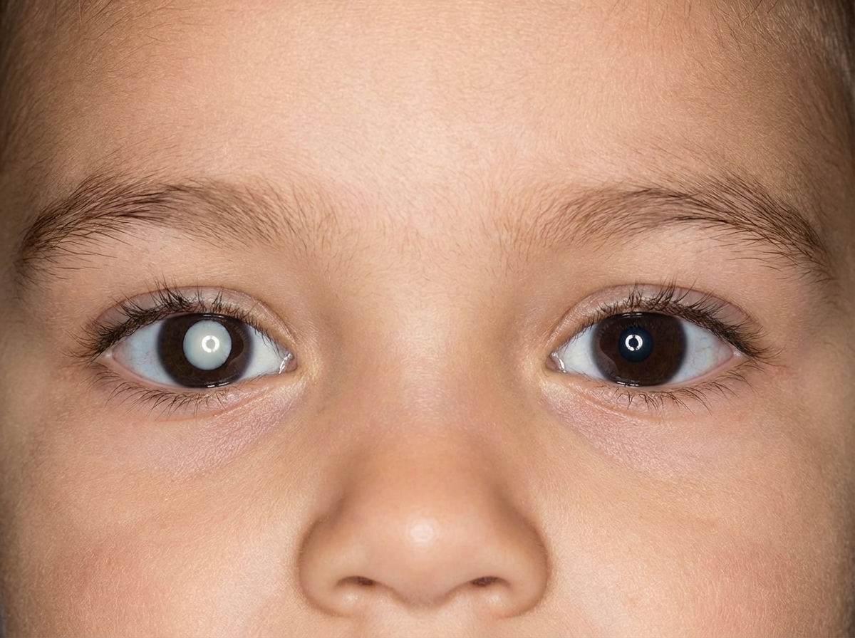
Amaurotic cat eye reflex is seen in which condition?
What is the preferred cycloplegic drug for a 1-year-old child?
Loss of heterozygosity is associated with which of the following conditions?
Duane syndrome involves dysfunction of which extraocular muscle?
Conjugate fixation reflex is established by the age of:
Extraretinal fibrovascular proliferation at the ridge between normal and avascular retina is characteristic of which grade of Retinopathy of Prematurity (ROP)?
A 2-year-old child presented with a specific abnormality, and there is a history of a similar illness in his father. What is the most likely underlying condition responsible?

Weakness of both adduction and abduction is seen in which condition?
Explanation: ### Explanation **1. Why the Correct Answer is Right:** In **paralytic squint**, there is a limitation of ocular movement due to the paralysis of one or more extraocular muscles. Because the eyes are no longer aligned (especially when looking in the direction of the paralyzed muscle), the images of an object fall on non-corresponding retinal points. This leads to **diplopia** (double vision), which is the hallmark clinical feature of recent-onset paralytic squint. To avoid this, patients often adopt a compensatory head posture. **2. Why the Incorrect Options are Wrong:** * **A. Congenital:** Paralytic squints are most commonly **acquired** (due to trauma, diabetes, hypertension, or intracranial tumors). While congenital nerve palsies exist, they are less common and often present as "concomitant" over time. * **B. Amblyopia is present:** Amblyopia ("lazy eye") typically occurs in children during the visual development period (under age 7-9) due to constant suppression of an image. In adult-onset paralytic squint, the brain cannot easily suppress the image, leading to diplopia rather than amblyopia. * **D. Secondary deviation is equal to primary deviation:** This is a characteristic of *non-paralytic (concomitant)* squint. In paralytic squint, **secondary deviation is always greater than primary deviation**. This occurs because of **Hering’s Law of Equal Innervation**: the increased effort required by the paralyzed eye to move results in an excessive nerve impulse being sent to the yoke muscle of the sound eye. **3. High-Yield Clinical Pearls for NEET-PG:** * **Primary Deviation:** Measured when the normal eye fixes. * **Secondary Deviation:** Measured when the paralyzed eye fixes. * **False Orientation (Past-pointing):** The patient points beyond the object in the direction of the paralyzed muscle. * **Bielschowsky’s Head Tilt Test:** Used specifically to diagnose **4th Nerve (Superior Oblique) palsy**. * **Hering’s Law:** Governs yoke muscles (e.g., Right LR and Left MR). * **Sherrington’s Law:** Governs reciprocal innervation of antagonist muscles in the *same* eye.
Explanation: **Explanation:** **Knudson’s "Two-Hit" Hypothesis** is the fundamental genetic principle used to explain the development of **Retinoblastoma**, the most common primary intraocular malignancy in children. According to this hypothesis, both alleles of the **RB1 gene** (a tumor suppressor gene located on chromosome **13q14**) must be inactivated for a tumor to develop: * **Hereditary Retinoblastoma (40%):** The "first hit" is a germline mutation (inherited), and the "second hit" is a somatic mutation. This typically leads to **bilateral**, multifocal tumors at an earlier age. * **Sporadic Retinoblastoma (60%):** Both "hits" occur as somatic mutations in a single retinal cell. This usually results in **unilateral**, unifocal tumors at a later age. **Why other options are incorrect:** * **Glaucoma:** While some forms (like Juvenile Open Angle Glaucoma) have genetic links (e.g., MYOC gene), they do not follow the specific two-hit kinetic model described by Knudson. * **Cataract:** Pediatric cataracts are often idiopathic, metabolic (Galactosemia), or associated with intrauterine infections (TORCH), rather than a specific two-hit tumor suppressor mutation. * **Melanoma:** Uveal melanoma is the most common primary intraocular tumor in adults. While it involves genetic mutations (e.g., GNAQ/GNA11), Knudson’s hypothesis is classically taught and defined in the context of Retinoblastoma. **High-Yield Clinical Pearls for NEET-PG:** * **Most common presenting sign:** Leukocoria (White pupillary reflex). * **Second most common sign:** Strabismus. * **Characteristic Calcification:** Retinoblastoma is known for "fine, gritty" calcification (chalky white appearance). * **Pathology:** Flexner-Wintersteiner rosettes are pathognomonic. * **Trilateral Retinoblastoma:** Bilateral retinoblastoma associated with a pinealoblastoma.
Explanation: **Explanation:** **Amaurotic Cat’s Eye Reflex**, clinically known as **Leukocoria** (white pupillary reflex), is the most common presenting sign of **Retinoblastoma**. In this condition, the normal red reflex is replaced by a whitish-yellow reflection caused by light bouncing off the vascularized, intraocular tumor mass (usually an endophytic growth) located behind the lens. **Analysis of Options:** * **Retinoblastoma (Correct):** It is the most common primary intraocular malignancy of childhood. Leukocoria is seen in approximately 60% of cases. Other common signs include strabismus and secondary glaucoma. * **Cri-du-chat Syndrome:** This is a chromosomal deletion (5p-) characterized by a high-pitched "cat-like" cry, microcephaly, and intellectual disability. It does not typically present with an abnormal pupillary reflex. * **Trachoma:** A chronic keratoconjunctivitis caused by *Chlamydia trachomatis*. It affects the lids (entropion/trichiasis) and cornea (pannus/scarring) but does not cause a white reflex from the posterior segment. * **Retinitis Pigmentosa:** A degenerative disease of the photoreceptors characterized by "bone-spicule" pigmentation in the mid-periphery and a constricted visual field. It does not produce leukocoria. **High-Yield Clinical Pearls for NEET-PG:** * **Differential Diagnosis of Leukocoria:** Retinoblastoma (most common), Congenital Cataract, Coats' Disease, Persistent Fetal Vasculature (PFV), and Retinopathy of Prematurity (ROP). * **Calcification:** Pathognomonic feature of Retinoblastoma on CT/Ultrasound (chalky white appearance). * **Flexner-Wintersteiner Rosettes:** The most characteristic histopathological finding in Retinoblastoma. * **Genetics:** Mutation in the **RB1 gene** on chromosome **13q14**. Knudson’s "Two-hit" hypothesis explains the hereditary vs. sporadic patterns.
Explanation: **Explanation:** In pediatric ophthalmology, the goal of cycloplegic refraction is to completely paralyze the powerful ciliary muscle (accommodation) to uncover the true refractive error, especially latent hyperopia. **Why Atropine (Eye Ointment) is the Correct Choice:** Atropine is the most potent cycloplegic agent available. In children under 5–7 years of age, the accommodative reflex is extremely strong, and weaker agents like Tropicamide often fail to provide complete cycloplegia. For a **1-year-old child**, **1% Atropine ointment** applied twice daily for three days prior to examination is the gold standard. The **ointment form** is preferred over drops in infants because it minimizes systemic absorption through the nasolacrimal duct, reducing the risk of systemic toxicity (tachycardia, flushing, fever). **Analysis of Incorrect Options:** * **Atropine (oral):** Atropine is administered topically for ophthalmic purposes. Oral administration is not used for refraction and would cause significant systemic side effects. * **Tropicamide:** This is a short-acting cycloplegic. While useful in adults, it is too weak to overcome the active accommodation of a 1-year-old, leading to an underestimation of hyperopia. * **Phenylephrine:** This is a pure sympathomimetic mydriatic (dilates the pupil) but has **no cycloplegic action** (does not paralyze the ciliary muscle). It is never used alone for refraction. **High-Yield Clinical Pearls for NEET-PG:** 1. **Drug of Choice by Age:** * < 5 years: Atropine (Ointment preferred). * 5–12 years: Homatropine or Cyclopentolate. * > 12 years/Adults: Tropicamide. 2. **Atropine Toxicity:** Look for the "Mad as a hatter, red as a beet, dry as a bone" mnemonic. The antidote is **Physostigmine**. 3. **Contraindication:** Atropine is strictly avoided in children with **Down Syndrome** due to an exaggerated hypertensive/tachycardic response.
Explanation: **Explanation:** **Retinoblastoma** is the classic example of the **"Knudson Two-Hit Hypothesis."** The disease is caused by a mutation in the **RB1 gene** (a tumor suppressor gene) located on chromosome **13q14**. * In the **hereditary form**, a child inherits one defective allele (the first "hit") in all somatic cells. * Tumorigenesis occurs when the remaining functional allele in a retinal cell undergoes a somatic mutation or deletion (the second "hit"). * This transition from a heterozygous state (one normal, one mutant allele) to a state where no functional allele remains is termed **Loss of Heterozygosity (LOH)**. **Analysis of Incorrect Options:** * **A & B (AML and ALL):** While these leukemias involve complex genetic alterations (like translocations or point mutations), they are not primarily defined by the LOH mechanism seen in tumor suppressor gene-driven pediatric malignancies like Retinoblastoma. * **D (APL):** Acute Promyelocytic Leukemia is characteristically associated with a specific **balanced reciprocal translocation t(15;17)**, involving the PML and RAR-alpha genes, rather than LOH. **High-Yield Clinical Pearls for NEET-PG:** * **Most common primary intraocular tumor** in children. * **Most common presenting sign:** Leukocoria (White pupillary reflex), followed by strabismus. * **Pathology:** Flexner-Wintersteiner rosettes (highly specific) and Homer-Wright rosettes. * **Calcification:** Dystrophic calcification within the tumor is a hallmark feature on CT scans. * **Trilateral Retinoblastoma:** Bilateral retinoblastoma associated with a pinealoma (pineoblastoma).
Explanation: **Explanation:** **Duane Retraction Syndrome (DRS)** is a congenital cranial dysinnervation disorder characterized by the **agenesis or hypoplasia of the Abducens nerve (CN VI) nucleus**. Because the Abducens nerve normally supplies the **Lateral Rectus (LR)**, this muscle is primarily affected. In DRS, the Lateral Rectus fails to receive its normal innervation. Instead, it is paradoxically innervated by branches from the **Oculomotor nerve (CN III)**. This leads to "co-contraction" of both the Medial Rectus and the Lateral Rectus during adduction, causing the classic clinical sign of **globe retraction** and narrowing of the palpebral fissure. **Analysis of Incorrect Options:** * **A & B (Superior/Inferior Oblique):** These muscles are involved in vertical and torsional movements. While "upshoots" or "downshoots" can occur in DRS due to the leash effect, the primary muscle dysfunction and the underlying neurogenic defect involve the LR. * **D (Superior Rectus):** This muscle is supplied by the Oculomotor nerve. While CN III is involved in the aberrant innervation of the LR in Duane syndrome, the Superior Rectus itself functions normally. **High-Yield Clinical Pearls for NEET-PG:** * **Huber’s Classification:** * **Type I (Most Common):** Limited abduction (most frequent). * **Type II:** Limited adduction. * **Type III:** Limited both abduction and adduction. * **Key Signs:** Globe retraction on adduction, narrowing of palpebral fissure, and compensatory head tilt. * **Association:** Often associated with **Goldenhar Syndrome**. * **Inheritance:** Mostly sporadic; if familial, it is usually Autosomal Dominant (DURS1/DURS2 genes).
Explanation: **Explanation:** The **Conjugate Fixation Reflex** is the ability of both eyes to move together synchronously to maintain focus on an object. In pediatric ophthalmology, the development of visual reflexes follows a specific chronological timeline as the macula and neurological pathways mature. * **Correct Answer (B - 6 weeks):** By the age of **6 weeks**, the conjugate fixation reflex is established. At this stage, a healthy infant should be able to follow a moving object (like a bright toy or the mother's face) with both eyes moving in unison. This is a critical milestone; failure to achieve this may indicate sensory deprivation or neurological delay. **Analysis of Incorrect Options:** * **A. 3 weeks:** At this age, fixation is rudimentary and inconsistent. The infant may momentarily look at a light source, but the coordinated conjugate movement is not yet established. * **C. 3 months:** By this age, **Binocular Single Vision (BSV)** and stereopsis begin to develop. While fixation is well-established by now, 6 weeks is the recognized onset for the reflex itself. * **D. 6 months:** By 6 months, the macula is fully developed, and the child achieves adult-like levels of accommodation and convergence. **High-Yield Clinical Pearls for NEET-PG:** * **Fixation Reflex:** Starts at birth, becomes steady by **6 weeks**, and is fully developed by **6 months**. * **Accommodation:** Reaches adult levels by **3–4 months**. * **Pseudo-strabismus:** Common in infants due to a wide epicanthal fold; it must be differentiated from true strabismus using the Hirschberg (corneal light reflex) test. * **Critical Period:** The most sensitive period for visual development (and risk of amblyopia) is from birth to **age 7–8 years**.
Explanation: **Explanation:** Retinopathy of Prematurity (ROP) is a vasoproliferative disorder of the retina in preterm infants. The International Classification of Retinopathy of Prematurity (ICROP) defines the severity of the disease through five distinct stages: * **Stage 3 (Correct Answer):** This stage is characterized by **extraretinal fibrovascular proliferation**. The hallmark is the growth of new, abnormal vessels (neovascularization) extending from the ridge into the vitreous. This is a critical stage as it indicates a high risk of progression to retinal detachment. **Analysis of Incorrect Options:** * **Stage 1:** Characterized by a thin, flat, white **demarcation line** separating the vascularized retina from the avascular retina. There is no height or thickness to this line. * **Stage 2:** The demarcation line progresses into a **ridge** that has height, width, and volume. However, the proliferation remains intraretinal (within the ridge). * **Stage 4:** Defined by **subtotal retinal detachment**. It is further divided into 4A (extra-foveal) and 4B (foveal involvement). Stage 5 represents total retinal detachment. **High-Yield Clinical Pearls for NEET-PG:** * **Screening Criteria (India):** Birth weight **<1750g** or Gestational Age **<34 weeks**, or infants with an unstable clinical course. * **Screening Timing:** First screening should be done at **4 weeks** post-natal age (or 3 weeks if born <28 weeks). * **Plus Disease:** Characterized by arterial tortuosity and venous dilation in the posterior pole; it indicates active, severe disease. * **Treatment:** Laser photocoagulation or Anti-VEGF (Ranibizumab/Bevacizumab) is typically indicated for "Type 1 ROP" (e.g., Stage 3 with Plus disease in Zone I or II).
Explanation: ***Retinoblastoma*** - The **hereditary pattern** (autosomal dominant inheritance of **RB1 gene** mutations) explains the similar illness in the father, making familial retinoblastoma highly likely. - Presents with **leukocoria** (white pupillary reflex/cat's eye reflex) as the classic sign in young children, typically before age 5. *Retinitis pigmentosa* - This condition causes **progressive night blindness** and **peripheral vision loss**, not the acute presentation described. - While it can be hereditary, it typically manifests later in childhood or adulthood with **bone spicule pigmentation** on fundoscopy. *Acute Leukemia* - Leukemia is primarily a **hematological malignancy** affecting blood cells, not typically causing specific ocular abnormalities. - The **hereditary pattern** mentioned strongly suggests a genetic condition rather than acquired malignancy. *Coloboma* - Coloboma is a **congenital structural defect** of the eye (iris, retina, or optic nerve) present from birth. - While it can be hereditary, it presents as a **keyhole-shaped pupil** or **visual field defects**, not the white reflex typical of retinoblastoma.
Explanation: **Explanation:** Duane’s Retraction Syndrome (DRS) is a congenital cranial dysinnervation disorder caused by the absence or hypoplasia of the abducens nerve (CN VI) and aberrant innervation of the lateral rectus (LR) muscle by the oculomotor nerve (CN III). **Why Option C is Correct:** Duane’s Retraction Syndrome is classified into three types based on Huber’s classification: * **Type 1 (Most common):** Limited **abduction** (due to CN VI palsy). * **Type 2 (Least common):** Limited **adduction** (due to LR being fired by CN III fibers during adduction, creating a mechanical block). * **Type 3:** Limited **both abduction and adduction**. This occurs because the lateral rectus and medial rectus muscles co-contract significantly, neutralizing movement in both directions. **Analysis of Incorrect Options:** * **Option A (Type 1):** Characterized by marked limitation of abduction with normal or near-normal adduction. * **Option B (Type 2):** Characterized by marked limitation of adduction with normal or near-normal abduction. * **Option C (Double Elevator Palsy):** This is a vertical gaze palsy affecting both the superior rectus and inferior oblique muscles. It limits elevation, not horizontal movements (abduction/adduction). **High-Yield Clinical Pearls for NEET-PG:** 1. **Hallmark Sign:** Globe retraction and narrowing of the palpebral fissure on attempted adduction (due to co-contraction of the recti). 2. **Epidemiology:** More common in females and usually affects the left eye. 3. **Associated Syndrome:** Wildervanck syndrome (DRS + Klippel-Feil anomaly + Deafness). 4. **Pathophysiology:** It is a myogenic/neurogenic paradox where the lateral rectus is "miswired."
Amblyopia
Practice Questions
Esotropia
Practice Questions
Exotropia
Practice Questions
Vertical Deviations
Practice Questions
Special Forms of Strabismus
Practice Questions
Nystagmus in Children
Practice Questions
Pediatric Cataract
Practice Questions
Retinopathy of Prematurity
Practice Questions
Pediatric Glaucoma
Practice Questions
Pediatric Neuro-ophthalmology
Practice Questions
Genetic Eye Diseases in Children
Practice Questions
Pediatric Ocular Trauma
Practice Questions
Get full access to all questions, explanations, and performance tracking.
Start For Free