What condition presents with a white pupillary reflex?
In concomitant squint, which of the following is true regarding primary and secondary deviations?
A 3-year-old child presents with suspected squint. What is the drug of choice for refraction in this case?
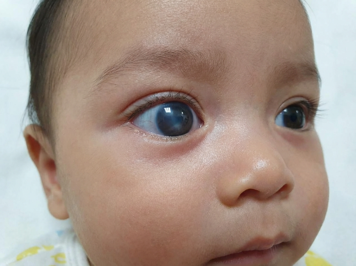
Comment on the diagnosis?

Which of the following drug classes is used in the management of accommodative esotropia?
Increased lactic dehydrogenase (LDH) in aqueous humor suggests which diagnosis?
Which childhood malignancy commonly presents with proptosis?
The Hirschberg test measures which of the following conditions?
Which pair of extraocular muscles is known as the 'yolk muscle pair'?
Blindness in a child is most commonly due to which of the following conditions?
Explanation: **Explanation:** The clinical term for a white pupillary reflex is **Leukocoria**. Normally, the pupil appears black, and a red reflex is seen on ophthalmoscopy due to the vascularity of the choroid. Leukocoria occurs when any intraocular pathology reflects light back through the pupil before it reaches the retina or when a white mass occupies the vitreous chamber. **Analysis of Options:** * **Retinoblastoma:** This is the most common primary intraocular malignancy in children and the most common cause of leukocoria. The white reflex is caused by the calcified tumor mass itself. * **Complete Retinal Detachment:** In conditions like Persistent Fetal Vasculature (PFV) or advanced Retinopathy of Prematurity (ROP), the detached retina bunches up behind the lens, creating a white retrolental membrane. * **Endophthalmitis:** Severe inflammation or infection leads to the accumulation of inflammatory debris and pus in the vitreous (vitreous abscess), which presents as a yellowish-white pupillary reflex. Since all three conditions can obstruct the normal red reflex and present with a white pupil, **Option D** is the correct answer. **High-Yield Clinical Pearls for NEET-PG:** * **Differential Diagnosis of Leukocoria:** Includes Congenital Cataract (most common cause overall), Retinoblastoma (most life-threatening), Coats’ disease, Toxocariasis, and ROP. * **Initial Investigation:** The first step in a child with leukocoria is a **Brückner test** (Red reflex test). * **Imaging:** B-scan Ultrasound is vital to look for **calcification**, which is a hallmark of Retinoblastoma (seen in 90% of cases). * **Management:** Any case of leukocoria in a child is a medical emergency until Retinoblastoma is ruled out.
Explanation: ### Explanation In ophthalmology, understanding the relationship between primary and secondary deviations is crucial for differentiating types of strabismus (squint). **1. Why the Correct Answer is Right (Primary = Secondary):** In **concomitant (non-paralytic) squint**, the angle of deviation remains constant regardless of which eye is fixing or the direction of gaze. This occurs because there is no paralysis or mechanical restriction of the extraocular muscles. According to **Hering’s Law of Equal Innervation**, the nerve impulse sent to the muscles of both eyes is equal. Since the muscle strength is normal in both eyes in concomitant squint, the amount of deviation measured when the normal eye fixes (**Primary Deviation**) is exactly equal to the deviation measured when the squinting eye fixes (**Secondary Deviation**). **2. Why Other Options are Incorrect:** * **Option B (Secondary > Primary):** This is the hallmark of **Incomitant (Paralytic) Squint**. In paralytic cases, when the paretic eye attempts to fixate, it requires a much stronger neural impulse to overcome the weakness. By Hering’s Law, this "excessive" innervation is also sent to the yoke muscle of the healthy eye, resulting in a larger secondary deviation. * **Option A (Primary > Secondary):** This scenario does not typically occur in clinical strabismus and is physiologically incorrect based on innervation laws. **3. Clinical Pearls for NEET-PG:** * **Hering’s Law:** Governs equal innervation to **yoke muscles** (e.g., Right Lateral Rectus and Left Medial Rectus). * **Sherrington’s Law:** Governs reciprocal innervation to **antagonistic muscles** of the *same* eye (e.g., as Right LR contracts, Right MR relaxes). * **Synoptophore:** The clinical instrument used to measure these deviations accurately. * **Key Differentiator:** If a question mentions "Secondary deviation > Primary deviation," immediately think of **Paralytic Squint** or **A/V patterns**.
Explanation: **Explanation:** In pediatric ophthalmology, the gold standard for refraction is **Cycloplegic Refraction**. Children have a very strong accommodative reflex due to a highly active ciliary muscle. To obtain an accurate refractive error (especially to uncover latent hyperopia in cases of accommodative esotropia), this accommodation must be completely paralyzed. **Why Atropine is the Correct Answer:** * **Mechanism:** Atropine is a potent muscarinic antagonist that provides the most powerful and prolonged cycloplegia (paralysis of the ciliary muscle). * **Indication:** It is the **drug of choice for children under 5–7 years of age**, particularly those with suspected strabismus (squint). In younger children, weaker agents often fail to overcome the strong ciliary tone, leading to an underestimation of hyperopia. * **Administration:** Usually applied as 1% ointment or drops twice daily for 3 days prior to the examination. **Why Other Options are Incorrect:** * **Pilocarpine:** This is a miotic (parasympathomimetic) used to constrict the pupil and stimulate accommodation. It is used in glaucoma, not for refraction. * **Tropicamide:** While it is a cycloplegic, it has the shortest duration and weakest action. It is the drug of choice for **adult fundus examination** but is insufficient for cycloplegic refraction in young children. * **Lidocaine:** This is a local anesthetic used for topical anesthesia (e.g., before tonometry or minor procedures); it has no effect on accommodation or pupil size. **High-Yield Clinical Pearls for NEET-PG:** * **Drug of choice by age:** * < 5-7 years: **Atropine** (1%) * 7–20 years: **Homatropine** or **Cyclopentolate** (Cyclopentolate is the DOC for school-aged children). * > 20 years: **Homatropine** or **Tropicamide**. * **Atropine Toxicity:** Look for signs like flushing, fever, tachycardia, and dry mouth ("Red as a beet, dry as a bone, hot as a hare, blind as a bat, and mad as a hatter"). * **Refractive Correction:** When using Atropine, a "deduction" of **1.00 Diopter** is typically made from the objective retinoscopy finding to account for the "tonal allowance" of the ciliary muscle.
Explanation: ***Buphthalmos*** - **Enlarged globe and cornea** in a child is pathognomonic of **congenital glaucoma**, causing the characteristic "ox-eye" appearance. - Results from **increased intraocular pressure** during early development when the sclera is still pliable and can stretch. *Retinoblastoma* - Typically presents with **leukocoria** (white pupillary reflex) rather than globe enlargement. - Most common **intraocular malignancy** in children but doesn't cause corneal or globe enlargement. *Esotropia* - Characterized by **inward deviation** of one or both eyes, not globe enlargement. - A form of **strabismus** with normal globe size and corneal dimensions. *Anterior uveitis* - Presents with **ciliary injection**, **photophobia**, and **decreased vision** without globe enlargement. - Inflammatory condition affecting the **iris and ciliary body**, not associated with corneal or scleral changes.
Explanation: **Explanation:** **Accommodative Esotropia** occurs due to an over-convergence of the eyes triggered by the effort to accommodate (usually to compensate for hyperopia). The management of choice is typically full cycloplegic refraction and corrective glasses. However, **Miotics** (Option A) serve as a pharmacological alternative or adjunct. **Why Miotics are Correct:** Miotics, such as **Echothiophate iodide** or **Pilocarpine**, act by stimulating the ciliary muscle directly. This produces "peripheral accommodation," which reduces the need for central accommodative effort (mediated by the 3rd cranial nerve). By decreasing the central drive to accommodate, the associated **accommodative convergence** is also reduced, thereby straightening the eyes. They are particularly useful in "High AC/A ratio" esotropia. **Why Other Options are Incorrect:** * **B. Mydriatics:** These drugs (like Atropine) paralyze accommodation (cycloplegia). While used for diagnosis (refraction), chronic use would blur near vision and potentially worsen the sensory adaptation to the squint. * **C. Steroids:** These are used for inflammatory conditions (uveitis, episcleritis) and have no role in correcting ocular alignment or innervation. * **D. Antibiotics:** These treat ocular infections and do not affect the extraocular muscles or the accommodative reflex. **High-Yield Clinical Pearls for NEET-PG:** * **Drug of Choice:** Long-acting cholinesterase inhibitors like **Echothiophate iodide (0.125%)** are the most common miotics used for this condition. * **Side Effect:** A classic exam-favorite side effect of long-term miotic use in children is the formation of **Iris Cysts** (at the pupillary margin). These can be prevented by co-administering **Phenylephrine (2.5%)**. * **Surgical Note:** Surgery is only indicated in accommodative esotropia if there is a "residual deviation" despite full hyperopic correction.
Explanation: **Explanation:** The correct answer is **Retinoblastoma**. **Why Retinoblastoma is correct:** Retinoblastoma is the most common primary intraocular malignancy of childhood. The tumor cells exhibit high glycolytic activity and rapid cell turnover, leading to the release of intracellular enzymes into the surrounding fluids. In cases of retinoblastoma, the **Aqueous LDH levels** are significantly elevated compared to serum levels. A ratio of **Aqueous LDH to Serum LDH > 1.0** is highly suggestive of retinoblastoma, serving as a useful biochemical marker when the diagnosis is clinically challenging. **Why the other options are incorrect:** * **Galactosemia:** This metabolic disorder is associated with "Oil droplet cataracts" due to the accumulation of dulcitol in the lens, but it does not typically alter aqueous LDH levels. * **Glaucoma:** While intraocular pressure is elevated, there is no specific correlation with increased aqueous LDH. * **Gyrate atrophy:** This is an autosomal recessive chorioretinal degeneration caused by a deficiency of the enzyme ornithine aminotransferase. It is characterized by high **serum ornithine** levels, not LDH. **High-Yield Clinical Pearls for NEET-PG:** * **Most common sign:** Leukocoria (60%), followed by Strabismus (20%). * **Pathognomonic Histology:** Flexner-Wintersteiner rosettes (true rosettes with a central lumen). * **Calcification:** Dystrophic calcification is a hallmark feature seen on B-scan USG or CT scan (helpful for differentiating from Coats' disease). * **Genetics:** Mutation in the **RB1 gene** on Chromosome **13q14**. * **Aqueous LDH Ratio:** If >1.5, it is considered strongly diagnostic for Retinoblastoma.
Explanation: **Explanation:** Proptosis (protrusion of the eyeball) in children is a clinical emergency that often signals an underlying malignancy. The correct answer is **All of the above** because each of these conditions is a well-recognized cause of pediatric orbital masses. * **Neuroblastoma:** This is the most common extracranial solid tumor of childhood. It frequently metastasizes to the orbit (specifically the orbital bones). A classic NEET-PG presentation is **"Raccoon eyes"** (periorbital ecchymosis) accompanied by rapid-onset, often bilateral, proptosis. * **Chloroma (Granulocytic Sarcoma):** This is a localized collection of primitive granulocytic cells. It is a common extramedullary manifestation of **Acute Myeloid Leukemia (AML)**. It often presents as a rapidly progressing, painless proptosis in a child, sometimes preceding systemic blood involvement. * **Optic Nerve Glioma:** This is a slow-growing benign tumor (usually a juvenile pilocytic astrocytoma) that causes **axial proptosis** and early vision loss. It has a strong association with **Neurofibromatosis Type 1 (NF-1)**. **Clinical Pearls for NEET-PG:** * **Most common primary malignant orbital tumor in children:** Rhabdomyosarcoma (presents with sudden, explosive proptosis). * **Most common benign orbital tumor in children:** Capillary Hemangioma. * **Most common cause of both bilateral and unilateral proptosis in children:** Orbital Cellulitis (inflammatory, not neoplastic). * **Differential Diagnosis Tip:** If proptosis is pulsatile, consider orbital encephalocoele or NF-1 (due to sphenoid wing dysplasia).
Explanation: **Explanation:** The **Hirschberg test**, also known as the **Corneal Light Reflex test**, is a simple, non-invasive clinical method used to estimate the presence and magnitude of **strabismus** (ocular misalignment). **Why Strabismus is the Correct Answer:** The test is performed by shining a light source (penlight) into the patient's eyes from a distance of about 33 cm while the patient fixates on the light. The clinician observes the position of the light reflection (reflex) on the cornea relative to the pupil: * **Normal (Orthophoria):** The reflex is centered or slightly nasal in both pupils. * **Abnormal:** If the reflex is displaced, it indicates a deviation. For every **1 mm** of displacement from the center, there is approximately **7 degrees (or 15 prism diopters)** of ocular deviation. * *Temporal displacement* indicates Esotropia (inward turn). * *Nasal displacement* indicates Exotropia (outward turn). **Why Other Options are Incorrect:** * **Cataract:** Diagnosed via slit-lamp biomicroscopy or distant direct ophthalmoscopy (looking for a red reflex defect). * **Refractive Error:** Measured using retinoscopy or automated refractometry. * **Glaucoma:** Assessed via tonometry (IOP), gonioscopy (angle), and fundoscopy (optic disc cupping). **High-Yield Clinical Pearls for NEET-PG:** * **Krimsky Test:** A modification of the Hirschberg test where prisms are used to center the displaced reflex to quantify the deviation more accurately. * **Pseudo-strabismus:** A common "distractor" in exams; a wide epicanthal fold may make a child look cross-eyed, but a **normal Hirschberg test** (central reflex) confirms the eyes are actually aligned. * **Bruckner Test:** Uses a direct ophthalmoscope to view red reflexes simultaneously; asymmetry suggests strabismus or anisometropia.
Explanation: ### Explanation The concept of **Yoke Muscles** (often referred to as 'yolk' in some texts) is based on **Hering’s Law of Equal Innervation**. This law states that during any conjugate eye movement, equal and simultaneous innervation is sent to the muscles of both eyes that are responsible for moving the eyes in a specific direction. **1. Why the Correct Answer is Right:** * **Option B (Right Medial Rectus & Left Lateral Rectus):** These are yoke muscles for **left gaze** (levoversion). When looking to the left, the Left Lateral Rectus (LLR) contracts to abduct the left eye, and the Right Medial Rectus (RMR) contracts to adduct the right eye. Because they work together to achieve a single direction of gaze, they are considered a yoke pair. **2. Analysis of Incorrect Options:** * **Option A:** These are **antagonists** in the same eye (ipsilateral). When one contracts, the other must relax (**Sherrington’s Law**). * **Option C:** These are not a yoke pair. The yoke muscle for the Right Superior Oblique (RSO) is the **Left Inferior Rectus** (LIR), as both are the primary muscles for **gaze down and to the left**. * **Option D:** While both move the eyes upward, they are not the primary yoke pair for a specific cardinal position of gaze. The yoke muscle for the Right Superior Rectus (RSR) is the **Left Inferior Oblique** (LIO) for **gaze up and to the right**. **3. High-Yield Facts for NEET-PG:** * **Hering’s Law:** Applies to **yoke muscles** (bilateral/binocular). * **Sherrington’s Law:** Applies to **antagonist muscles** (unilateral/monocular). * **Clinical Application:** Hering’s law explains "secondary deviation" in paralytic squint, where the deviation is greater when the paralyzed eye fixes, due to excessive innervation sent to the yoke muscle of the sound eye. * **Cardinal Positions of Gaze:** There are 6 cardinal positions, each associated with a specific yoke muscle pair.
Explanation: **Explanation:** **Keratomalacia (Option A)** is the correct answer because Vitamin A deficiency remains the leading cause of preventable childhood blindness worldwide, particularly in developing countries like India. Keratomalacia represents the most advanced stage of Xerophthalmia (X3B), characterized by rapid liquefactive necrosis of the cornea. This leads to perforation, endophthalmitis, or phthisis bulbi, resulting in irreversible bilateral blindness. **Analysis of Incorrect Options:** * **Congenital Cataract (Option B):** While it is the most common cause of *treatable* or *surgical* blindness in children, it ranks second to Vitamin A deficiency in overall prevalence in the Indian subcontinent. * **Glaucoma (Option C):** Congenital glaucoma is a significant cause of visual impairment but is statistically less common than nutritional deficiencies or cataracts. * **Injuries (Option D):** Ocular trauma is a major cause of *unilateral* blindness in children but does not surpass the systemic impact of nutritional blindness on a population level. **High-Yield Clinical Pearls for NEET-PG:** * **WHO Classification of Xerophthalmia:** Remember the sequence: X1A (Conjunctival xerosis) → X1B (Bitot’s spots) → X2 (Corneal xerosis) → X3A (Ulceration <1/3 cornea) → X3B (Keratomalacia >1/3 cornea). * **First Sign:** Night blindness (Nyctalopia/XN) is the earliest clinical symptom. * **First Objective Sign:** Conjunctival xerosis. * **Treatment:** The WHO schedule for Vitamin A (2 lakh IU on days 0, 1, and 14) is a frequent exam favorite. * **Global Context:** In developed nations, retinopathy of prematurity (ROP) and genetic conditions are more common causes, but for NEET-PG (Indian context), Keratomalacia remains the top choice.
Amblyopia
Practice Questions
Esotropia
Practice Questions
Exotropia
Practice Questions
Vertical Deviations
Practice Questions
Special Forms of Strabismus
Practice Questions
Nystagmus in Children
Practice Questions
Pediatric Cataract
Practice Questions
Retinopathy of Prematurity
Practice Questions
Pediatric Glaucoma
Practice Questions
Pediatric Neuro-ophthalmology
Practice Questions
Genetic Eye Diseases in Children
Practice Questions
Pediatric Ocular Trauma
Practice Questions
Get full access to all questions, explanations, and performance tracking.
Start For Free